. 2021 Oct 7;28(10):1758-1774.e8.
doi: 10.1016/j.stem.2021.07.001. Epub 2021 Jul 27.

Establishment, maintenance, and recall of inflammatory memory

Affiliations

Establishment, maintenance, and recall of inflammatory memory

Samantha B Larsen et al. Cell Stem Cell. .

Abstract

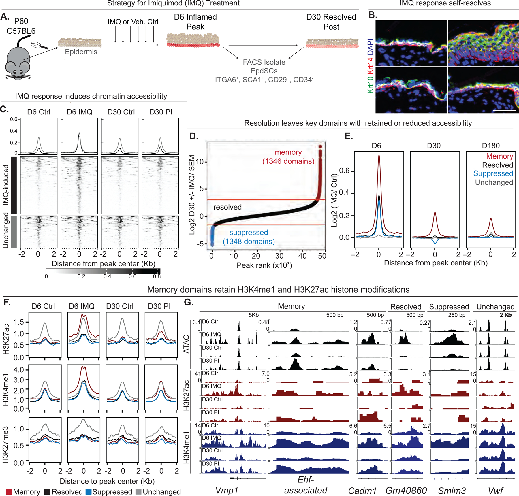
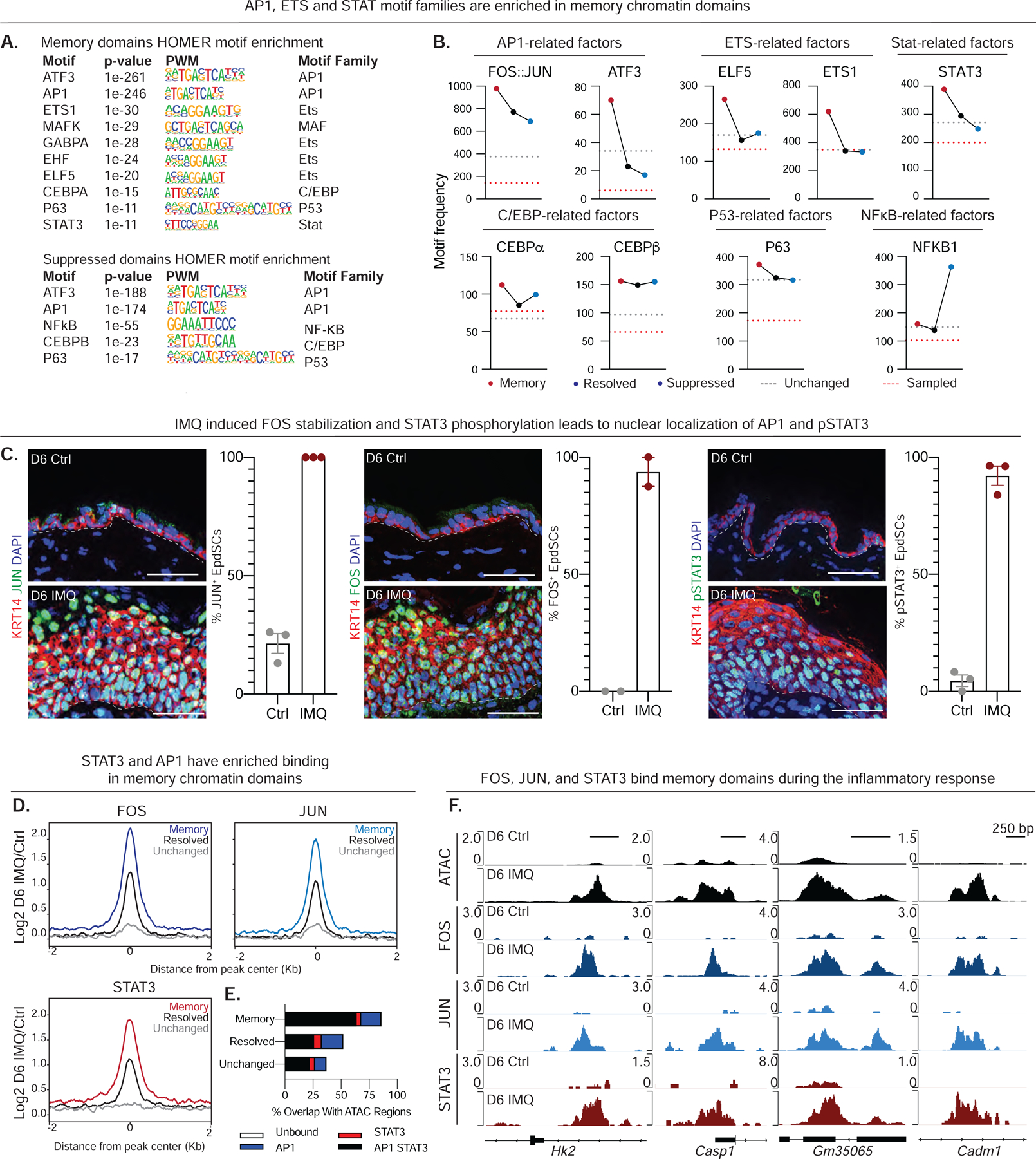
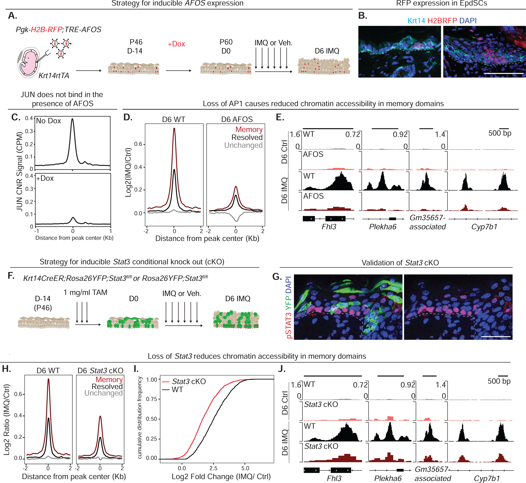
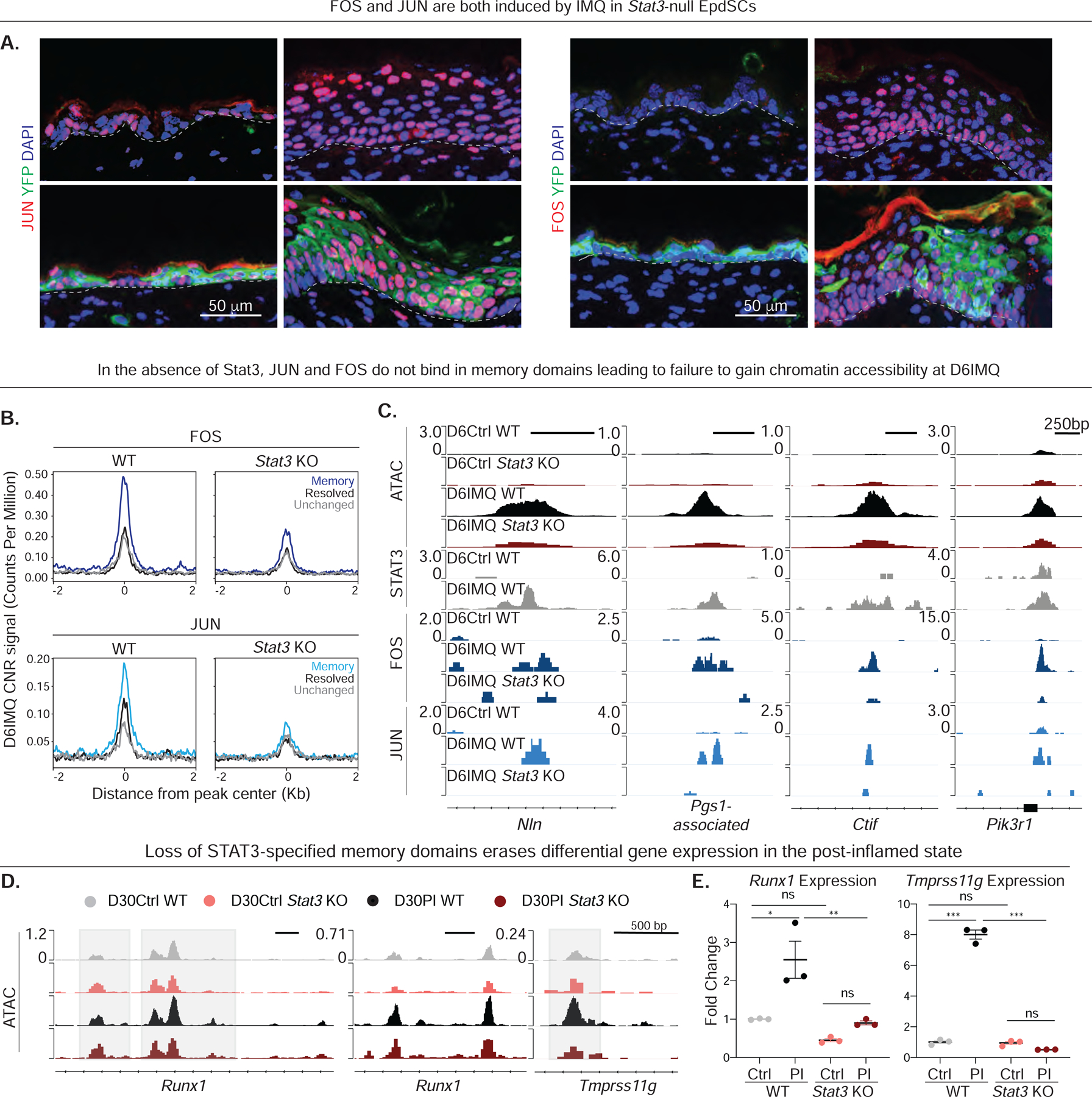
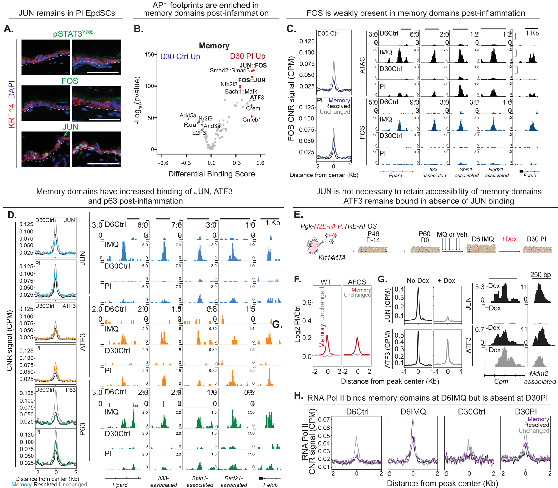
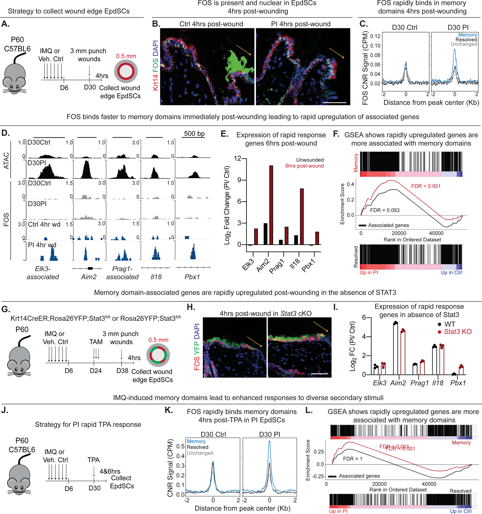
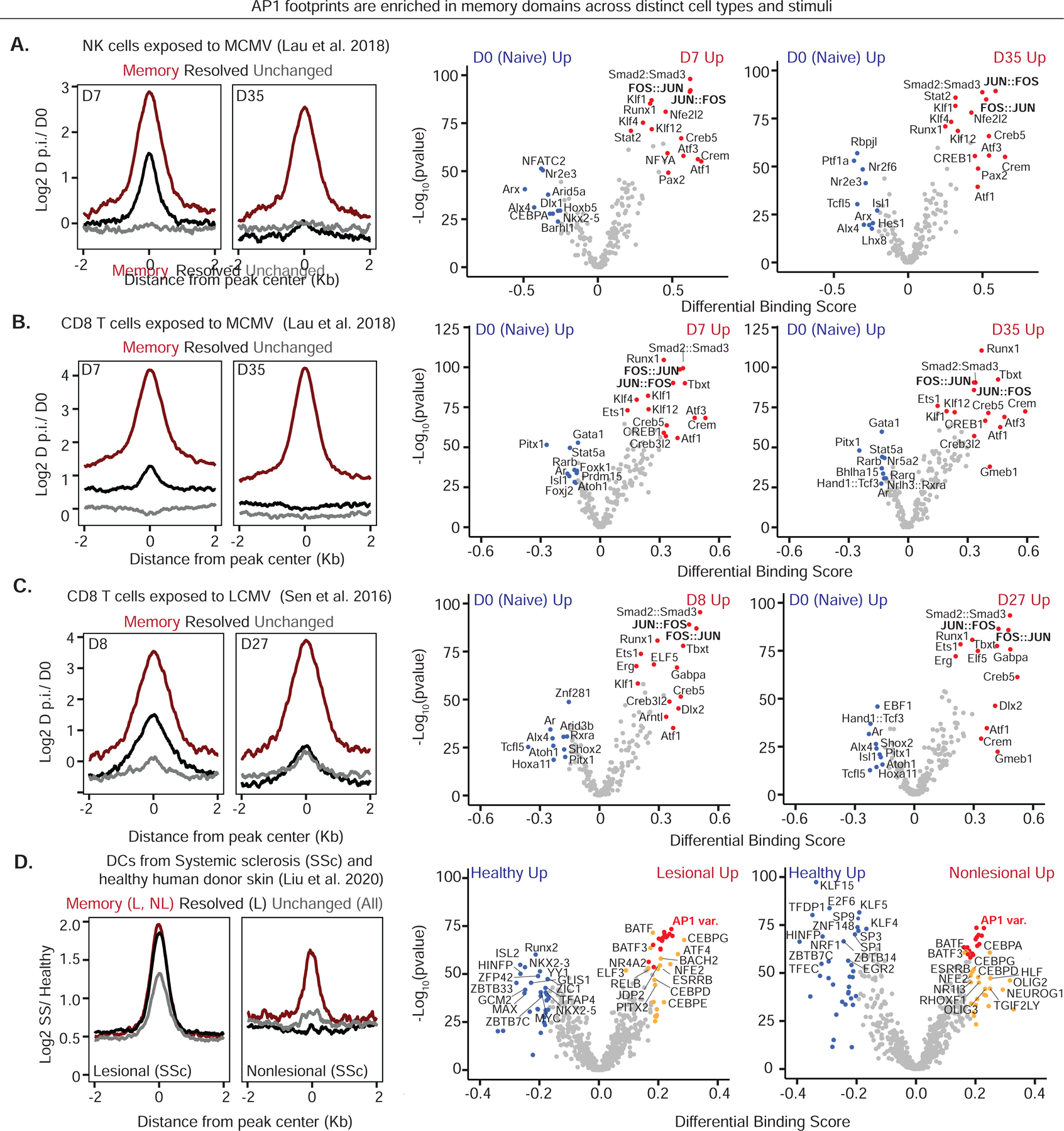
Known for nearly a century but through mechanisms that remain elusive, cells retain a memory of inflammation that equips them to react quickly and broadly to diverse secondary stimuli. Using murine epidermal stem cells as a model, we elucidate how cells establish, maintain, and recall inflammatory memory. Specifically, we landscape and functionally interrogate temporal, dynamic changes to chromatin accessibility, histone modifications, and transcription factor binding that occur during inflammation, post-resolution, and in memory recall following injury. We unearth an essential, unifying role for the general stress-responsive transcription factor FOS, which partners with JUN and cooperates with stimulus-specific STAT3 to establish memory; JUN then remains with other homeostatic factors on memory domains, facilitating rapid FOS re-recruitment and gene re-activation upon diverse secondary challenges. Extending our findings, we offer a comprehensive, potentially universal mechanism behind inflammatory memory and less discriminate recall phenomena with profound implications for tissue fitness in health and disease.

Keywords: AP1 transcription factors; ATAC sequencing; CUT&RUN; ChIP sequencing; FOS; FOS:JUN; STAT3; broadened immune protection; epigenetic memory; histone modifications; inflammation; inflammatory disorders; inflammatory memory; tissue stem cells; trained immunity.


Conflict of interest statement

Declaration of interests The authors declare no competing financial interests. E.F. serves as an advisory board member for Cell Stem Cell and serves on the scientific advisory boards of L’Oreal and Arsenal Biosciences.

Figures

Figure 1.
Figure 1.. Memory domains acquire chromatin accessibility and histone modifications during inflammation and retain them post-inflammation.
(A) Schematic of strategy to induce psoriatic-like inflammation. (B) Imiquimod (IMQ) induces epidermal hyperthickening of Keratin 14 (Krt14) and Krt10 layers. Ctrl, naïve skin control; PI, post-inflammation skin; D, day; DAPI, 4′, 6-diamidino-2-phenylindole. (C) Heatmap of ATAC-Seq signal intensity over IMQ-induced or unchanged (IMQ-insensitive) EpdSC chromatin peaks. n = 2–3 mice pooled per replicate, 4 replicates per group [together with (Naik et al., 2017)]. Kb, kilobase pairs. (D) Elbow plot of IMQ-induced chromatin regions ranked by their D30 Wald statistic (Log2 Fold Change/SEM). SEM, Standard error of the mean. (E) Line plots depicting Log2 Ratio of IMQ vs Ctrl ATAC-Seq signal intensity of chromatin domains at D6, D30 and D180 post-inflammation. (F) Line plots of ChIP-seq signals over different chromatin domains as indicated. n = 2–3 mice pooled per replicate, 4 replicates per group (see also Figure S1G). (G) Representative Integrative Genomics Viewer (IGV) images of genomic loci with ATAC, H3K27ac and H3K4me1 signals. Genes associated with these chromatin regions are as indicated. Genomic regions listed in Supplemental Table 1. See also Figures S1 and S2.
Figure 2.
Figure 2.. FOS-JUN and STAT3 bind preferentially to memory domains during the inflammatory response
(A) HOMER known motif analysis of IMQ-sensitive memory and suppressed chromatin domains post-inflammation. PWM, position weight matrix. (B) Frequencies of JASPAR TF motifs within memory, resolved and suppressed regions over randomly sampled genomic background and IMQ-unchanged regions. (C) Immunofluorescence images of JUN, FOS and pSTAT3Y705 in naïve (control, Ctrl) and IMQ-inflamed skin (n=2–3). Quantifications are for immunolabeled nuclei within the basal layer of EpdSCs. Grey dotted line denotes epidermal-dermal border. (D) Log2 ratio of IMQ over Ctrl CUT&RUN signals for FOS, JUN, and STAT3 binding specifically to memory, resolved or unchanged chromatin regions. n = 2–3 mice pooled per replicate, 2 replicates per group. (E) Percentages of chromatin domains that are physically bound by AP1 and/or STAT3. (F) IGV images showing ATAC peaks along with FOS, JUN and STAT3 CUT&RUN binding profiles of representative D6 memory domains of chromatin isolated from FACS-purified Ctrl and IMQ-inflamed EpdSCs. Genomic regions listed in Supplemental Table 1. See also Figure S3.
Figure 3.
Figure 3.. AP1 and STAT3 are necessary to establish inflammatory memory
(A) Schematic for inducible expression of dominant negative FOS (AFOS) in EpdSCs. TRE, tetracycline response element; RFP, red fluorescent protein; Dox, doxycycline. D, day post IMQ treatment; P, postnatal day; Veh, vehicle control. (B) RFP immunofluorescence indicative of successful lentiviral transduction of EpdSCs. Grey dotted line denotes epidermal-dermal interface. (C) JUN CUT&RUN signal in stably transduced TRE-AFOS keratinocytes with and without doxycycline treatment. CPM, counts per million. (D) Log2 Ratio of D6 IMQ over Ctrl EpdSC ATAC-Seq signal over chromatin domains in TRE-AFOS and WT mice. n=3–4 mice pooled per replicate, 2 replicates per group. (E) IGV images of genomic loci of ATAC signal from TRE-AFOS and WT mice in Ctrl and inflamed EpdSCs. Genomic regions listed in Supplemental Table 1. (F) Schematic for CreER inducible Stat3 conditional ablation specific in EpdSCs. TAM, 4-hydroxytamoxifen. (G) pSTAT3Y705 and YFP are mutually exclusive in TAM treated mosaic skin. Grey dotted line denotes epidermal-dermal border. (H) Log2 Ratio (IMQ vs naïve) of ATAC-Seq signals in chromatin regions from Stat3 null and WT EpdSCs purified from D6 IMQ inflamed and D6 naïve (Ctrl) skins. n = 2–3 mice per replicate, 2 replicates per group. (I) Cumulative distribution frequencies show a loss of chromatin accessibility in memory domains of Stat3 null relative to WT EpdSCs. (J) IGV images of genomic loci of ATAC signals from representative memory domains in chromatin of Stat3 null and WT EpdSCs FACS-purified from skins of Stat3 cKO and WT mice ± IMQ. See also Figures S4 and S5.
Figure 4.
Figure 4.. STAT3 is required for JUN and FOS accessibility, binding and establishment of memory domains
(A) Immunofluorescence images of JUN and FOS in WT and Stat3 cKO skins. YFP, yellow fluorescent protein; WT, Rosa26YFP;Stat3fl/fl TAM-treated. Grey dotted lines denote the epidermal-dermal border. (B) CUT&RUN signals for FOS and JUN binding to WT and Stat3-null EpdSC chromatin from D6 IMQ skins. n = 2–3 mice pooled per replicate, 2 replicates per group. (C) IGV images of representative genomic loci, which in D6 WT EpdSCs show IMQ-induced ATAC peaks accompanied by STAT3, FOS and JUN binding to memory domains. (D, E) IGV images and qPCR analysis. (D) IGV images of ATAC signals over D30 memory domains in the chromatin of WT and Stat3 null EpdSCs. Grey boxes denote memory domains that lost significant accessibility at D30 in the Stat3 KO relative to WT: Runx1 region 1 p-adj = 0.00373; Runx1 region 2 p-adj = 3.26e-6; for Tmprss11g p-adj = 0.01833. Data are similar to (C) in that STAT3 loss results in diminished accessibility of memory domains. (E) qPCR reveals that without memory domains, the differential transcription of memory-associated genes is abolished. Statistics were calculated using DESeq2. See also Figure S6.
Figure 5.
Figure 5.. JUN, ATF3 and P63, but not FOS, remain bound in memory domains following resolution of inflammation
(A) Immunofluorescence images of pSTAT3Y705, FOS and JUN in D30 Ctrl and PI skin. Grey dotted lines denote epidermal-dermal borders. (B) TOBIAS-predicted enriched TF footprints in memory domains post-inflammation. (C) FOS CUT&RUN signals (left) and IGV images (right) over chromatin domains in D30 Ctrl and PI EpdSCs showing that AP1 sites within memory domains remain open in the post-inflamed state, even though FOS is absent. n = 2–3 mice pooled per replicate, 2 replicates per group. (D) JUN, ATF3, and p63 CUT&RUN signals (left) and IGV images (right) at memory vs resolved and unchanged domains in D6 and D30 EpdSCs from Ctrl and IMQ-treated skins (see Figure 2). (E) Schematic for inducing expression of dominant negative FOS (AFOS) specifically in EpdSCs of post-inflamed skin. TRE, tetracycline response element; RFP, red fluorescent protein; Dox, doxycycline. D, day post IMQ treatment; P, postnatal day; Veh, vehicle control. (F,G) Log2 Ratio of PI/ Ctrl ATAC signal over memory and unchanged regions in WT and AFOS EpdSCs (F) and JUN and ATF3 CUT&RUN (G). Log2Ratio of CUT&RUN signals and representative genomic loci illustrate that ATF3 is not affected by AFOS and remains bound to open memory domains in JUN’s absence. For (F) n = 3–4 mice pooled per replicate, 2 replicates per group, and (G) from stably transduced Tre-AFOS keratinocytes. (H) Pol II CUT&RUN signals in memory vs resolved and unchanged regions in D6 and D30 EpdSCs from Ctrl and IMQ-treated skin. n = 2–3 mice pooled per replicate. D6 – 1 replicate, D30 – 2 replicates. See also Figure S6.
Figure 6.
Figure 6.. FOS is rapidly induced and binds inflammation-experienced memory domains whose associated genes become transcribed within minutes after a secondary challenge
(A) Schematic of wounding experiment performed on resolved skin 30D post-IMQ treatment. (B) Immunofluorescence images of FOS in D30 Ctrl and PI skin 4 hours after wounding. w.b, wound bed; asterisk denotes autofluorescence. Grey dotted lines represent the epidermal-dermal interface. Yellow arrow denotes direction of wound bed. (C) FOS CUT&RUN signal over chromatin domains in D30 Ctrl and PI wound-edge EpdSCs taken from skin 4 hours after injury. n = 2–3 mice per replicate, 2 replicates per group. CNR, CUT&RUN; CPM, counts per million. (D) IGV images of ATAC-seq signal and FOS TF binding profiles over representative memory domains associated with wound-induced rapid response genes. Shown are data relative to Ctrl. (E) Log2 fold change of PI over Ctrl transcripts of the same rapid response genes as in (D), analyzed 6 hours after wounding. (F) Gene set enrichment analysis (GSEA) of memory-associated (top, red) or resolved-associated (bottom, black) genes along the continuum of genes differentially expressed in PI and Ctrl EpdSCs 6 hours post-wounding. (G) Schematic of Stat3 ablation after resolution of IMQ pathology and prior to wounding and collection of wound-edge EpdSCs. (H) Immunofluorescence images reveals FOS induction in Stat3 null EpdSCs at 4 hr after wounding of skins which were either naïve or IMQ-experienced prior to Stat3 conditional targeting. Yellow arrow denotes direction of wound bed (w.b.). (I) qPCR of memory-associated genes from wound-edge EpdSCs purified from IMQ-experienced skins, treated at D24 with tamoxifen and wounded 4 hr prior to analysis. WT, Rosa26YFP;Stat3fl/fl TAM treated. (J) Schematic of TPA treatment of D30 IMQ-experienced and naïve skin and collection of EpdSCs 4–6hr later. (K) FOS CUT&RUN signal over chromatin domains in D30 Ctrl and IMQ-experienced (PI) EpdSCs taken from skin 4 hours after TPA treatment. n = 2 mice per replicate, 2 replicates per group. (L) GSEA of memory-associated (top) or resolved-associated (bottom) genes along the continuum of genes differentially expressed in PI and Ctrl EpdSCs 6 hours after TPA treatment. See also Figure S6.
Figure 7.
Figure 7.. FOS-JUN as a potential universal mediator of inflammatory memory and recall
All analyses were performed on existing databases as indicated. Left, Line plots depicting Log2 Ratios of inflammation-exposed versus naïve ATAC-seq signals over chromatin domains. Kb, kilobase pairs. D, day; p.i. post-infection. Right, TOBIAS-predicted enriched TF footprints specific to memory domains during or following resolution of inflammation. In all cases, TOBIAS predicts that FOS-JUN play a specific role in memory domains in inflammation-experienced cells relative to their naïve counterparts. (A) Chromatin of MCMV-exposed versus naïve NK cells analyzed at D0, D7 and D35 post-infection. (B) Chromatin of MCMV-exposed versus naïve CD8+ T lymphocytes at D0, D7 and D35 post-infection. (C) Chromatin of LCMV-exposed versus naïve CD8+ T cells at D0, D8 and D27 post-infection. (D) Chromatin of human dendritic cells (DCs) isolated from lesional (L) and nonlesional (NL) skin from systemic sclerosis (SSc) patients and compared to normal skin from healthy volunteers. We consider the peak of inflammation as L skin, inflammation-experienced as NL skin and ‘memory’ domains as those common between L and NL of DCs compared to skin from healthy volunteers. All AP1 variants are shown as red dots with other enriched motifs in yellow.

Comment in

Similar articles

Cited by

References

    1. Adam RC, Yang H, Ge Y, Infarinato NR, Gur-Cohen S, Miao Y, Wang P, Zhao Y, Lu CP, Kim JE, et al. (2020). NFI transcription factors provide chromatin access to maintain stem cell identity while preventing unintended lineage fate choices. Nat Cell Biol 22, 640–650. 10.1038/s41556-020-0513-0. - DOI - PMC - PubMed
    1. Adam RC, Yang H, Ge Y, Lien WH, Wang P, Zhao Y, Polak L, Levorse J, Baksh SC, Zheng D, and Fuchs E (2018). Temporal Layering of Signaling Effectors Drives Chromatin Remodeling during Hair Follicle Stem Cell Lineage Progression. Cell Stem Cell 22, 398–413.e397. 10.1016/j.stem.2017.12.004. - DOI - PMC - PubMed
    1. Adam RC, Yang H, Rockowitz S, Larsen SB, Nikolova M, Oristian DS, Polak L, Kadaja M, Asare A, Zheng D, and Fuchs E (2015). Pioneer factors govern super-enhancer dynamics in stem cell plasticity and lineage choice. Nature 521, 366–370. papers3://publication/doi/10.1038/nature14289. - PMC - PubMed
    1. Ahn S, Olive M, Aggarwal S, Krylov D, Ginty DD, and Vinson C (1998). A dominant-negative inhibitor of CREB reveals that it is a general mediator of stimulus-dependent transcription of c-fos. Mol Cell Biol 18, 967–977. 10.1128/mcb.18.2.967. - DOI - PMC - PubMed
    1. Angel P, Szabowski A, and Schorpp-Kistner M (2001). Function and regulation of AP-1 subunits in skin physiology and pathology. Oncogene 20, 2413–2423. 10.1038/sj.onc.1204380. - DOI - PubMed

Publication types