. 2021 Aug 4;143(30):11435-11448.
doi: 10.1021/jacs.1c03270. Epub 2021 Jul 26.

Nonrefoldability is Pervasive Across the E. coli Proteome

Affiliations

Nonrefoldability is Pervasive Across the E. coli Proteome

Philip To et al. J Am Chem Soc. .

Abstract

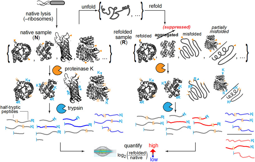
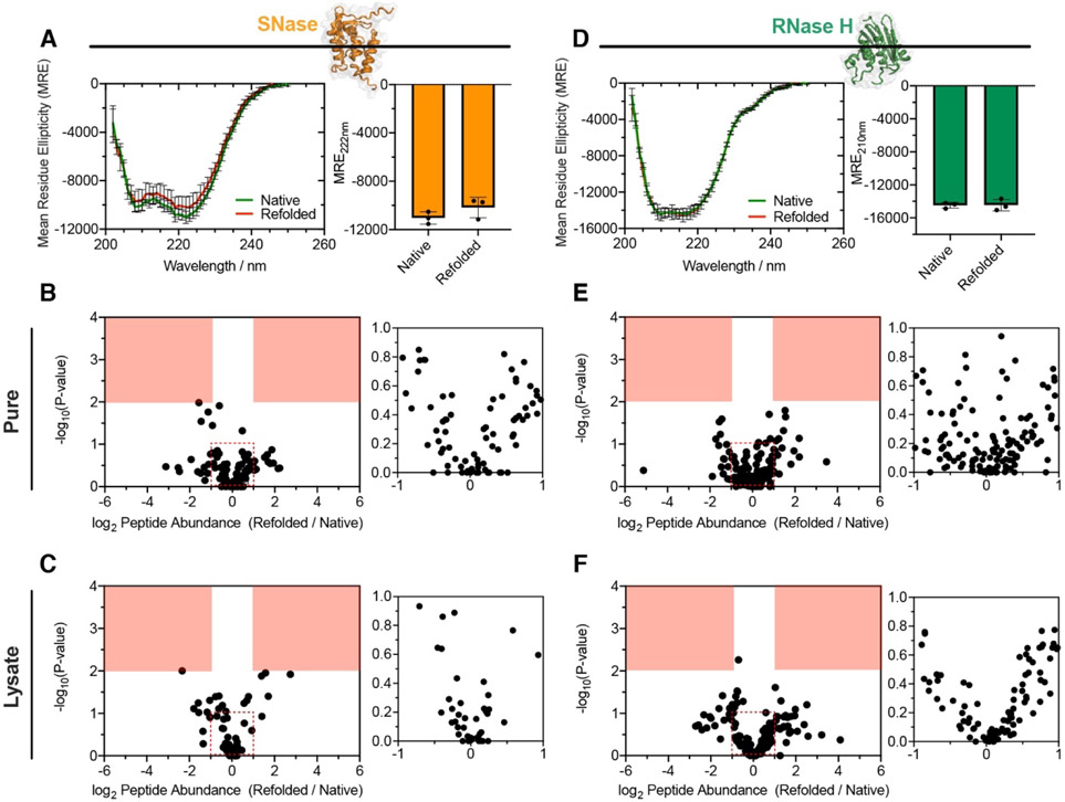
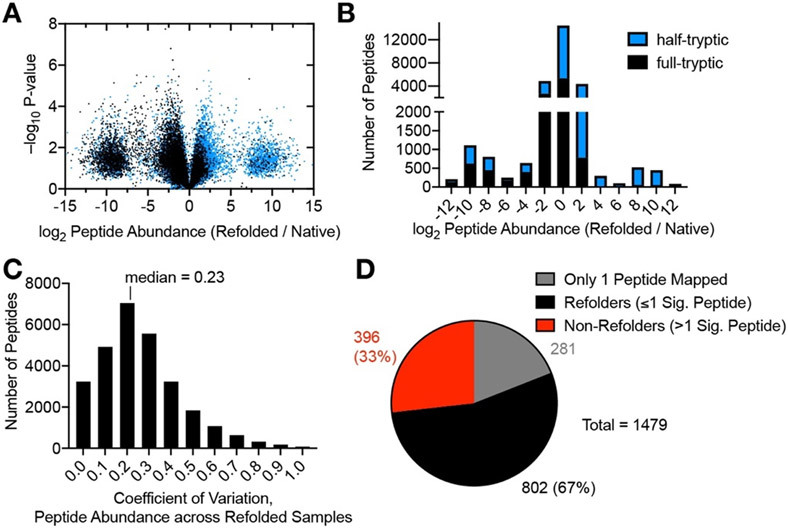
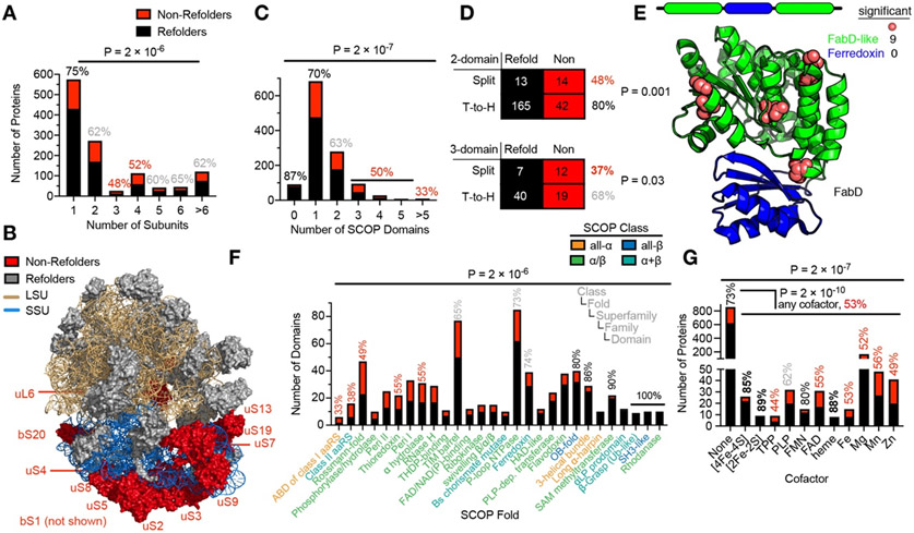
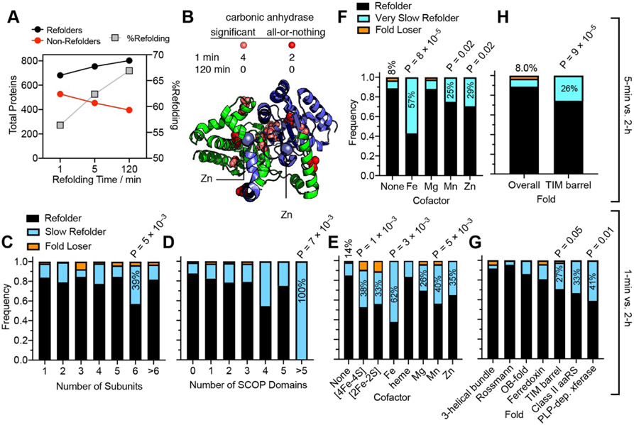
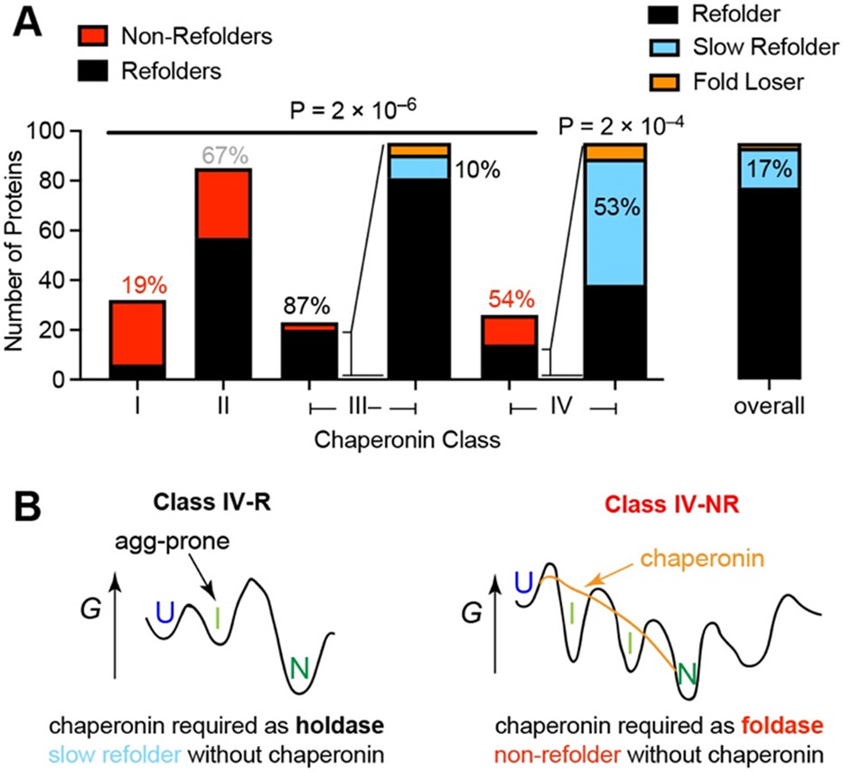
Decades of research on protein folding have primarily focused on a subset of small proteins that can reversibly refold from a denatured state. However, these studies have generally not been representative of the complexity of natural proteomes, which consist of many proteins with complex architectures and domain organizations. Here, we introduce an experimental approach to probe protein refolding kinetics for whole proteomes using mass spectrometry-based proteomics. Our study covers the majority of the soluble E. coli proteome expressed during log-phase growth, and among this group, we find that one-third of the E. coli proteome is not intrinsically refoldable on physiological time scales, a cohort that is enriched with certain fold-types, domain organizations, and other biophysical features. We also identify several properties and fold-types that are correlated with slow refolding on the minute time scale. Hence, these results illuminate when exogenous factors and processes, such as chaperones or cotranslational folding, might be required for efficient protein folding.


Figures

Fig. 1.
Fig. 1.. Limited Proteolysis Mass Spectrometry (LiP-MS) to Interrogate Refolding of the Proteome.
Lysates from E. coli are prepared under native conditions, globally unfolded and refolded. The structures of the refolded proteins are probed by pulse proteolysis with proteinase K (PK) and compared to that of their native forms. Label free quantification (LFQ) of half-tryptic peptides reveals sites across the proteome where local conformation differs between a protein’s native and refolded forms.
Fig. 2.
Fig. 2.. Refolding of Small Model Proteins.
(A, D) Circular Dichroism (CD) spectrum of (A), Staphylococcal nuclease (SNase) or (D), Ribonuclease H from Thermus thermophilus (TtRNase H), natively expressed from E. coli (green), and following unfolding in 8 M urea and 50-fold dilution (red). Bar chart shows no significant difference in MRE at 222 nm (SNase) or 210 nm (RNase H) (n = 3). (B) Volcano plot comparing peptide abundances from native and refolded SNase (n = 3). Effect sizes reported as ratio of averages, and P-values are based on Welch’s t-test. Red regions designate significance (effect-size > 2, P-value < 0.01). Inset shows large number of points clustered near the origin. The data suggest no significant difference in the structure of native SNase and the conformation produced when it is diluted out of urea. (C) As in B, except SNase was spiked into E. coli lysate, providing a complex background. (E) As in B, except for the purified protein, TtRnaseH. (F) As in C, except for TtRnaseH spiked into E. coli lysate.
Fig. 3.
Fig. 3.. Refoldability of the E. coli Proteome.
(A) Volcano plot comparing peptide abundances from 3 native and 3 refolded E. coli lysates normalized for protein abundance after 2 h of refolding. Effect sizes reported as ratio of averages, and P-values are based on Welch’s t-test. Replicates are from separate bacterial cultures. “All or nothing” peptides form the two lobes centered at ±10 of the abscissa, and to be counted were detected in all 3 replicates of one sample-type (refolded or native) and zero out of 3 of the other. (B) Histogram of abundance ratios for half-tryptic and full-tryptic peptides. Half-tryptic peptides (denoting sites that are susceptible to Proteinase K) are enriched in the refolded lysate. (C) Histogram of coefficients of variation for peptide abundances detected in 3 independent proteome-wide refolding reactions, after 2 h of refolding. (D) Overall number of refolding proteins out of 1198 E. coli proteins after 2 h of refolding. 281 proteins only furnished one peptide and hence too little data to make an assessment.
Fig. 4.
Fig. 4.. Characteristics of (Non-)Refolding Proteins.
(A) Proteins that are part of complexes with multiple subunits – but especially trimers and tetramers – are less refoldable than monomeric proteins (P = 2 × 10⁻⁶ by chi-square test). Percentages denote percent of proteins that are refoldable (red, more non-refolding than average; black, more refolding than average; gray, average level of refoldability). (B) Structure of the 70S ribosome, showing the distribution of non-refolding ribosomal proteins, which are highly enriched on the small-subunit (15.6-fold, P = 4 × 10⁻⁵ by Fisher’s exact test). (C) Proteins with many domains are more non-refoldable than proteins with zero or one domain (P = 2 × 10⁻⁷ by chi-square test). (D) Contingency tables showing that proteins with split domain architecture (i.e., in which one domain is non-contiguous with respect to sequence) are more non-refoldable than proteins in which domains are organized in a tail-to-head (T-to-H) manner. (E) X-ray structure of FabD (PDB: 2G2O), a non-refoldable 2-domain protein in which a FabD-like domain is interrupted with a ferredoxin-like domain. Typical for this class, the interrupted domain is non-refoldable whilst the intervening domain is refoldable. (F) 28 SCOP fold-types, ranked in order of refoldability, and colored by their SCOP class (see legend). Anticodon binding domains of class I aminoacyl-tRNA synthetase (aaRS) domains and class II aaRS domains are the least refoldable (33%, 38%), whilst some fold-types are 100% refoldable from the attested examples (P = 2 × 10⁻⁶ by chi-square test). Several folds discussed in the text are labeled with their refolding frequency. (G) Overall, proteins that host cofactors are less refoldable than apo-proteins (P = 2 × 10⁻¹⁰ by chi-square test), though the effect is non-uniform across cofactor types. Iron-sulfur cluster-containing proteins and heme proteins are more refoldable than apo-proteins, whilst, Fe-, Mg-, Mn-, and Zn- metalloproteins are less refoldable.
Fig. 5.
Fig. 5.. Proteome Refolding Kinetics.
(A) From 1 min to 2 h, the number of refolding proteins increases. (B) X-ray structure of carbonic anhydrase (PDB: 1T75) illustrating the proximity to Zn cations of sites that are distinct from the native structure after 1 min but return to native-like after 2 h. At 1 minute of refolding, carbonic anhydrase admitted 6 characteristic peptides, of which two were not detected in the native sample (“all-or-nothing” peptides), shown in red. (C, D, E) Frequency bars showing the fraction of the proteins of a given classification that are refolders (refoldable at 1 min and 2 h), slow refolders (non-refoldable at 1 min, refoldable at 2 h; 125 proteins, 16.8%), or fold losers (refoldable at 1 min, non-refoldable at 2 h; 15 examples, 2.0%). Note that these analyses only cover proteins that were identified at both time points with two more peptides each, and all P-values are based on chi-square tests. (C) Hexamers are significantly enriched in the population of slow refolders (2.3-fold), but not other subunit-counts. (D) Proteins with >5 domains are significantly enriched in the population of slow refolders (6-fold), but not proteins with other domain counts. (E) Many cofactor-containing proteins are significantly enriched in the population of slow refolders. (F) Frequency bars showing the fraction of proteins divided by cofactors that are refolders (refoldable at 5 min and 2 h), very slow refolders (non-refoldable at 5 min, refoldable at 2 h; 71 proteins, 9.4%), or fold losers (refoldable at 5min, non-refoldable at 2 h; 22 examples, 2.9%). Metalloproteins are significantly enriched in the population of very slow refolders. (G) Frequency bars showing the fraction of domains for various SCOP folds that are refolders, slow refolders, or fold losers. Some folds have few slow refolders, such as 3-helical bundles. TIM barrels, class II aaRS domains, and PLP-dependent transferase domains are enriched for slow refolders. (H) TIM barrels are disproportionately represented amongst very slow refolding domains (require more than 5 min).
Fig. 6.
Fig. 6.. Relationship between Chaperonins and Refoldability.
(A) Class I proteins (those which are de-enriched in the fraction of proteins that co-precipitate with GroEL/GroES) are highly non-refoldable, whereas class III proteins (those which are enriched in the fraction of proteins that co-precipitate with GroEL/GroES, but do not require it to stay soluble in the cytosol) are highly refoldable (4.6-fold more; P = 2 × 10⁻⁶ by chi-square test). Class IV proteins (those which are enriched in the fraction of proteins that co-precipitate with GroEL/GroES, and require it to stay soluble in the cytosol) are split half-half; the portion which is refoldable is 3.1-fold enriched for slow-refolders (53% vs. 17% overall; P = 2 × 10⁻⁴ by chi-square test). (B) Refoldable class IV proteins (class IV-R) are those which can intrinsically refold under low-aggregation conditions but would require chaperonin to act as a holdase to prevent their aggregation in the cytosol. Non-refoldable class IV proteins (class IV-NR) use chaperonins to smooth their free energy landscape (as a foldase), and therefore are unable to refold intrinsically.

Similar articles

Cited by

References

    1. Dobson CM; Evans PA The folding of hen lysozyme involves partially structured intermediates and multiple pathways. Nature 1992, 358, 302–307. - PubMed
    1. Hughson FM; Wright PE; Baldwin RL Structural Characterization of a Partly Folded Apomyoglobin Intermediate. Science 1990, 249, 1544–1548. - PubMed
    1. Lipman EA; Schuler B; Bakajin O; Eaton WA Single-Molecule Measurement of Protein Folding Kinetics. Science 2003, 301, 1233–1235. - PubMed
    1. Matouschek A; Kellis J; Serrano L; Fersht A Mapping the Transition State and Pathway of Protein Folding by Protein Engineering. Nature 1989, 340, 122–126. - PubMed
    1. Padmanabhan S; Marqusee S; Ridgeway T; Laue TM; Baldwin RL Relative Helix-Forming Tendencies of Nonpolar Amino Acids. Nature 1990, 344, 268–270. - PubMed

Publication types