. 2021 Jul 23;19(7):e3001344.
doi: 10.1371/journal.pbio.3001344. eCollection 2021 Jul.

Structure of the human C9orf72-SMCR8 complex reveals a multivalent protein interaction architecture

Affiliations

Structure of the human C9orf72-SMCR8 complex reveals a multivalent protein interaction architecture

Julia Nörpel et al. PLoS Biol. .

Abstract

A major cause of familial amyotrophic lateral sclerosis (ALS) and frontotemporal dementia (FTD) spectrum disorder is the hexanucleotide G4C2 repeat expansion in the first intron of the C9orf72 gene. Many underlying mechanisms lead to manifestation of disease that include toxic gain-of-function by repeat G4C2 RNAs, dipeptide repeat proteins, and a reduction of the C9orf72 gene product. The C9orf72 protein interacts with SMCR8 and WDR41 to form a trimeric complex and regulates multiple cellular pathways including autophagy. Here, we report the structure of the C9orf72-SMCR8 complex at 3.8 Å resolution using single-particle cryo-electron microscopy (cryo-EM). The structure reveals 2 distinct dimerization interfaces between C9orf72 and SMCR8 that involves an extensive network of interactions. Homology between C9orf72-SMCR8 and Folliculin-Folliculin Interacting Protein 2 (FLCN-FNIP2), a GTPase activating protein (GAP) complex, enabled identification of a key residue within the active site of SMCR8. Further structural analysis suggested that a coiled-coil region within the uDenn domain of SMCR8 could act as an interaction platform for other coiled-coil proteins, and its deletion reduced the interaction of the C9orf72-SMCR8 complex with FIP200 upon starvation. In summary, this study contributes toward our understanding of the biological function of the C9orf72-SMCR8 complex.


Conflict of interest statement

The authors have declared that no competing interests exist.

Figures

Fig 1
Fig 1. Reconstitution of C9orf72-SMCR8 complex.
(A) Domain organization of the full-length C9orf72, SMCR8, and WDR41 proteins (top). Gel filtration profile of the full-length C9orf72 ternary complex is shown (bottom). SDS-PAGE analysis of the peak fraction is shown. (B) Domain organization of the full-length C9orf72 and design of the chimeric SMCR8 construct (top). Gel filtration profile of the C9orf72-SMCR8N-C binary complex is shown (bottom). SDS-PAGE analysis of the peak fraction is shown. All fractions analyzed by SDS-PAGE are indicated on the profile and shown in S1A and S1C Fig, respectively. The raw images can be found in S1 Raw Images. cDenn, central Denn; dDenn, downstream Denn; Denn, differently expressed in normal and neoplastic cells; Fl, full-length; uDenn, upstream Denn.
Fig 2
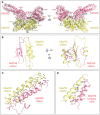
Fig 2. Structure of the C9orf72-SMCR8N-C complex.
(A) Overview of the cryo-EM map and structure of the C9orf72-SMCR8N-C complex in 2 orientations. C9orf72 (yellow orange) and SMCR8 (salmon pink) are shown as cartoons, and the interfaces between these proteins are boxed and shown in B, C, and D. Dashed lines indicate the 2 separate structural modules of both proteins. (B) Interaction interface 1 between the uDenn domains of C9orf72 (yellow orange) and SMCR8 (salmon pink). The β-sheet extension occurring due to the interaction of C9orf72 and SMCR8 is displayed (left). The interacting residues contributing to the formation of the hydrophobic core are labeled and displayed as sticks (right). (C) Region of interaction interface 2 between the cDenn domains of C9orf72 (yellow orange) and SMCR8 (salmon pink). The interacting residues are labeled and displayed as sticks. (D) Region of interaction interface 2 between the cDenn domain of C9orf72 (yellow orange) and the bridging helix of SMCR8 (salmon pink). The interacting residues are labeled and displayed as sticks. The corresponding cryo-EM map densities at the interaction interfaces are shown in S6 Fig. cDenn, central Denn; cryo-EM, cryo-electron microscopy; dDenn, downstream Denn; Denn, differently expressed in normal and neoplastic cells; uDenn, upstream Denn.
Fig 3
Fig 3. Interaction of C9orf72-SMCR8N-C complex with WDR41.
(A) Pull-down assay of purified WDR41 with the binary complex containing C9orf72 along with various constructs of SMCR8. The raw images can be found in S1 Raw Images. (B) Cross-linking of C9orf72-SMCR8-WDR41 ternary complex with DSSO and subsequent analysis of the cross-linked sample by mass spectrometry to identify cross-linked fragments. For clarity, only intermolecular cross-links are represented by green lines. Dead-end cross-linker–modified residues (Type 0 links [35] or monolinks) are shown as purple flags (top). The identified cross-linked fragments between the cDenn and dDenn domains of SMCR8 and WDR41 are displayed (bottom). cDenn, central Denn; Denn, differently expressed in normal and neoplastic cells; dDenn, downstream Denn; DSSO, disuccinimidyl sulfoxide; Fl, full-length.
Fig 4
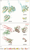
Fig 4. Comparison of C9orf72-SMCR8N-C complex with FLCN-FNIP2 complex.
(A) Superposition of full-length (left top) and the individual uDenn (left bottom), cDenn (right top) and dDenn (right bottom) domains of C9orf72 (yellow orange) protein on the full-length and the individual uDenn, cDenn, and dDenn domains of FNIP2 (cyan) protein (PDB: 6ULG) [28]. (B) Superposition of full-length (left top) and the individual uDenn (left bottom), cDenn (right top), and dDenn (right bottom) domains of SMCR8N-C (salmon pink) protein on the full-length and the individual uDenn, cDenn, and dDenn domains of FLCN (green) protein (PDB: 6ULG) [28]. (C) Structural-based sequence alignment of a region in the uDenn domains of SMCR8 and FLCN. The residue numbers are indicated. The blue triangle indicates the conserved arginine that is suggested to be crucial for GAP activity. cDenn, central Denn; dDenn, downstream Denn; Denn, differently expressed in normal and neoplastic cells; FLCN, folliculin; FNIP2, Folliculin Interacting Protein 2; GAP, GTPase activating protein; PDB, Protein Data Bank; uDenn, upstream Denn.
Fig 5
Fig 5. Structural and biochemical analysis of the SMCR8 coiled-coil region.
(A) Structure of an artificial 4 helix bundle generated by 2 monomers (PDB: 6DLC) (blue and purple) [39]. (B) Model of a 4-helix bundle formed by SMCR8N-C coiled coil (salmon pink) and FIP200 coiled-coil dimer (brown, PDB: 6GMA) [40] based on the artificial 4 helix bundle (PDB: 6DLC) [39]. (C) Model of SMCR8 coiled coil bound to FIP200 coiled-coil dimer along with the uDenn domain of SMCR8. Arg 147 of SMCR8 is shown in sphere representation. (D) Reduction in interaction between C9orf72-SMCR8ΔCC with FIP200 upon starvation. Fold change in the LFQ values of C9orf72 and SMCR8 Fl (WT, blue) or SMCR8ΔCC (ΔCC, orange) and FIP200 (normalized to the Fed condition) are shown. Standard error of the mean is shown. (E) Increased interaction between C9orf72-SMCR8ΔCC and WDR41. Fold change in the LFQ values of WDR41 (normalized to the WT Fed condition) is shown for both SMCR8 Fl (WT, blue) or SMCR8ΔCC (ΔCC, orange) pull-down experiments. Standard error of the mean is shown. Student t test: WDR41 Fed WT/ΔCC, p = 0.091; WDR41 Starved WT/ΔCC, p = 0.004; N = 3. The LFQ intensities and the corresponding analysis can be found in S2 Data. CC, coiled coil; Denn, differently expressed in normal and neoplastic cells; LFQ, label-free quantification; PDB, Protein Data Bank; uDenn, upstream Denn; WT, wild-type.
Fig 6
Fig 6. Schematic of C9orf72-SMCR8 complex with the position of the interaction proteins in cells.
Potential active site residue is situated behind the coiled coil and is indicated in blue. cDenn, central Denn; dDenn, downstream Denn; Denn, differently expressed in normal and neoplastic cells; uDenn, upstream Denn.

Similar articles

Cited by

References

    1. DeJesus-Hernandez M, Mackenzie IR, Boeve BF, Boxer AL, Baker M, Rutherford NJ, et al.. Expanded GGGGCC hexanucleotide repeat in noncoding region of C9ORF72 causes chromosome 9p-linked FTD and ALS. Neuron. 2011;72:245–56. doi: 10.1016/j.neuron.2011.09.011 - DOI - PMC - PubMed
    1. Renton AE, Majounie E, Waite A, Simón-Sánchez J, Rollinson S, Gibbs JR, et al.. A hexanucleotide repeat expansion in C9ORF72 is the cause of chromosome 9p21-linked ALS-FTD. Neuron. 2011;72:257–68. doi: 10.1016/j.neuron.2011.09.010 - DOI - PMC - PubMed
    1. Mori K, Weng S-M, Arzberger T, May S, Rentzsch K, Kremmer E, et al.. The C9orf72 GGGGCC repeat is translated into aggregating dipeptide-repeat proteins in FTLD/ALS. Science (New York, NY). 2013;339:1335–8. doi: 10.1126/science.1232927 - DOI - PubMed
    1. Ash PEA, Bieniek KF, Gendron TF, Caulfield T, Lin W-L, DeJesus-Hernandez M, et al.. Unconventional translation of C9ORF72 GGGGCC expansion generates insoluble polypeptides specific to c9FTD/ALS. Neuron. 2013;77:639–46. doi: 10.1016/j.neuron.2013.02.004 - DOI - PMC - PubMed
    1. Balendra R, Isaacs AM. C9orf72-mediated ALS and FTD: multiple pathways to disease. Nat Rev Neurol. 2018;14:544–58. doi: 10.1038/s41582-018-0047-2 - DOI - PMC - PubMed

Publication types

Supplementary concepts

Grants and funding

The Novartis Research Foundation (J.A.C), the Swiss National Science Foundation grant 31003A_182314 (J.A.C), the SNF-NCCR RNA & Disease (J.A.C) and the Synapsis Foundation 2017 CDA 01 (V.B.) supported this work. The funders had no role in study design, data collection and analysis, decision to publish, or preparation of the manuscript.