. 2021 Jun 29;12(3):e0022721.
doi: 10.1128/mBio.00227-21. Epub 2021 Jun 1.

A BMPR2/YY1 Signaling Axis Is Required for Human Cytomegalovirus Latency in Undifferentiated Myeloid Cells

Affiliations

A BMPR2/YY1 Signaling Axis Is Required for Human Cytomegalovirus Latency in Undifferentiated Myeloid Cells

Emma Poole et al. mBio. .

Abstract

Human cytomegalovirus (HCMV) presents a major health burden in the immunocompromised and in stem cell transplant medicine. A lack of understanding about the mechanisms of HCMV latency in undifferentiated CD34+ stem cells, and how latency is broken for the virus to enter the lytic phase of its infective cycle, has hampered the development of essential therapeutics. Using a human induced pluripotent stem cell (iPSC) model of HCMV latency and patient-derived myeloid cell progenitors, we demonstrate that bone morphogenetic protein receptor type 2 (BMPR2) is necessary for HCMV latency. In addition, we define a crucial role for the transcription factor Yin Yang 1 (YY1) in HCMV latency; high levels of YY1 are maintained in latently infected cells as a result of BMPR2 signaling through the SMAD4/SMAD6 axis. Activation of SMAD4/6, through BMPR2, inhibits TGFbeta receptor signaling, which leads to the degradation of YY1 via induction of a cellular microRNA (miRNA), hsa-miR-29a. Pharmacological targeting of BMPR2 in progenitor cells results in the degradation of YY1 and an inability to maintain latency and renders cells susceptible to T cell killing. These data argue that BMPR2 plays a role in HCMV latency and is a new potential therapeutic target for maintaining or disrupting HCMV latency in myeloid progenitors. IMPORTANCE Understanding the mechanisms which regulate HCMV latency could allow therapeutic targeting of the latent virus reservoir from where virus reactivation can cause severe disease. We show that the BMPR2/TGFbeta receptor/YY1 signaling axis is crucial to maintain HCMV latency in undifferentiated cells and that pharmacological reduction of BMPR2 in latently infected cells leads to reactivation of the viral lytic transcription program, which renders the infected cell open to immune detection and clearance in infected individuals. Therefore, this work identifies key host-virus interactions which regulate HCMV latent infection. It also demonstrates a potential new therapeutic approach to reduce HCMV reactivation-mediated disease by the treatment of donor stem cells/organs prior to transplantation, which could have a major impact in the transplant disease setting.

Keywords: BMPR2; YY1; human cytomegalovirus; latency; stem cells.


Figures

FIG 1
FIG 1
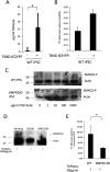
Cellular YY1 is required for HCMV latency. Wild-type iPSCs (WT-iPSCs), iPSCs which overexpressed Cas9 (Cas9-iPSCs), or which overexpressed Cas9 and were treated by CRISPR to remove YY1 (YY1-KO iPSCs) were stained for YY1 protein by indirect immunofluorescence staining (red), and the nuclei were costained with Hoechst (blue) (A). Alternatively, lysates from the same cells were analyzed by Western blotting for actin and YY1 proteins (B). (C) Undifferentiated iPSCs (- Differentiation) described for panels A and B) were infected with TB40E-IE2YFP and 4 days later analyzed by fluorescence for the presence of IE expression (green). This also included cells treated with PMA to induce differentiation (+ Differentiation) as a positive control for HCMV permissiveness. Bright-field images are also shown. Studies whose results are shown in panel C were performed in triplicate and enumerated (graph in panel C). Cells described for panel C were also harvested for RT-qPCR analysis and analyzed for IE expression and UL138 RNA relative to the housekeeping GAPDH gene (D). The supernatants from panel C were also transferred to fresh fibroblasts, and IE2-YFP-positive foci of infection in triplicate wells were counted after 9 days and enumerated (E). Wild-type THP1 cells (WT), THP1 cells which overexpressed Cas9 (Cas9), or which overexpressed Cas9 and a guide RNA to YY1 (YY1-KO) were harvested and analyzed by Western blotting for the housekeeping actin gene or YY1 as indicated (F). The experiment carried out for panel C was repeated using the THP1 cells described for panel F, and results are presented with graphical enumeration (G). The THP1 cells described for panel G were also harvested for RT-qPCR analysis of viral IE and UL138 genes alongside the housekeeping GAPDH gene (H). The supernatants from panel G were transferred to fresh fibroblasts, and IE2-YFP foci of infection in triplicate wells were counted after 9 days (I). The iPSCs described for panel A were also stained for the pluripotency markers Oct4 and Nanog (red) alongside a nuclear stain, Hoechst (blue) as indicated (J). The cells described for panel J were also differentiated prior to staining for Oct4 and Nanog (K). Finally, THP1 cells described for panel F were analyzed for the indicated differentiation markers by FACS (L). All graphs represent average values with standard deviation error bars.
FIG 1
FIG 1
Cellular YY1 is required for HCMV latency. Wild-type iPSCs (WT-iPSCs), iPSCs which overexpressed Cas9 (Cas9-iPSCs), or which overexpressed Cas9 and were treated by CRISPR to remove YY1 (YY1-KO iPSCs) were stained for YY1 protein by indirect immunofluorescence staining (red), and the nuclei were costained with Hoechst (blue) (A). Alternatively, lysates from the same cells were analyzed by Western blotting for actin and YY1 proteins (B). (C) Undifferentiated iPSCs (- Differentiation) described for panels A and B) were infected with TB40E-IE2YFP and 4 days later analyzed by fluorescence for the presence of IE expression (green). This also included cells treated with PMA to induce differentiation (+ Differentiation) as a positive control for HCMV permissiveness. Bright-field images are also shown. Studies whose results are shown in panel C were performed in triplicate and enumerated (graph in panel C). Cells described for panel C were also harvested for RT-qPCR analysis and analyzed for IE expression and UL138 RNA relative to the housekeeping GAPDH gene (D). The supernatants from panel C were also transferred to fresh fibroblasts, and IE2-YFP-positive foci of infection in triplicate wells were counted after 9 days and enumerated (E). Wild-type THP1 cells (WT), THP1 cells which overexpressed Cas9 (Cas9), or which overexpressed Cas9 and a guide RNA to YY1 (YY1-KO) were harvested and analyzed by Western blotting for the housekeeping actin gene or YY1 as indicated (F). The experiment carried out for panel C was repeated using the THP1 cells described for panel F, and results are presented with graphical enumeration (G). The THP1 cells described for panel G were also harvested for RT-qPCR analysis of viral IE and UL138 genes alongside the housekeeping GAPDH gene (H). The supernatants from panel G were transferred to fresh fibroblasts, and IE2-YFP foci of infection in triplicate wells were counted after 9 days (I). The iPSCs described for panel A were also stained for the pluripotency markers Oct4 and Nanog (red) alongside a nuclear stain, Hoechst (blue) as indicated (J). The cells described for panel J were also differentiated prior to staining for Oct4 and Nanog (K). Finally, THP1 cells described for panel F were analyzed for the indicated differentiation markers by FACS (L). All graphs represent average values with standard deviation error bars.
FIG 2
FIG 2
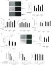
Latency-induced levels of TGFbeta stimulate SMAD2 phosphorylation, leading to a decrease in YY1 levels, only in the absence of BMPR2. iPSCs were either infected with TB40E-IE2YFP or left uninfected for 4 days before supernatants were harvested and analyzed for TGFbeta content by ELISA (A). Cells described for panel A were also analyzed by RT-qPCR for cellular YY1 transcript relative to the housekeeping GAPDH gene (B). Recombinant TGFbeta was added to either the WT-iPSCs (top) or BMPR2 KO-iPSCs (bottom) at the indicated concentrations for 1 h before harvesting for Western blotting for SMAD2 phosphorylation and the housekeeping actin gene (C). CD14+ monocytes were pretreated with SD208 (SD) or SB431542 (SB) for 30 min before addition of recombinant TGFbeta. Cells were then harvested for Western blotting for phosphorylated SMAD2 or the housekeeping actin gene (D). Finally, WT- or BMPR2 KO-iPSCs were treated with 30 pg/ml of TGFbeta for 1 h before harvesting by RT-qPCR for cellular GAPDH and YY1 genes as indicated (E). All graphs represent average values with standard deviations. Statistical significance was determined using Student’s t test.
FIG 3
FIG 3
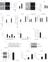
Latency cannot be established in the absence of BMPR2 but can be recovered if TGFbeta is also removed using neutralizing antibodies. WT-iPSCs or BMPR2−/+ or BMPR2+/+ iPSCs, as indicated, were infected with TB40E-IE2YFP for 4 days before analysis by immunofluorescence (IF; (right) and bright-field microscopy (left) (A). Cells from panel A were enumerated (10 fields of view of 100 cells each) alongside PMA-treated positive controls, and graphs represent triplicate experiments (B). Cells from panel B were also harvested for RNA analysis for the presence of viral lytic IE gene (left graph) and latency-associated gene UL138 (right graph) relative to the housekeeping GAPDH gene (C). WT-iPSCs and BMPR2−/+ and BMPR2−/− iPSCs, as indicated, were pretreated with BMP4 (50 ng/ml) prior to infection with TB40E-IE2YFP. IE2-YFP-positive cells were then enumerated in triplicate with 10 fields of view of 100 cells each (D). WT-iPSCs and BMPR2−/+ and BMPR2−/− iPSCs were infected with TB40E-IE2YFP for 4 days before supernatants were assayed for secreted TGFbeta by ELISA. Uninfected WT cells are also shown, and statistical significance determined using Student’s t test (E). Finally, BMPR2−/− and BMPR2−/+ or YY1-KO iPSCs, as indicated, were pretreated with the indicated concentrations of TGFbeta neutralizing antibody before infection with TB40E-IE2YFP. Medium was changed with fresh neutralizing antibody daily and IE2-YFP-positive cells were scored by IF after 4 days. Triplicate experiments were carried out with 10 fields of view of 100 cells each (F). WT and BMPR2−/+ cells were differentiated along the myeloid lineage into monocyte-like cells and then either infected with TB40E-IE2YFP for 4 days before analysis by IF (right) and bright-field microscopy (left) as well as being enumerated alongside a PMA-positive control in triplicate with 10 fields of view of 100 cells each (right), with standard deviation error bars shown (G). All graphs show mean values with standard deviations.
FIG 4
FIG 4
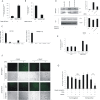
BMPR2 inhibitor LDN prevents the establishment of HCMV latency in iPSCs and monocytes, but this can be rescued with TGFbeta inhibitors and BMPR2-stimulated SMAD6 aids maintenance of latency. Monocytes or iPSCs, as indicated, were infected with TB40E-IE2YFP for 4 days in the presence or absence of LDN (medium was changed daily and replaced with fresh LDN). Cells were then analyzed by IF for IE2-YFP and bright-field microscopy (left) (A). These cells were enumerated alongside a PMA-positive control, and the graph represents triplicate experiments in 5 fields of view of 100 cells each (right) (A). Monocytes, WT-iPSCs, BMPR2−/+ iPSCs, or BMPR2−/− iPSCs were infected with TB40E-IE2YFP in the presence or absence of LDN, SB431542, SD208, or PMA as indicated, with medium and drug changes every day for 4 days before enumeration of IE2-YFP-positive cells in 6 fields of view from 100 cells each from triplicate experiments. The graph shows mean values, with standard deviation error bars (B). iPSCs were infected with TB40E-GATA2mCherry for 4 days prior to sorting and analysis by single-cell RNA-seq analysis. Six cells were analyzed for numbers of transcripts of the UL122, YY1, SMAD6, SMAD7, and BMPR2 genes. Cells in the left graph were selected on the basis of the ability to detect SMAD6 transcript. Cells in the right graph represent cells which were BMPR2 negative. Graphs show mean values with standard deviation error bars (C). BMPR2−/− iPSCs and WT-iPSCs were infected with TB40E-IE2YFP and then either treated with LDN or left untreated as indicated prior to harvesting for RNA analysis. Levels of GAPDH and SMAD6 were determined by RT-qPCR. The data are from triplicate samples, and average delta delta CT values for SMAD6 relative to GAPDH are shown with standard deviation error bars. WT-iPSCs, BMPR2−/− iPSCs, or monocytes that had been treated with LDN were harvested for RT-qPCR analysis of SMAD6 RNA relative to the housekeeping GAPDH gene (D). WT-iPSCs and BMPR2−/+ and BMPR2−/− iPSCs were either left uninfected or infected with TB40E-IE2GFP for 4 days before harvesting for Western blotting and analysis for the presence of SMAD6 and actin proteins (E). The latently infected cells described for panel E were analyzed for total SMAD2 and phosphorylated SMAD2 by Western blotting (F). THP1 cells were nucleofected with an siRNA to SMAD6 and then left for 48 h before analysis of SMAD6 and actin proteins by Western blotting (G) and RNA by RT-qPCR; the graph represents delta delta CT with standard errors shown (H). Finally, CD14+ monocytes or THP1 cells were infected with IE2YFP-TB40E and left for 3 days (I). After 3 days, the cells were nucleofected with control siRNA or SMAD6 siRNA or, as a control, reactivated with GM-CSF/interleukin 4 (IL-4) and lipopolysaccharide (LPS) (I, left) or PMA (I, right). Cells were then enumerated for IE positivity. The graph represents 6 fields of view of 100 cells, with standard deviations and significance determined with Student’s t test (I).
FIG 5
FIG 5
The absence of BMPR2 leads to a decrease in levels of YY1 mRNA during HCMV latency and a lack of YY1 occupancy on the MIEP. WT-iPSCs and BMPR2−/− iPSCs were latently infected with HCMV for 4 days before analysis for miRNA hsa-miR-29a and the housekeeping miRNA hsa-miR-16. Graphs represent triplicate mean delta delta CT values with standard deviation error bars (A). WT-iPSCs and BMPR2−/− iPSCs were either left uninfected or infected with TB40E-IE2YFP for 4 days before the RNA was harvested and analyzed for YY1 and housekeeping gene GAPDH mRNAs (B). WT-iPSCs and BMPR2−/− iPSCs were infected with TB40E-IE2YFP for 4 days before harvesting for ChIP analysis to detect YY1 association with the MIEP. Input samples were diluted 1:10, and the MIEP was detected by qPCR (C). Graphs represent triplicate mean values with standard deviation error bars. Finally, BMPR2 KO cells or WT cells (D and E) were transfected with control siRNA (control) or an antagomir to hsa-miR-29a (anti-hsa-miR-29a) in the presence or absence of 30 pg/ml of TGFbeta. After 48 h, cells were harvested for Western blotting (left, D) for actin and YY1 and bands were then analyzed by densitometry (right, D). Alternatively, cells were harvested for RNA. (E) Analysis of YY1 and GAPDH transcripts. The graph represents delta delta CT, with standard deviations shown. Finally, CD14+ monocytes were isolated from an HCMV-seronegative or HCMV-seropositive individual and remaining PBMCs were stored. The CD14+ cells were then infected with TB40E-SV40GFP and latency was established for 3 days. After this time, LDN was added to the cells as indicated for 24 h before addition of PBMCs at physiological effector-to-target (E:T) ratios (F and G). After 24 h, cells were analyzed by light and fluorescence microscopy (F) and GFP-positive cells were enumerated and presented graphically; standard deviation error bars are shown from 6 fields of view (**, significance value of <0.005 by Student’s t test; NS, not significant) (G).
FIG 6
FIG 6
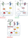
Mechanism for the role of BMPR2 in supporting HCMV latency and inhibition of SMAD2/3 signaling. In the presence of BMPR2 (A), there is expression of the inhibitory SMAD protein SMAD6. This prevents activation of the TGFbeta receptor by TGFbeta, which is secreted during latency, and therefore, the YY1 present in the cell is associated with the MIEP, enhancing the maintenance of HCMV latency. In the absence of BMPR2, by either direct removal of BMPR2 or inhibition of BMPR2 signaling using LDN (B), there is a lack of inhibitory protein SMAD6, and this allows the TGFbetaR to respond to the TGFbeta in the secretome, stimulating SMAD2/3 phosphorylation and leading to the expression of the miRNA hsa-miR-29a, which, in turn, causes degradation of the protein YY1. This protein is then unable to associate with the MIEP, and lytic IE gene expression can occur. The activation of lytic gene expression can be inhibited using specific inhibitors SB431542 and SD208. Finally, TGFbeta signaling is upregulated during latency (31) by viral miRNA miR-US5-2a (49), and the virus has a number of mechanisms in place to prevent TGFbeta- and activin-mediated SMAD2/3 signaling. These mechanisms include the expression of viral miRNAs US22A (49) and miR-UL148D (53) as well as the presence of BMPR2 (C).

Similar articles

Cited by

References

    1. Avery RK. 2007. Ganciclovir-resistant cytomegalovirus disease in heart transplant recipients: the dilemma of donor-positive/recipient-negative serostatus. Clin Infect Dis 45:448–449. doi:10.1086/519940. - DOI - PubMed
    1. Autmizguine J, Theoret Y, Launay E, Duval M, Rousseau C, Tapiero B, Boivin G, Ovetchkine P. 2011. Low systemic ganciclovir exposure and preemptive treatment failure of cytomegalovirus reactivation in a transplanted child. J Popul Ther Clin Pharmacol 18:e257–e260. - PubMed
    1. Fishman JA. 2011. Infections in immunocompromised hosts and organ transplant recipients: essentials. Liver Transpl 17:S34–S37. doi:10.1002/lt.22378. - DOI - PubMed
    1. Sinclair J, Poole E. 2014. Human cytomegalovirus latency and reactivation in and beyond the myeloid lineage. Future Virol 9:557–563. doi:10.2217/fvl.14.34. - DOI
    1. Poole E, Wills M, Sinclair J. 2014. Human cytomegalovirus latency: targeting differences in the latently infected cell with a view to clearing latent infection. New J Sci 2014:1–10. doi:10.1155/2014/313761. - DOI

Publication types

MeSH terms

Substances