HDAC6 regulates primordial follicle activation through mTOR signaling pathway
- PMID: 34052832
- PMCID: PMC8164630
- DOI: 10.1038/s41419-021-03842-1
HDAC6 regulates primordial follicle activation through mTOR signaling pathway
Abstract
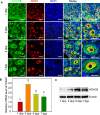
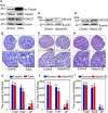
Primordial follicle pool established perinatally is a non-renewable resource which determines the female fecundity in mammals. While the majority of primordial follicles in the primordial follicle pool maintain dormant state, only a few of them are activated into growing follicles in adults in each cycle. Excessive activation of the primordial follicles accelerates follicle pool consumption and leads to premature ovarian failure. Although previous studies including ours have emphasized the importance of keeping the balance between primordial follicle activation and dormancy via molecules within the primordial follicles, such as TGF-β, E-Cadherin, mTOR, and AKT through different mechanisms, the homeostasis regulatory mechanisms of primordial follicle activation remain unclear. Here, we reported that HDAC6 acts as a key negative regulator of mTOR in dormant primordial follicles. In the cytoplasm of both oocytes and granulosa cells of primordial follicles, HDAC6 expressed strong, however in those activated primordial follicles, its expression level is relatively weaker. Inhibition or knockdown of HDAC6 significantly promoted the activation of limited primordial follicles while the size of follicle pool was not affected profoundly in vitro. Importantly, the expression level of mTOR in the follicle and the activity of PI3K in the oocyte of the follicle were simultaneously up-regulated after inhibiting of HDAC6. The up-regulated mTOR leads to not only the growth and differentiation of primordial follicles granulosa cells (pfGCs) into granulosa cells (GCs), but the increased secretion of KITL in these somatic cells. As a result, inhibition of HDAC6 awaked the dormant primordial follicles of mice in vitro. In conclusion, HDAC6 may play an indispensable role in balancing the maintenance and activation of primordial follicles through mTOR signaling in mice. These findings shed new lights on uncovering the epigenetic factors involved physiology of sustaining female reproduction.
Conflict of interest statement
The authors declare no competing interests.
Figures


Similar articles
-
Somatic cells initiate primordial follicle activation and govern the development of dormant oocytes in mice.Curr Biol. 2014 Nov 3;24(21):2501-8. doi: 10.1016/j.cub.2014.09.023. Epub 2014 Oct 23. Curr Biol. 2014. PMID: 25438940
-
HDAC6-dependent deacetylation of NGF dictates its ubiquitination and maintains primordial follicle dormancy.Theranostics. 2024 Mar 25;14(6):2345-2366. doi: 10.7150/thno.95164. eCollection 2024. Theranostics. 2024. PMID: 38646645 Free PMC article.
-
Dormancy and activation of human oocytes from primordial and primary follicles: molecular clues to oocyte regulation.Hum Reprod. 2017 Aug 1;32(8):1684-1700. doi: 10.1093/humrep/dex238. Hum Reprod. 2017. PMID: 28854595
-
Cellular and molecular regulation of the activation of mammalian primordial follicles: somatic cells initiate follicle activation in adulthood.Hum Reprod Update. 2015 Nov-Dec;21(6):779-86. doi: 10.1093/humupd/dmv037. Epub 2015 Jul 30. Hum Reprod Update. 2015. PMID: 26231759 Review.
-
Crosstalk between PTEN/PI3K/Akt Signalling and DNA Damage in the Oocyte: Implications for Primordial Follicle Activation, Oocyte Quality and Ageing.Cells. 2020 Jan 14;9(1):200. doi: 10.3390/cells9010200. Cells. 2020. PMID: 31947601 Free PMC article. Review.
Cited by
-
Signaling pathway intervention in premature ovarian failure.Front Med (Lausanne). 2022 Nov 25;9:999440. doi: 10.3389/fmed.2022.999440. eCollection 2022. Front Med (Lausanne). 2022. PMID: 36507521 Free PMC article. Review.
-
Enhanced glycolysis in granulosa cells promotes the activation of primordial follicles through mTOR signaling.Cell Death Dis. 2022 Jan 27;13(1):87. doi: 10.1038/s41419-022-04541-1. Cell Death Dis. 2022. PMID: 35087042 Free PMC article.
-
The metallic compound promotes primordial follicle activation and ameliorates fertility deficits in aged mice.Theranostics. 2023 May 21;13(10):3131-3148. doi: 10.7150/thno.82553. eCollection 2023. Theranostics. 2023. PMID: 37351158 Free PMC article.
-
Effects and potential mechanism of Ca2+/calmodulin‑dependent protein kinase II pathway inhibitor KN93 on the development of ovarian follicle.Int J Mol Med. 2022 Oct;50(4):121. doi: 10.3892/ijmm.2022.5177. Epub 2022 Aug 5. Int J Mol Med. 2022. PMID: 35929517 Free PMC article.
-
Epigenetic regulation in premature ovarian failure: A literature review.Front Physiol. 2023 Jan 4;13:998424. doi: 10.3389/fphys.2022.998424. eCollection 2022. Front Physiol. 2023. PMID: 36685174 Free PMC article. Review.
References
Publication types
MeSH terms
Substances
LinkOut - more resources
Full Text Sources
Other Literature Sources
Molecular Biology Databases
Research Materials
Miscellaneous

