

3D Printed Zn-doped Mesoporous Silica-incorporated Poly-L-lactic Acid Scaffolds for Bone Repair
- PMID: 33997435
- PMCID: PMC8114096
- DOI: 10.18063/ijb.v7i2.346
3D Printed Zn-doped Mesoporous Silica-incorporated Poly-L-lactic Acid Scaffolds for Bone Repair
Abstract
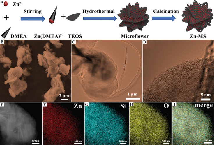
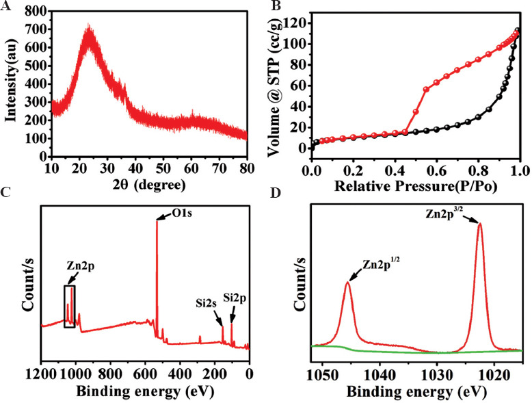
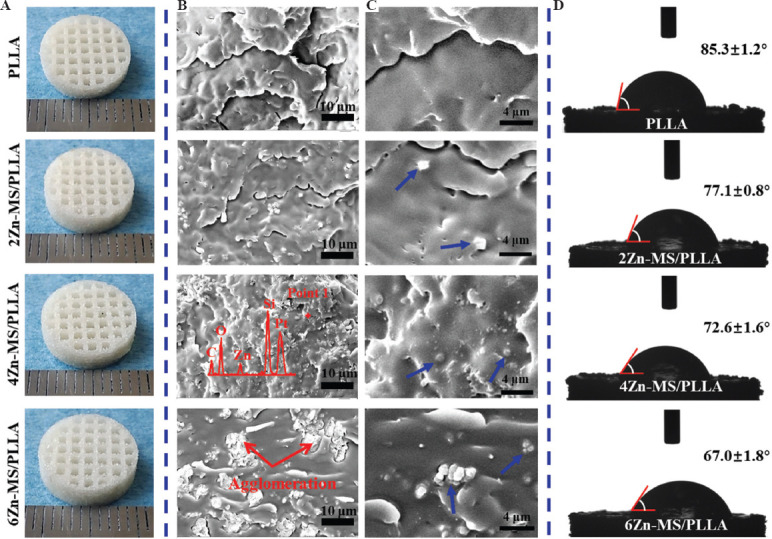
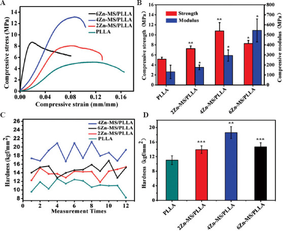
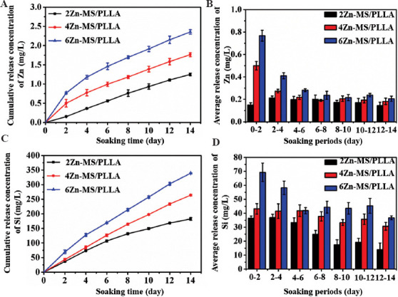
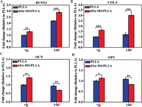
Poly-L-lactic acid (PLLA) lacks osteogenic activity, which limits its application in bone repair. Zinc (Zn) is widely applied to strengthen the biological properties of polymers due to its excellent osteogenic activity. In the present study, Zn-doped mesoporous silica (Zn-MS) particles were synthesized by one-pot hydrothermal method. Then, the particles were induced into PLLA scaffolds prepared by selective laser sintering technique, aiming to improve their osteogenic activity. Our results showed that the synthesized particles possessed rosette-like morphology and uniform mesoporous structure, and the composite scaffold displayed the sustained release of Zn ion in a low concentration range, which was attributed to the shield effect of the PLLA matrix and the strong bonding interaction of Si-O-Zn. The scaffold could evidently promote osteogenesis differentiation of mouse bone marrow mesenchymal stem cells by upregulating their osteogenesis-related gene expression. Besides, Zn-MS particles could significantly increase the compressive strength of the PLLA scaffold because of their rosette-like morphology and mesoporous structure, which can form micromechanical interlocking with the PLLA matrix. The Zn-MS particles possess great potential to improve various polymer scaffold properties due to their advantageous morphology and physicochemical properties.
Keywords: Bone repair; Poly-L-lactic acid; Zinc doped mesoporous silica.
Copyright: © 2021 Qian, et al.
Conflict of interest statement
The authors declare no conflicts of interest.
Figures
Similar articles
-
Electrophoretic Deposition of Dexamethasone-Loaded Mesoporous Silica Nanoparticles onto Poly(L-Lactic Acid)/Poly(ε-Caprolactone) Composite Scaffold for Bone Tissue Engineering.ACS Appl Mater Interfaces. 2016 Feb 17;8(6):4137-48. doi: 10.1021/acsami.5b11879. Epub 2016 Feb 5. ACS Appl Mater Interfaces. 2016. PMID: 26736029
-
Poly-l-lactic acid scaffold incorporated chitosan-coated mesoporous silica nanoparticles as pH-sensitive composite for enhanced osteogenic differentiation of human adipose tissue stem cells by dexamethasone delivery.Artif Cells Nanomed Biotechnol. 2019 Dec;47(1):4020-4029. doi: 10.1080/21691401.2019.1658594. Artif Cells Nanomed Biotechnol. 2019. PMID: 31595797
-
Three-Dimensional Printing of Poly-L-Lactic Acid Composite Scaffolds with Enhanced Bioactivity and Controllable Zn Ion Release Capability by Coupling with Carbon-ZnO.Bioengineering (Basel). 2023 Feb 28;10(3):307. doi: 10.3390/bioengineering10030307. Bioengineering (Basel). 2023. PMID: 36978698 Free PMC article.
-
Dual-functional scaffolds of poly(L-lactic acid)/nanohydroxyapatite encapsulated with metformin: Simultaneous enhancement of bone repair and bone tumor inhibition.Mater Sci Eng C Mater Biol Appl. 2021 Jan;120:111592. doi: 10.1016/j.msec.2020.111592. Epub 2020 Nov 12. Mater Sci Eng C Mater Biol Appl. 2021. PMID: 33545810
-
Towards resorbable 3D-printed scaffolds for craniofacial bone regeneration.Orthod Craniofac Res. 2023 Dec;26 Suppl 1:188-195. doi: 10.1111/ocr.12645. Epub 2023 Mar 13. Orthod Craniofac Res. 2023. PMID: 36866957 Review.
Cited by
-
A spatiotemporal drug release scaffold with antibiosis and bone regeneration for osteomyelitis.J Adv Res. 2023 Dec;54:239-249. doi: 10.1016/j.jare.2023.01.019. Epub 2023 Jan 24. J Adv Res. 2023. PMID: 36706987 Free PMC article.
-
Laser Additively Manufactured Iron-Based Biocomposite: Microstructure, Degradation, and In Vitro Cell Behavior.Front Bioeng Biotechnol. 2021 Dec 2;9:783821. doi: 10.3389/fbioe.2021.783821. eCollection 2021. Front Bioeng Biotechnol. 2021. PMID: 34926428 Free PMC article.
-
Laser-Sintered Mg-Zn Supersaturated Solid Solution with High Corrosion Resistance.Micromachines (Basel). 2021 Nov 6;12(11):1368. doi: 10.3390/mi12111368. Micromachines (Basel). 2021. PMID: 34832780 Free PMC article.
-
In situ synthesis of hydroxyapatite nanorods on graphene oxide nanosheets and their reinforcement in biopolymer scaffold.J Adv Res. 2021 Apr 5;35:13-24. doi: 10.1016/j.jare.2021.03.009. eCollection 2022 Jan. J Adv Res. 2021. PMID: 35024192 Free PMC article.
-
Personalized bioceramic grafts for craniomaxillofacial bone regeneration.Int J Oral Sci. 2024 Oct 31;16(1):62. doi: 10.1038/s41368-024-00327-7. Int J Oral Sci. 2024. PMID: 39482290 Free PMC article. Review.
References
-
- Wang G, Qian G, Zan J, et al. A Co-dispersion Nanosystem of Graphene Oxide@Silicon-doped Hydroxyapatite to Improve Scaffold Properties. Mater Design. 2020;2020:109399. https://doi.org/10.1016/j.matdes.2020.109399.
-
- Shuai C, Yu L, Feng P, et al. Organic Montmorillonite Produced an Interlayer Locking Effect in a Polymer Scaffold to Enhance Interfacial Bonding. Mater Chem Front. 2020;4:2398–408. https://doi.org/10.1039/d0qm00254b.
-
- Shuai C, Yang W, Feng P, et al. Accelerated Degradation of HAP/PLLA Bone Scaffold by PGA Blending Facilitates Bioactivity and Osteoconductivity. Bioact Mater. 2020;6:490–502. https://doi.org/10.1016/j.bioactmat.2020.09.001. - PMC - PubMed
-
- Qi F, Wang C, Peng S, et al. A Co-dispersed Nanosystem from Strontium-Anchored Reduced Graphene Oxide to Enhance Bioactivity and Mechanical Property in Polymer Scaffolds. Mater Chem Front. 2021;2021:958. https://doi.org/10.1039/d0qm00958j.
-
- King JC, Shames DM, Woodhouse LR. Zinc Homeostasis in Humans. J Nutr. 2020;130:1360, S–6S. - PubMed
LinkOut - more resources
Full Text Sources
Other Literature Sources