. 2022 Jun;27(6):2879-2900.
doi: 10.1038/s41380-021-01124-y. Epub 2021 May 14.

Enkephalin release from VIP interneurons in the hippocampal CA2/3a region mediates heterosynaptic plasticity and social memory

Affiliations

Enkephalin release from VIP interneurons in the hippocampal CA2/3a region mediates heterosynaptic plasticity and social memory

Felix Leroy et al. Mol Psychiatry. 2022 Jun.

Abstract

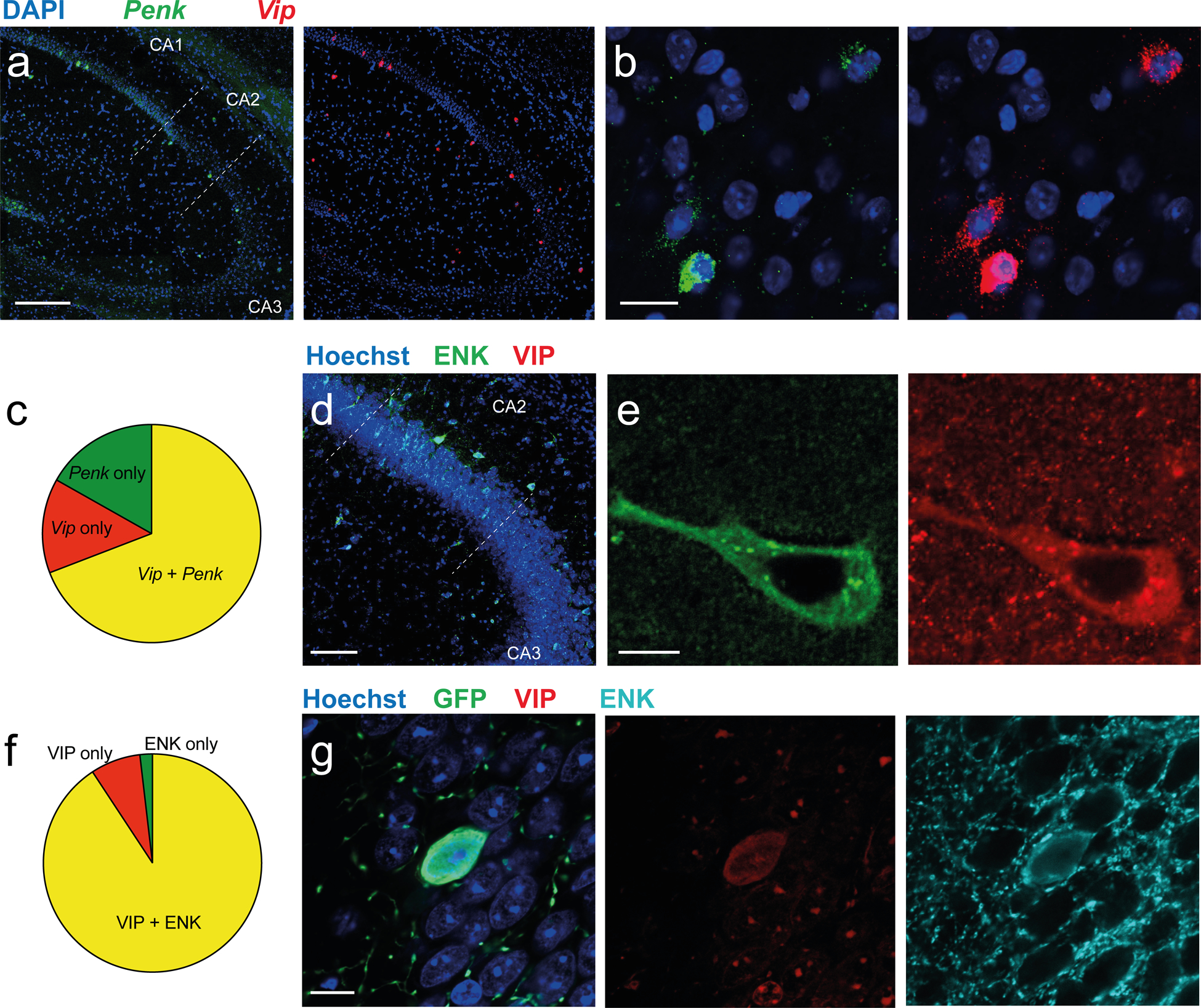
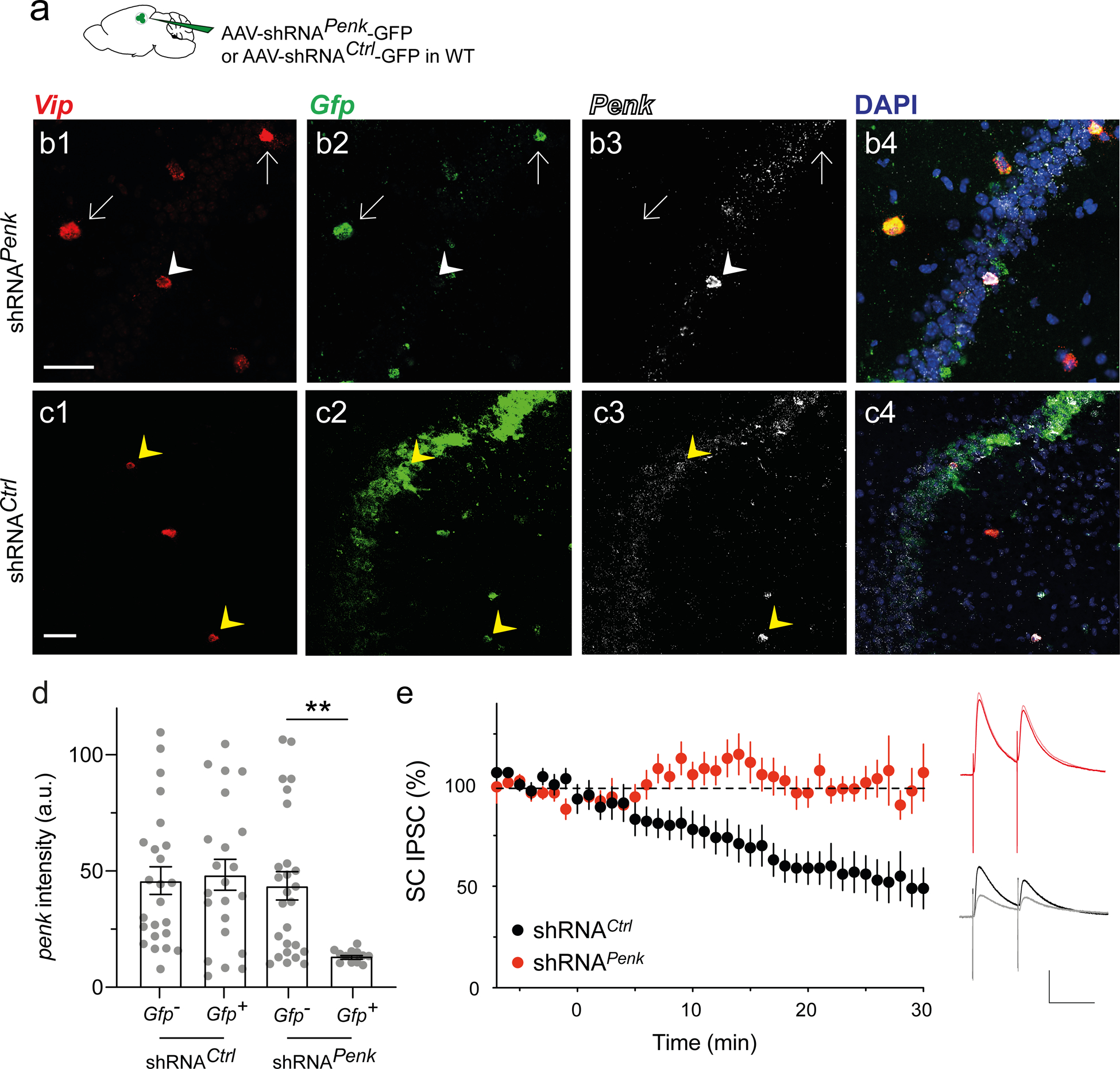
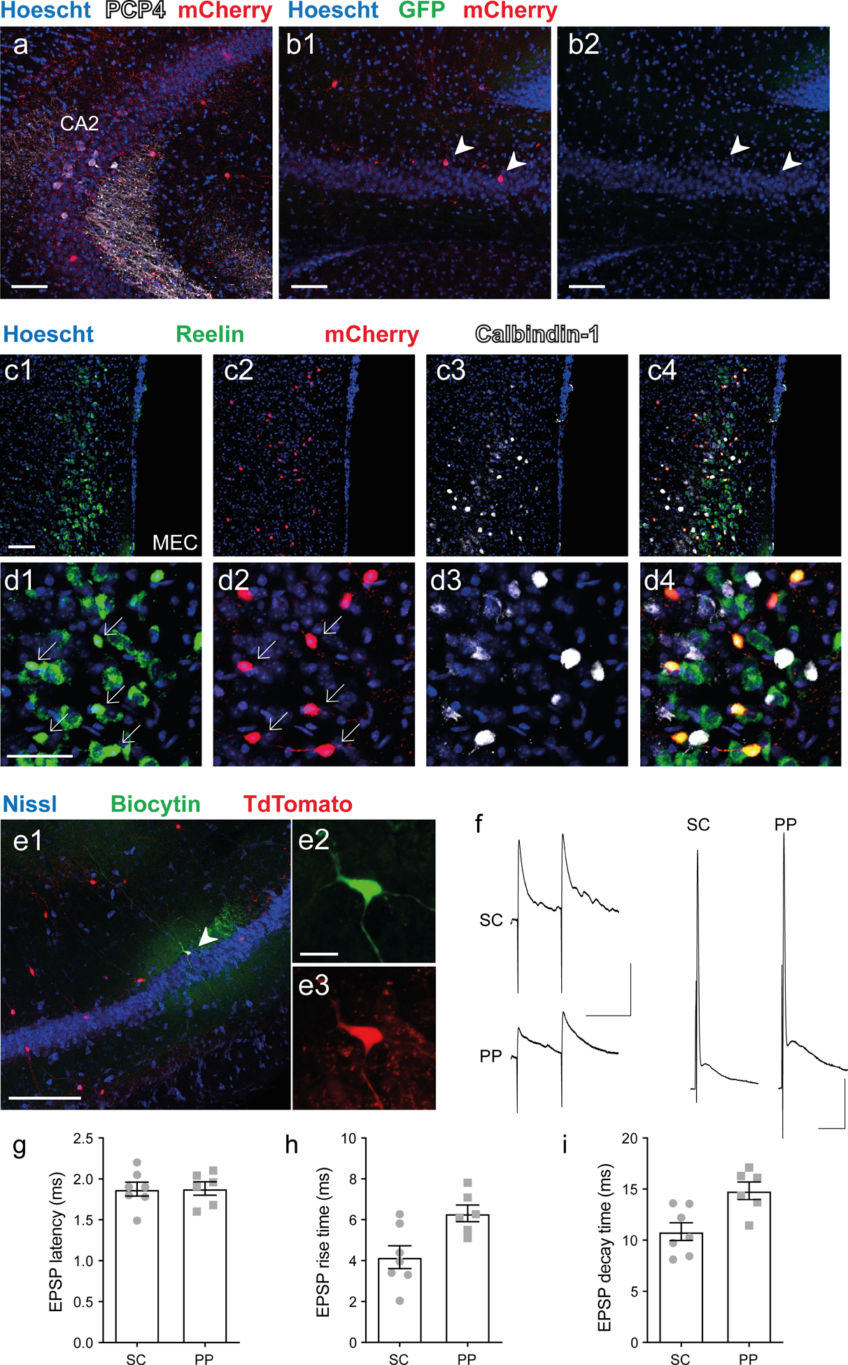
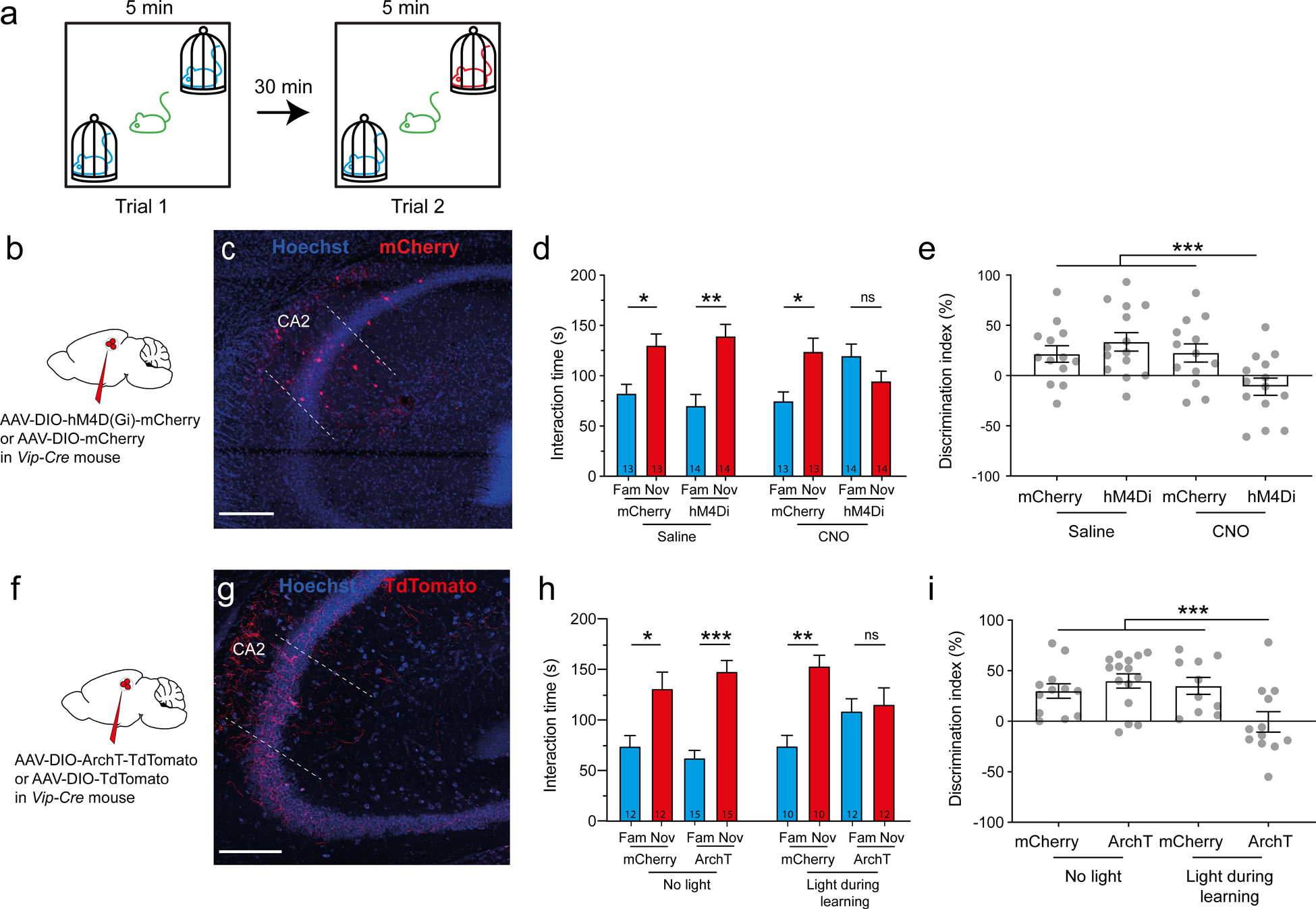
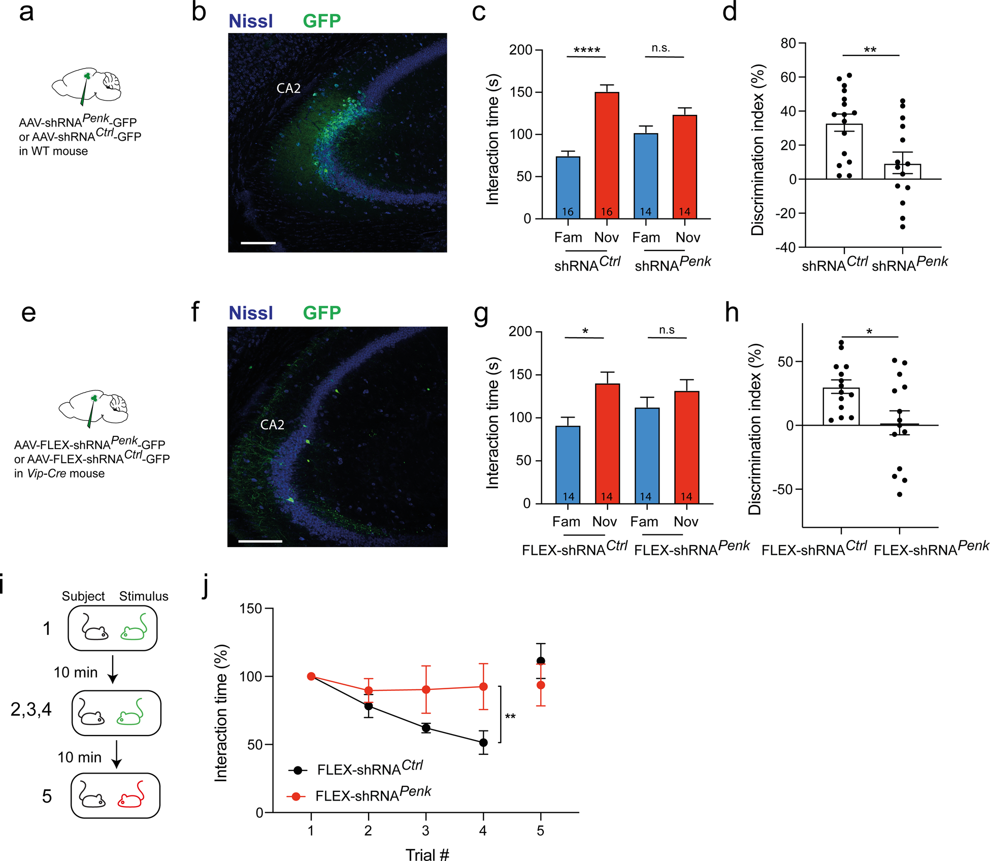
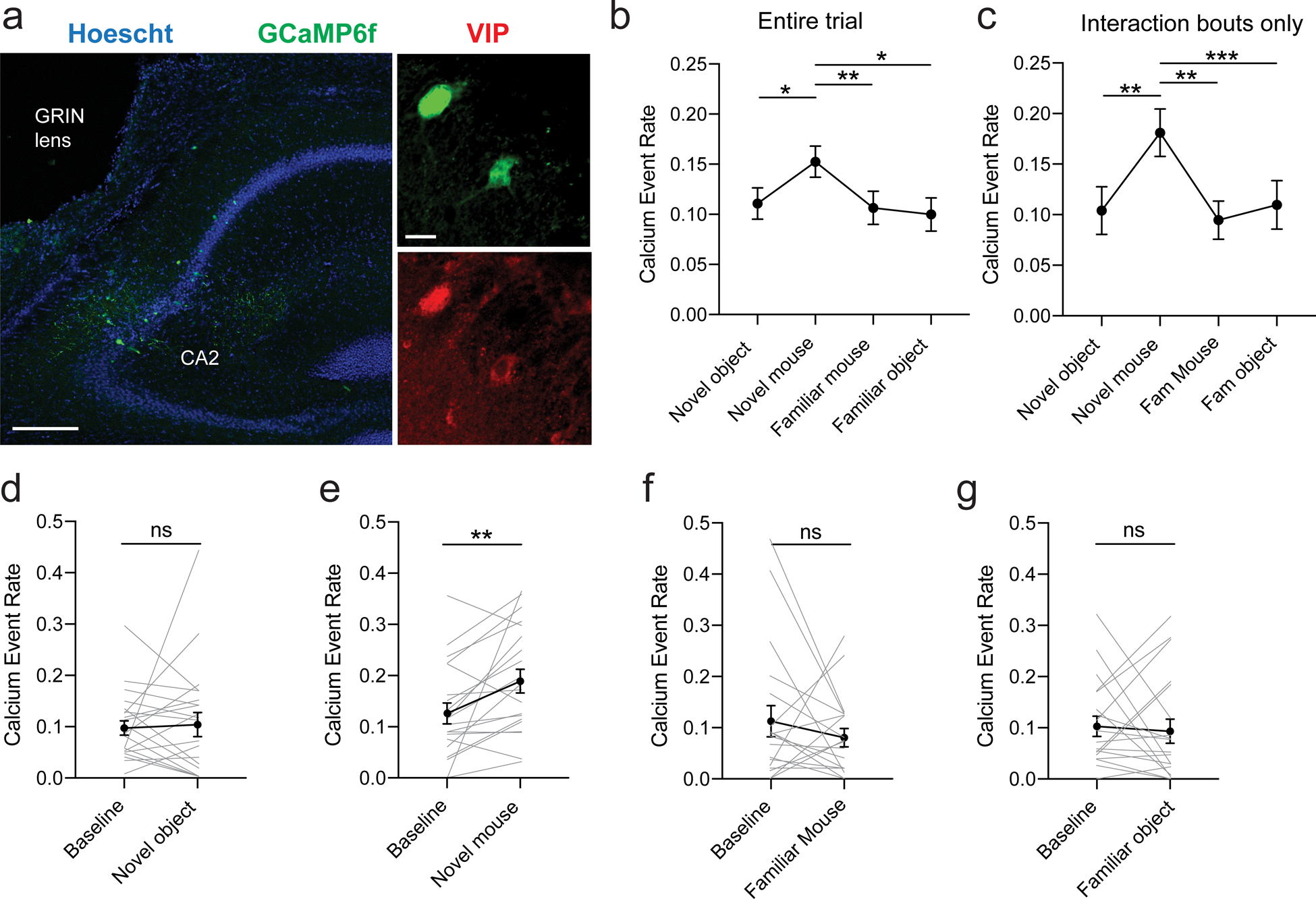
The hippocampus contains a diverse array of inhibitory interneurons that gate information flow through local cortico-hippocampal circuits to regulate memory storage. Although most studies of interneurons have focused on their role in fast synaptic inhibition mediated by GABA release, different classes of interneurons express unique sets of neuropeptides, many of which have been shown to exert powerful effects on neuronal function and memory when applied pharmacologically. However, relatively little is known about whether and how release of endogenous neuropeptides from inhibitory cells contributes to their behavioral role in regulating memory formation. Here we report that vasoactive intestinal peptide (VIP)-expressing interneurons participate in social memory storage by enhancing information transfer from hippocampal CA3 pyramidal neurons to CA2 pyramidal neurons. Notably, this action depends on release of the neuropeptide enkephalin from VIP neurons, causing long-term depression of feedforward inhibition onto CA2 pyramidal cells. Moreover, VIP neuron activity in the CA2 region is increased selectively during exploration of a novel conspecific. Our findings, thus, enhance our appreciation of how GABAergic neurons can regulate synaptic plasticity and mnemonic behavior by demonstrating that such actions can be mediated by release of a specific neuropeptide, rather than through classic fast inhibitory transmission.


Conflict of interest statement

Conflict of interest statement

The authors declare no conflicts of interest.

Figures

Figure 1:
Figure 1:. VIP and enkephalin expression overlaps in the hippocampal CA2 region.
a-b. In situ hybridization images of dorsal hippocampal slices (a) and CA2 (b) showing staining for Penk and Vip mRNAs. c. Quantification of the fraction of neurons positive for Penk alone (green), for Vip alone (red) and for both Penk and Vip (yellow) across all layers of the CA2 region. d-e. Immunohistochemistry images of dorsal hippocampal slices with antibodies against ENK (Santa Cruz antibody) and VIP. f. Quantification of the fraction of hippocampal cells positive for ENK alone, VIP alone, and co-labeled for both ENK (Fitzgerald antibody) and VIP in the CA2 region. g. Immunohistochemistry for ENK and VIP in the CA2 region of a Vip-Cre mouse injected in dorsal CA2 with AAV2/5 EF1a.DIO.eYFP. Scale bars: a 200 μm, b 20 μm, d 100 μm, e and g 10 μm.
Figure 2:
Figure 2:. VIP neurons are necessary and sufficient to induce CA2 ITDP through DOR activation.
a. Vip-Cre mice injected in dorsal CA2 with AAV2/9 CBA.FLEX.Arch3.0-GFP.WPRE.SV40 to express Arch-3.0-GFP or with AAV2/9 CAG.FLEX.eGFP.WPRE to express eGFP as a control (top). Image shows immunohistochemistry in acute hippocampal slice labelling a VIP neuron expressing GFP (green) next to a CA2 PN filled with biocytin during a patch-clamp recording (red). b. Average normalized SC-evoked PSP amplitude in CA2 PNs before and after electrical pairing of EC and SC inputs to induce ITDP in presence of continuous illumination with yellow light (which activates Arch3.0). The PSP showed a significant progressive potentiation following pairing, indicative of ITDP, in slices in which GFP was expressed in VIP neurons (black). In contrast, ITDP was absent in slices expressing Arch3.0 (red). Inset shows example PSPs before and after pairing. c. Vip-Cre mice injected in dorsal CA2 with AAV2/5 EF1a.DIO.hChR2(E123T/T159C)-eYFP (top). Immunohistochemistry of acute hippocampal slice showing labelling of VIP neurons expressing GFP next to a recorded CA2 PN filled with biocytin (bottom). d. Average normalized SC PSPs in CA2 PNs evoked by electrical stimulation before and after photostimulation of VIP neurons expressing ChR2, with or without 0.1 μM naltrindole in bath. Photostimulation of VIP neurons was sufficient to enhance the PSP, suggestive of ITDP, in the absence (red) but not presence (black) of naltrindole. Inset shows example PSPs before and after photostimulation. e. Photostimulation of VIP neurons expressing ChR2 induces long-term depression of SC-evoked IPSC recorded from CA2 PNs voltage-clamped at +10 mV (same viral injection than panel d). Scale bars: a 100 μm, c 200 μm, b and d 50 ms / 2 mV, e 100 ms / 100 pA.
Figure 3:
Figure 3:. Enkephalin is necessary for ITDP induction.
a. WT mice injected in dorsal CA2 with AAV2/8 U6.shRNAPenk.hSyn.GFP.WPRE to downregulate Penk or control AAV2/8 U6.shRNCtrl.hSyn.GFP.WPRE. b-c. In situ hybridization images of one hippocampal slice expressing shRNAPenk (b) and one expressing shRNCtrl (c) labelled for Vip, Gfp, and Penk mRNAs. White arrows denote Vip+ neurons that express shRNAPenk, showing reduced levels of Penk. White arrowheads denote a Vip+ neuron that does not express shRNAPenk and expresses Penk. Yellow arrowheads denote Vip+ neurons that express shRNACtrl and Penk. d. Quantification of Penk expression in Vip+ cells using in situ hybridization in slices from mice injected with AAV expressing control of Penk shRNAs. In each slice neurons were classified as to whether they were uninfected or infected with virus based on Gfp expression (3 mice per group; each point is from a different slice). Note reduction in Penk expression in neurons infected with Penk shRNA. e. Time course of SC IPSCs recorded in CA2 PNs before and after ITDP induction using paired electrical stimulation from slices expressing control or Penk shRNAs. Insets show sample IPSCs before and after ITDP induction in the two groups. Scale bars: b and c 50 μm, e 100 ms / 200 pA.
Figure 4:
Figure 4:. VIP neurons receive excitatory inputs from both CA3 and EC layer II stellate cells.
a-d. Vip-Cre mouse injected in dorsal CA2 with AAV2/8 syn.DIO.TVA.2A.GFP.2A.B19G and rabies virus SAD.B19.EnvA.ΔG.mCherry to label monosynaptic inputs to VIP neurons. Immunohistochemistry of hippocampal and MEC slices showing: (a) CA2 PN marker PCP4 and rabies-based mCherry-labeled VIP neurons in CA2 region; (b) mCherry-labeled pyramidal neurons in CA3 (arrowheads); (c) Low- and (d) high-magnification images of MEC showing mCherry labeling of reelin-expressing LII stellate cells but not of calbindin-expressing LII pyramidal cells. e. Low (e1) and high (e2–3) magnification immunohistochemistry images of an acute hippocampal slice from a Vip-Cre × Ai14 mouse showing biocytin (e2) and TdTomato labeling (e3) of a patch-clamped VIP neuron. f. Representative PSPs recorded from VIP neuron following electrical stimulation of PP and SC inputs using low intensity stimulation that evoked subthreshold PSPs (left) or high intensity stimulation that evoked a spike (right). g-i, PSP parameters elicited by PP or SC stimulation. g. PSP latency. h. PSP rise time. i. PSP decay time. Scale bars: a, b1, b3, and c 100 μm; d 50 μm; e1 200 μm; e2 20 μm; f left 100 ms / 5 mV; f right 10 ms / 10 mV.
Figure 5:
Figure 5:. Silencing VIP neurons impairs social memory formation.
a. Schematic of the social memory test. In trial 1 a subject mouse explored two novel mice for 5 min. The subject was then removed from the arena. In trial 2 (memory recall trial), after a 30 min intertrial interval the subject mouse was reintroduced into the arena for 5 min, during which time it was allowed to explore one of the two mice encountered in trial 1 and a new novel mouse. b. Vip-Cre mouse injected in dorsal CA2 with AAV2/8 hSyn.DIO.hM4D(Gi)-mCherry to express the inhibitory DREADD (hM4Di) or AAV2/8 hSyn.DIO.mCherry to express mCherry in the control group. c. Immunohistochemistry showing mCherry expression in hippocampal slice. d. Interaction time with novel or familiar mouse during trial 2 in mice expressing mCherry or hM4Di; both groups were injected with saline (left groups) or 5 mg/kg of the DREADD agonist CNO (right group). Number of mice indicated in each bar. Paired t-tests: p = 0.02, p = 0.0001, p = 0.004 and p = 0.8. e. Social memory assessed by discrimination index of four groups during recall trial. ANOVA F(3,50) = 4.991, p = 0.004; Dunnett’s multiple comparisons tests: hM4Di + CNO vs. mCherry + saline, p = 0.03; hM4Di + CNO vs. mCherry + CNO, p = 0.002; hM4Di + CNO vs. mCherry + CNO, p = 0.02. f. Vip-Cre mouse injected in dorsal CA2 with AAV2/2 CAG.DIO.ArchT-TdTomato or AAV2/2 CAG.DIO.TdTomato. g. Immunohistochemistry of hippocampal slice. h. Interaction time with novel or familiar mouse during recall trial in mice expressing mCherry or ArchT in absence (left groups) or present (right groups) of yellow light. Paired t-tests: p = 0.01, p = 0.005, p = 0.03 and p = 0.2. i. Discrimination index during recall. ANOVA F(3,45) = 5.007, p = 0.004; Dunnett’s multiple comparisons tests: ArchT + light vs. mCherry + no-light, p = 0.03; ArchT + light vs. ArchT + no-light, p = 0.002; ArchT + light vs. mCherry + light, p = 0.02. Scale bars: a and b 200 μm.
Figure 6:
Figure 6:. Enkephalin expression in CA2 region is necessary for social memory.
a-d. WT mice injected in dCA2 with AAV2/8 U6.shRNAPenk.hSyn.GFP or AA2/8 U6.shRNACtrl.hSyn.GFP. b. Immunohistochemistry of hippocampal slice. c. Interaction time with novel or familiar mouse during recall trial in mice expressing shRNAPenk or control shRNA. Number of mice indicated in each bar. Paired t-test, p < 0.0001 and p = 0.2. d. Discrimination index during recall trial. e-k. Vip-Cre mice injected in dCA2 with AAV2/8 U6.FLEX.shRNAPenk.hSyn.GFP or AA2/8 U6.FLEX.shRNACtrl.hSyn.GFP. f. Immunohistochemistry of hippocampal slice. g. Interaction time with novel or familiar mouse during recall trial. Paired t-test, p = 0.03 and p = 0.4. h. Discrimination index during recall trial. i. Interaction times during repetitive presentation of the same ovariectomized female (trials 1–4) and a novel ovariectomized female (trial 5), a test of social memory. j. Normalized interaction times during presentations. 5 mice per group. Two-way ANOVA; trial × virus F(3,52) = 1.9, p = 0.1; trial F(3,52) = 3.6, p = 0.02; virus F(1,52) = 9.7, p = 0.002. Scale bars: b and f 200 μm.
Figure 7:
Figure 7:. Activity of VIP neurons in CA2 increases specifically during interaction with a novel mouse.
a. Immunohistochemistry of hippocampal slices in Vip-Cre mouse injected with AAV2/1.syn.FLEX.GCaMP6f.WPRE.SV40 in dorsal CA2. Scale bars: left 200 μm, right 20 μm. b. Calcium event rate (events/s) during each session (novel object, novel mouse, familiar mouse and familiar object, 21 cells, 4 mice). ANOVA: F(2.358, 41.16) = 7.054, p = 0.001. Dunnett’s multiple comparisons tests: novel mouse vs. novel object, p = 0.02; novel mouse vs. familiar mouse, p = 0.006; novel mouse vs. familiar object, p = 0.01. c. Calcium event rate during each interaction. ANOVA F(2.519, 50.39) = 6.961, p = 0.001. Dunnett’s multiple comparisons tests: novel mouse vs. novel object, p = 0.007; novel mouse vs. familiar mouse, p = 0.002; novel mouse vs. familiar object, p = 0.0002. d. Calcium event rate during novel object exploration compared to exploration of the arena within the same session (baseline). Individual lines represent cells. Paired t-test, p = 0.8. e. Calcium event rate during novel mouse exploration compared to baseline. Individual lines represent cells. Paired t-test, p = 0.007. f. Calcium event rate during familiar mouse exploration compared to baseline. Individual lines represent cells. Paired t-test, p = 0.3. g. Calcium event rate during familiar mouse exploration compared to baseline. Individual lines represent cells. Paired t-test, p = 0.8.

Similar articles

Cited by

References

    1. Basu J, Zaremba JD, Cheung SK, Hitti FL, Zemelman BV, Losonczy A, et al. Gating of hippocampal activity, plasticity, and memory by entorhinal cortex long-range inhibition. Science (80-). 2016;351:aaa5694–aaa5694. - PMC - PubMed
    1. Letzkus JJ, Wolff SBE, Meyer EMM, Tovote P, Courtin J, Herry C, et al. A disinhibitory microcircuit for associative fear learning in the auditory cortex. Nature. 2011;480:331–335. - PubMed
    1. Letzkus JJ, Wolff SBE, Lüthi A. Disinhibition, a Circuit Mechanism for Associative Learning and Memory. Neuron. 2015;88:264–276. - PubMed
    1. Turi GF, Li W-K, Chavlis S, Pandi I, O’Hare J, Priestley JB, et al. Vasoactive Intestinal Polypeptide-Expressing Interneurons in the Hippocampus Support Goal-Oriented Spatial Learning. Neuron. 2019;101:1150–1165.e8. - PMC - PubMed
    1. Krabbe S, Paradiso E, D’Aquin S, Bitterman Y, Courtin J, Xu C, et al. Adaptive disinhibitory gating by VIP interneurons permits associative learning. Nat Neurosci. 2019;22:1834–1843. - PubMed

Publication types

MeSH terms

LinkOut - more resources