. 2021 May 7;12(5):454.
doi: 10.1038/s41419-021-03728-2.

LINC-PINT impedes DNA repair and enhances radiotherapeutic response by targeting DNA-PKcs in nasopharyngeal cancer

Affiliations

LINC-PINT impedes DNA repair and enhances radiotherapeutic response by targeting DNA-PKcs in nasopharyngeal cancer

You-Hong Wang et al. Cell Death Dis. .

Abstract

Radioresistance continues to be the leading cause of recurrence and metastasis in nasopharyngeal cancer. Long noncoding RNAs are emerging as regulators of DNA damage and radioresistance. LINC-PINT was originally identified as a tumor suppressor in various cancers. In this study, LINC-PINT was significantly downregulated in nasopharyngeal cancer tissues than in rhinitis tissues, and low LINC-PINT expressions showed poorer prognosis in patients who received radiotherapy. We further identified a functional role of LINC-PINT in inhibiting the malignant phenotypes and sensitizing cancer cells to irradiation in vitro and in vivo. Mechanistically, LINC-PINT was responsive to DNA damage, inhibiting DNA damage repair through ATM/ATR-Chk1/Chk2 signaling pathways. Moreover, LINC-PINT increased radiosensitivity by interacting with DNA-dependent protein kinase catalytic subunit (DNA-PKcs) and negatively regulated the expression and recruitment of DNA-PKcs. Therefore, these findings collectively support the possibility that LINC-PINT serves as an attractive target to overcome radioresistance in NPC.


Conflict of interest statement

The authors declare no competing interests.

Figures
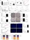
Fig. 1
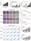
Fig. 1. LINC-PINT is downregulated in NPC and acts as a tumor suppressor.
a The fold change of LINC-PINT expression in NPC cells (HNE1, HONE1, CNE1, and CNE2) compared with that in immortalized nasopharyngeal epithelial cell line NP69 by qRT-PCR analysis. b The expression of LINC-PINT in NPC tumor tissues from our data (left) and the GDS3341 dataset (right). c In our data, the area under the ROC curve showing the discriminatory power of the LINC-PINT expression to predict the risk of NPC. d Correlation between LINC-PINT expression and treatment efficacy. CR complete response, PR partial response, SD stable disease, PD progressive disease. e Correlation between LINC-PINT expression and expressions of oncogenes (KRAS, MMP1, MYC, and TP53) in the NPC samples from the GDS3341 dataset. f HNE1 and HONE1 cells overexpressed LINC-PINT and verified by qRT-PCR. g LINC-PINT overexpression inhibited colony formation in NPC cells. Representative images (left) and quantitative analyses (right) are shown. h LINC-PINT overexpression suppressed cell proliferation in NPC cell lines, which are detected by CCK-8 assay. i Effect of LINC-PINT overexpression on the cell cycle progression. j Hoechst staining (green fluorescence) was also used to detect the effect of LINC-PINT on changes in apoptosis (up), Scale bar = 100 μm. Statistical diagrams show significant differences (bottom). Data represent the mean ± SEM from three independent experiments (aj). Significance calculated with Student’s T test. *P < 0.05, **P < 0.01, ***P < 0.001. Data shown are mean ± SEM.
Fig. 2
Fig. 2. LINC-PINT is transcriptionally induced by DNA damage and increases radiosensitivity in vitro.
a LINC-PINT was induced upon DNA damage with different DNA-damaging agents. HNE1 cells were treated with the DNA-damaging agents: Eto (10 μM), Bleo (1 μg/ml), TMZ (125 μM), and IR (8 Gy). LINC-PINT levels were measured by qRT-PCR. b Expression of LINC-PINT was determined by real-time RT-PCR after treating HNE1 cells with different DNA-damaging agents at different time points. RNA samples were analyzed by quantitative RT-PCR. c Representative images of clonogenic survival assay in HNE1 and HONE1 cells, which were transiently transfected with LINC-PINT overexpression plasmid or control plasmid, followed by a range of 0–8-Gy radiation doses. d It was shown that radiation survival curves were fitted by a linear quadratic equation. Up, HNE1 cells; Bottom, HONE1 cells. e Flow-cytometry analysis was performed to detect cell apoptosis for NPC cells, which were transfected with LINC-PINT vectors or negative controls and exposed to different doses of radiation. f Quantitative analyses of cell apoptosis in HNE1 (up) and HONE1 (bottom). Data represent the mean ± SEM from three independent experiments (af). Statistical significance was calculated using a two-sided t test. *P < 0.05, **P < 0.01, ***P < 0.001.
Fig. 3
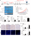
Fig. 3. LINC-PINT is a radiotherapy sensitizer for NPC in vivo.
a Treatment schedule in vivo. b Tumor images of xenografts in four groups at the end of the experiment. c Tumor growth curves of xenografts following initiated treatment with irradiation. Each tumor size was monitored and measured three times per week. d The volume (left), weight (middle), and the tumor inhibition rate (right) of xenograft tumors at sacrifice were analyzed. e Representative images of immunohistochemical staining for γH2AX (up) and TUNEL staining for apoptosis (bottom) in LINC-PINT-overexpressing tumors and control group with or without irradiation. Scale bar = 50 μm. f Quantification of γ-H2AX staining (up) and TUNEL staining (bottom) in four groups. Data represent the mean ± SEM. Statistical significance was calculated using a two-sided t test. *P < 0.05, **P < 0.01, ***P < 0.001.
Fig. 4
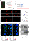
Fig. 4. LINC-PINT participates in DNA damage repair response.
a Heatmap of transcriptomic profiling in HNE1 cells transfected with LINC-PINT-expressing plasmid or control plasmid (green = downregulated; red = upregulated). b Gene ontology analysis of LINC-PINT-affected mRNA. The “Response to stress” network was among the top ten most differentially expressed. c, d IR-induced DNA damage in LINC-PINT-overexpressing cells and control group were analyzed by the comet assay at oh, 1, 2, 4, and 10 h. Representative images (c scale bars, 50 µm) and quantitative analysis (d) of tail DNA in each group. e, f Representative fluorescence images (e scale bars, 20 µm; green, γ-H2AX; blue, DAPI) and quantification (f) of γ-H2AX foci in cells with and without LINC-PINT overexpression. H2AX foci were counted at 0, 1, and 4 h post irradiation. g The expression of different DNA damage response proteins in HNE1 and HONE1 cells, which were transfected with LINC-PINT-overexpressing plasmid or control plasmid with and without irradiation. The protein level was analyzed by western blot more than three times. GAPDH was used as a loading control. Data represent the mean ± SEM from three independent experiments (cg). Statistical significance was calculated using a two-sided t test. *P < 0.05, **P < 0.01, ***P < 0.001.
Fig. 5
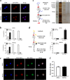
Fig. 5. DNA-PKcs is a direct target of LINC-PINT.
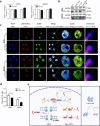
a Representative images of the FISH assay for detecting the subcellular localization of LINC-PINT in HNE1 and HONE1 cell lines (scale bar, 10 μm; blue, DAPI; red, LINC-PINT). b The expression of LINC-PINT in cytoplasmic and nuclear fractions of NPC cells. RNA samples were analyzed by qRT-PCR three times. Data represent the mean ± SEM. Statistical significance was calculated using a two-sided t test. *P < 0.05, **P < 0.01, ***P < 0.001. c Identification of the binding proteins of LINC-PINT in HNE1 and HONE1 cells. Left, Schematic diagram of the RNA pull-down strategy. Right, results of Silver staining. d RIP was performed using anti-DNA-PKcs antibodies. Left, schematic diagram of the RIP assay. Right, the levels of LINC-PINT and GAPDH in the coprecipitates were determined by qRT-PCR. e, f Representative fluorescence images (e scale bars, 10 µm; blue, DAPI; red, LINC-PINT; green, DNA-PKcs) and quantification (f n = 3 biologically independent samples) of colocalization between LINC-PINT and DNA-PKcs in HNE1 and HONE1 cells. Pearson’s R values were analyzed by Image J.
Fig. 6
Fig. 6. Mechanism of the radiosensitizing effect of LINC-PINT.
a, b LINC-PINT modulated the protein level of DNA-PKcs. After transfection, cells were treated with or without 8-Gy radiation and subjected to qRT-PCR (a) and western blot (b) for DNA-PKcs. Data represent the mean ± SEM from three independent experiments. Statistical significance was calculated using a two-sided t test. *P < 0.05, **P < 0.01, ***P < 0.001. c, d Representative fluorescence images (c scale bars, 10 µm; blue, DAPI; red, pDNA-PKcs; green, Ku80) and quantification (d n = 3 biologically independent samples) of colocalization between pDNA-PKcs (Ser2056) and Ku80 in HNE1 and HONE1 cells. Pearson’s R values were analyzed by Image J. e The molecular pathway of LINC-PINT-mediated radiosensitization. On the one hand, LINC-PINT was responsive to DNA damage, inhibiting DNA damage repair through ATM/ATR-Chk1/Chk2 signaling pathways. On the other hand, LINC-PINT increased radiosensitivity by interacting with DNA-PKcs and negatively regulated the expression and recruitment of DNA-PKcs. Therefore, LINC-PINT may confer radiosensitivity to NPC.

Similar articles

Cited by

References

    1. Ferlay J, et al. Estimating the global cancer incidence and mortality in 2018: GLOBOCAN sources and methods. Int. J. Cancer. 2019;144:1941–1953. doi: 10.1002/ijc.31937. - DOI - PubMed
    1. Chen YP, et al. Nasopharyngeal carcinoma. Lancet. 2019;394:64–80. doi: 10.1016/S0140-6736(19)30956-0. - DOI - PubMed
    1. Lee AW, Ma BB, Ng WT, Chan AT. Management of nasopharyngeal carcinoma: current practice and future perspective. J. Clin. Oncol. 2015;33:3356–3364. doi: 10.1200/JCO.2015.60.9347. - DOI - PubMed
    1. Curtin NJ. DNA repair dysregulation from cancer driver to therapeutic target. Nat. Rev. Cancer. 2012;12:801–817. doi: 10.1038/nrc3399. - DOI - PubMed
    1. Goldstein M, Kastan MB. The DNA damage response: implications for tumor responses to radiation and chemotherapy. Annu. Rev. Med. 2015;66:129–143. doi: 10.1146/annurev-med-081313-121208. - DOI - PubMed

Publication types

MeSH terms

Substances