. 2021 Apr 29;16(4):e0250454.
doi: 10.1371/journal.pone.0250454. eCollection 2021.

Castration causes an increase in lysosomal size and upregulation of cathepsin D expression in principal cells along with increased secretion of procathepsin D and prosaposin oligomers in adult rat epididymis

Affiliations

Castration causes an increase in lysosomal size and upregulation of cathepsin D expression in principal cells along with increased secretion of procathepsin D and prosaposin oligomers in adult rat epididymis

Lorena Carvelli et al. PLoS One. .

Abstract

In the epididymis, lysosomal proteins of the epithelial cells are normally targeted from the Golgi apparatus to lysosomes for degradation, although their secretion into the epididymal lumen has been documented and associated with sperm maturation. In this study, cathepsin D (CatD) and prosaposin (PSAP) were examined in adult epididymis of control, and 2-day castrated rats without (Ct) and with testosterone replacement (Ct+T) to evaluate their expression and regulation within epididymal epithelial cells. By light microscope-immunocytochemistry, a quantitative increase in size of lysosomes in principal cells of Ct animals was noted from the distal initial segment to the proximal cauda. Androgen replacement did not restore the size of lysosomes to control levels. Western blot analysis revealed a significant increase in CatD expression in the epididymis of Ct animals, which suggested an upregulation of its expression in principal cells; androgens restored levels of CatD to that of controls. In contrast, PSAP expression in Ct animals was not altered from controls. Additionally, an increase in procathepsin D levels was noted from samples of the epididymal fluid of Ct compared to control animals, accompanied by an increased complex formation with PSAP. Moreover, an increased oligomerization of prosaposin was observed in the epididymal lumen of Ct rats, with changes reverted to controls in Ct+T animals. Taken together these data suggest castration causes an increased uptake of substrates that are acted upon by CatD in lysosomes of principal cells and in the lumen by procathepsin D. These substrates may be derived from apoptotic cells noted in the lumen of proximal regions and possibly by degenerating sperm in distal regions of the epididymis of Ct animals. Exploring the mechanisms by which lysosomal enzymes are synthesized and secreted by the epididymis may help resolve some of the issues originating from epididymal dysfunctions with relevance to sperm maturation.


Conflict of interest statement

The authors have declared that no competing interests exist.

Figures

Fig 1
Fig 1. Immunostaining of epididymal tubules with cathepsin D of caput, corpus and cauda regions from control (Con), castrated (Ct) and castrated with testosterone supplementation (Ct+T) animals.
Small dense spherical reactive lysosomes (arrows with winged head) of principal cells (P) are evident in (Con) animals. In (Ct) animals, lysosomes (arrows with triangular head) appear to be larger in size and more abundant, with some remaining large in (Ct+T) animals. Some of these lysosomes show a dense homogeneous appearance, while others show a densely stained ring enveloped by a pale central core (triangular arrows). Lu, lumen; it, interstitial space; Sp, spermatozoa; reactive basal (arrowheads) and clear cells (C); n, nuclei of principal cells. Scale bars = 20 μm.
Fig 2
Fig 2. Quantification of lysosomal profile areas of principal cells.
Lysosomal profile areas were estimated from digital images obtained by immunocytochemical studies for control (Con), castrated (Ct) and castrated with testosterone supplementation (Ct+T) animals. Calculations were performed on principal cells of the distal IS and proximal caput designated as the IS/Caput region and the distal corpus and proximal cauda designated as the corpus/cauda region. Bars represent the means of lysosomal profile area per cell ± SEM from three independent experiments for IS/caput (A) and corpus/cauda (B) regions. a, b and c are significantly different from each other (p < 0.0001, obtained by Tukey’s Multiple Comparisons Test).
Fig 3
Fig 3. Immunostaining of epididymal tubules with cathepsin D and prosaposin of the initial segment from control (Con), castrated (Ct) and castrated with testosterone supplementation (Ct+T) animals.
Small dense reactive spherical lysosomes (winged arrows) are seen in principal cells (P) of (Con) animals. However, lysosomes appear to be more abundant and larger in size in (Ct) and (Ct+T) animals with both antibodies, with some revealing a pale central core surrounded by a ring of dense reactivity (triangular arrows). Few basally located apoptotic bodies (apo) are evident in epithelium of (Ct) and (Ct+T) animals. The lumen of a (Ct+T) animal contains cellular debris (curved arrows). Lu, lumen; it, interstitial space; Sp, spermatozoa; reactive basal (arrowheads), narrow (N) and apical (A) cells; n, nuclei of principal cells. Scale bars = 25 μm.
Fig 4
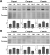
Fig 4
Immunodetection of cathepsin D in whole epididymal tissue (A) and luminal fluid (B) of the epididymis from control (Con), castrated (Ct) and castrated with testosterone supplementation (Ct+T) animals. Three regions of the epididymis were examined, i.e. IS/caput, corpus and cauda. Bands of 52 kDa and 48 kDa corresponding to immature and intermediate forms of CatD, respectively, are indicated. The figure shows a representative immunoblot for each region and experimental condition and the quantification of the bands in each case (intermediate CatD for tissues and immature form in fluids). Bars represent the means of relative optical density (ROD) ± SE from three independent experiments. (*) and (**) are significant differences from the control (p < 0.05 and p < 0.01 respectively, obtained by Tukey’s multiple comparisons Test). Ponceau staining was used as loading control.
Fig 5
Fig 5. Low power of epididymal tubules of caput, corpus and cauda regions from control (Con), castrated (Ct) and castrated with testosterone supplementation (Ct+T) animals immunostained with prosaposin.
While small reactive spherical lysosomes (winged arrows) appear in principal cells (P) of all 3 groups, in (Ct) animals, lysosomes (triangular head) appear to be larger in size and more abundant, with some remaining large in (Ct+T) animals. Some of these lysosomes show a dense homogeneous appearance, while others show a densely stained ring enveloped by a pale central core (triangular arrows). Apo, apoptotic basal cell; Lu, lumen; it, interstitial space; Sp, spermatozoa; reactive basal (arrowheads) and clear (C) cells; n, nuclei of principal cells. Scale bars = 20 μm.
Fig 6
Fig 6. High power of epididymal tubules of caput, corpus and cauda regions from castrated (Ct) and castrated with testosterone supplementation (Ct+T) animals immunostained with prosaposin.
In both (Ct) and (Ct+T) animals, lysosomes (arrows with triangular head) within principal cells (P) appear to be larger in size and more abundant. Some of these lysosomes show a dense homogeneous appearance, while others show a densely stained ring enveloped by a pale central core (triangular arrows). Small reactive spherical lysosomes (winged arrows) also appear in principal cells (P) of all 3 groups. Apo, apoptotic basal cell; Lu, lumen; it, interstitial space; reactive basal cells (arrowheads); n, nuclei of principal cells. Scale bars = 10 μm.
Fig 7
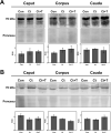
Fig 7
Immunodetection of prosaposin in whole epididymal tissue (A) and luminal fluid (B) of the epididymis from control (Con), castrated (Ct) and castrated with testosterone supplementation (Ct+T) animals. The figure shows a representative immunoblot of each region and experimental condition and the quantification of the bands in each case. Bars represent the means of relative optical density (ROD) ± SE from three independent experiments. No significant differences were noted between (C, Ct and Ct+T). Ponceau staining was used as loading control.
Fig 8
Fig 8. Apoptotic cells in the lumen of castrated animals with and without testosterone replacement.
Lumen and epithelium of the initial segment (a-c) and caput epididymidis (d-f) immunostained with PSAP of castrated with testosterone supplementation (a-c) and castrated (Ct) animals (d), and with CatD of Ct animals (e, f). In the lumen a plethora of abnormal structures with diverse shapes, sizes and content are noted, with some revealing a nucleus (curved arrows). Other small and larger structures had a more homogeneous appearance but without much content (asterisks). Many tiny fragments with a homogeneous appearance were also evident in the lumen (circled). Sperm were not evident in the initial segment, but some were seen in the caput region (Sp). In Fig 7F an apopotic cell appears at the base of the epithelium, while other cells appear to be sloughing from the epithelium into the lumen (rectangular box). Overall, the presence of these abnormal structures/cells suggests the apoptotic state of some of the epithelial cells after castration. Lu, lumen; it, interstitial space; reactive basal (arrowheads), narrow (N) and clear (C) cells; n, nuclei of principal cells. Scale bars = 20 μm.
Fig 9
Fig 9. Co-immunoprecipitation of cathepsin D with prosaposin.
Epididymal fluid from control versus castrated rats were mixed with beads preincubated with anti-PSAP antibody, as described in Materials and methods. After centrifugation and washing, cathepsin D was detected from the precipitates by immunoblot. Immature (52 kDa) bands of the enzyme were detected in the precipitates. Detection of IgG (55 kDa, heavy chain) was used as a loading control. The figure is representative of two independent experiments.
Fig 10
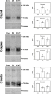
Fig 10. Detection of oligomeric forms of prosaposin in epididymal fluid from control (Con), castrated (Ct) and castrated with testosterone supplementation (Ct+T) animals.
Proteins from epididymal fluid were run on SDS-gel under non-reducing conditions and electrotransferred onto nitrocellulose membrane for detection with the specific antibody. Monomers (70 kDa) and oligomers (250 kDa) of PSAP were detected. The figure shows representative immunoblots from each epididymal region and the corresponding quantification of the bands, as indicated. Bars represent the means of relative optical density (ROD) ± SE from three independent experiments. (*) significant differences from the control (p < 0.05, obtained by Tukey’s Multiple Comparisons Test). Ponceau staining was used as loading control.

Similar articles

Cited by

References

    1. Robaire B, Hermo L. Efferent Ducts, epididymis and vas deferens: structure, functions, and their regulation. In: Knobil E, Neill J, editors. The Physiology of Reproduction. New York: Raven Press, Ltd.; 1988. pp. 999–1080.
    1. Cornwall GA. Role of posttranslational protein modifications in epididymal sperm maturation and extracellular quality control. Adv Exp Med Biol. 2014;759: 159–180. 10.1007/978-1-4939-0817-2_8 - DOI - PubMed
    1. Breton S, Nair A V., Battistone MA. Epithelial dynamics in the epididymis: role in the maturation, protection, and storage of spermatozoa. Andrology. 2019;7: 631–643. 10.1111/andr.12632 - DOI - PMC - PubMed
    1. Hamilton DW. Structure and function of the epithelium lining efferentes, ductus epididymidis and ductus deferens in the rat. In: Hamilton D, Greep R, editors. Handbook of Physiology. Washington DC: American Physiological Society; 1975. pp. 259–301. Available: https://ci.nii.ac.jp/naid/10025550105
    1. Adamali HI, Hermo L. Apical and narrow cells are distinct cell types differing in their structure, distribution, and functions in the adult rat epididymis. J Androl. 1996;17: 208–222. 10.1002/j.1939-4640.1996.tb01776.x - DOI - PubMed

Publication types

Grants and funding

Funded by Secretaría de Investigación, Internacionales y Posgrado, Universidad Nacional de Cuyo (SIIP). Grant number: M066. Recipient: Dr. Lorena Carvelli. http://www.uncuyo.edu.ar/ciencia_tecnica_y_posgrado/upload/rre414220191.pdf., and Natural Sciences and Engineering Research Council of Canadá (NSERC) Operating Grant Nr. 1153952 to Carlos R. Morales. The funders had no role in study design, data collection and analysis, decision to publish, or preparation of the manuscript.

LinkOut - more resources