. 2021 Apr 29;16(4):e0249876.
doi: 10.1371/journal.pone.0249876. eCollection 2021.

Effect of O-linked glycosylation on the antigenicity, cellular uptake and trafficking in dendritic cells of recombinant Ber e 1

Affiliations

Effect of O-linked glycosylation on the antigenicity, cellular uptake and trafficking in dendritic cells of recombinant Ber e 1

Nuzul N Jambari et al. PLoS One. .

Abstract

Ber e 1, a major Brazil nut allergen, has been successfully produced in the yeast Pichia pastoris expression system as homogenous recombinant Ber e 1 (rBer e 1) with similar physicochemical properties and identical immunoreactivity to its native counterpart, nBer e 1. However, O-linked glycans was detected on the P.pastoris-derived rBer e 1, which is not naturally present in nBer e 1, and may contribute to the allergic sensitisation. In this study, we addressed the glycosylation differences between P. pastoris-derived recombinant Ber e 1 and its native counterparts. We also determined whether this fungal glycosylation could affect the antigenicity and immunogenicity of the rBer e 1 by using dendritic cells (DC) as an immune cell model due to their role in modulating the immune response. We identified that the glycosylation occurs at Ser96, Ser101 and Ser110 on the large chain and Ser19 on the small polypeptide chain of rBer e 1 only. The glycosylation on rBer e 1 was shown to elicit varying degree of antigenicity by binding to different combination of human leukocyte antigens (HLA) at different frequencies compared to nBer e 1 when tested using human DC-T cell assay. However, both forms of Ber e 1 are weak immunogens based from their low response indexes (RI). Glycans present on rBer e 1 were shown to increase the efficiency of the protein recognition and internalization by murine bone marrow-derived dendritic cells (bmDC) via C-type lectin receptors, particularly the mannose receptor (MR), compared to the non-glycosylated nBer e 1 and SFA8, a weak allergenic 2S albumin protein from sunflower seed. Binding of glycosylated rBer e 1 to MR alone was found to not induce the production of IL-10 that modulates bmDC to polarise Th2 cell response by suppressing IL-12 production and DC maturation. Our findings suggest that the O-linked glycosylation by P. pastoris has a small but measurable effect on the in vitro antigenicity of the rBer e 1 compared to its non-glycosylated counterpart, nBer e 1, and thus may influence its applications in diagnostics and immunotherapy.


Conflict of interest statement

The authors have declared that no competing interests exist.

Figures

Fig 1
Fig 1. Detection of glycosylation on 2S albumin proteins by periodic acid Schiff’s (PAS) staining.
(A.) A positive glycoprotein control, CPDY (1.), rSFA8 (2.), rBer e 1 (3.), and nBer e 1 (4.) were separated on a 12% BisTris NuPAGE gel under denaturing conditions (300 mM DTT) and PAS stained to detect glycoproteins. Only the ~15 kDa band of rBer e 1 and the positive control, CPDY, reacted with the PAS stain, indicating the presence of glycan residues. nBer e 1 and rSFA8 did not react with PAS stain, thus indicate the absence of glycosylation according to this assay. (B.) An identical NuPAGE gel stained with Coomassie brilliant blue shows all of the proteins present. Raw images of Commassie- and PAS-stained gels are presented in (S3 Fig). (C.) rBer 1, nBer e 1 and rSFA8 protein bands were excised for peptide identification. Samples of rBer e 1(I and II), and nBer e 1 (III and IV) and rSFA8 (V) were trypsinised and analysed by LC-MSMS analysis. (D.) Amino acid sequence of precursor native Ber e 1 in planta. Italic fonts represent the N-terminal signal sequence, the underlined fonts represent the pre-prosignal sequences, and the italic and underlined fonts represent the short linker and C-terminal linker. These sequences were cleaved and removed to produce mature Ber e 1 in planta that consists of small subunit (yellow highlight) and large subunit. (E.) Amino acid sequence of mature recombinant Ber e 1 retained the spacer sequence (bold fonts) downstream of the Kex2 cleavage site, spacer, and C-terminal vacuolar targeting signal sequence (italic and underlined fonts) and it is made up of small subunit (yellow highlight) and large subunit [16, 24].
Fig 2
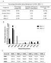
Fig 2. In vitro antigenicity of rBer e 1 and nBer e 1 by DC-T cell response assay.
(A.) Response Index (RI) values for rBer e 1, nBer e 1 and reference protein from percentage antigenicity and stimulation data (percent stimulation above background ≥ 0.02%, SEM = 2) (B.) Graph of responding donors with percentage stimulation above 0.02% (SEM = 2) after stimulating with nBer e 1 and rBer e 1. Dotted line indicates higher stimulation cut-off of 0.5% (C.) Table of DRB1, DQB1, and DPB1 allele profiles of donors responding at the higher stimulation cut off of ≥0.5% (SEM = 2) when stimulated with rBer e 1 or nBer e 1. Percentage stimulation above this cut-off indicates a greater significance of antigenic response.
Fig 3
Fig 3. Uptake of glycosylated Ber e 1 and non-glycosylated Ber e 1 and SFA8 by bmDC.
(A.) The contour plots represent cell populations where cells in the gating area are CD11c+ bmDC with A488-labelled proteins. DC uptake was performed with increasing concentration (1–50 μg/ml) of A488-labelled 2S albumin proteins for 45 min (B.). Data were normalised as a percentage of A488+ and PE+ gated cells relative to the untreated control. (C.) The contour plot showed the distribution of CD11c+/A488+ or FITC+ cell population in the gated region. Proteins were internalised as shown when bmDC were pulsed for 45 min with labelled proteins at 4°C (cell-surface bound) and 37°C (internalised). Data were normalised to a percentage of the A488/FITC+ bmDC population incubated with Dextran-FITC at 37°C and are presented as mean ±S.D. (n = 2) ****p<0.0001 (D.).
Fig 4
Fig 4. Mechanisms of endocytosis of 2S albumins in bmDC and their uptake in CHO and CHO-MR.
(A.) The effect of macropinocytosis inhibitors, DMA and (B.) rottlerin, and (C.) mannose competitive inhibitor, mannan, on the endocytosis of rBer e 1-A488, nBer e 1-A488 and rSFA8-A488 into bmDC was determined by flow cytometry. (D.) The flow cytometry histogram of uptake of A488-labelled proteins by CHO and CHO-MR cell line, as analysed using flow cytometry (E.) The graph showed differences in the uptake of fluorescent-labelled rBer e 1, nBer e 1 and rSFA8 by CHO and CHO-MR where data are presented as normalised median fluorescent intensity (MFI) of cells pulsed with labelled proteins to unpulsed cells and presented as mean ±SD. ** p<0.01, *** p<0.001, ****p<0.0001. Statistical differences are relative to the untreated control.
Fig 5
Fig 5. Confocal fluorescence imaging of CD11c+ bmDC for colocalization analysis.
The colocalization between (A.) rBer e 1-A488 (green) and nBer e 1-A647 (red), (B.) rBer e 1-A488 (green) and rSFA8-A647 (red) and (C.) rBer e 1-A488 (green) and rSFA8-A647 (red), were visualised at different chasing times (0 min, 15 min, 45 min, and 120 min). Gradual reduction of fluorescent-labelled rBer e 1, nBer e 1, and rSFA8 in bmDC were observed over time. Bright spots of colocalized ROI represent areas where both proteins co-occurred. Scale bar for all images = 10 μm. Colocalization after different chasing times were compared for co-occurred regions representing overlapped of the two fluorescent labelled proteins in puncta and measured as MCC. MCC for fractional overlap of A488-labelled proteins and A647-labelled proteins were compared in graphs (D.–F.).
Fig 6
Fig 6. Co-stimulatory molecular markers and Th1/Th2 cytokine production upon CD11c+ bmDC stimulation with 2S albumins.
(A.) Percentage population of CD80+ or CD86+ bmDC post stimulation with 50 μg/ml rBer e 1, nBer e 1, or rSFA8, or 10 ng/mL LPS (positive control) were normalised to non-stimulated bmDC. (B.) Level of Th1 (IL-12, and IFN-γ)—and Th2 (IL-2, IL-4, IL-5)-specific cytokines detected in bmDC stimulated with 50 μg/ml of nBer e 1, rBer e 1 rSFA8 or 10 ng/ml LPS were measured after 24 hours by Luminex assay. Data are from three biological replicates and presented as mean ± SD (****, p < 0.0001) relative to non-stimulated bmDC.

Similar articles

Cited by

References

    1. Schmid-grendelmeier P, Crameri R. Recombinant allergens for skin testing. Int Arch Allergy Immunol. 2001;125:96–111. 10.1159/000053803 - DOI - PubMed
    1. Menz G, Dolecek C, Ferreira F, Moser M, Suter T, M S, et al.. Serological and skin-test diagnosis of bireh pollen allergy with recombinant Bet v I, the major birch pollen allergen. Clin Exp Allergy. 1996;26(26):50–60. - PubMed
    1. Lin J, Shewry R, Archer DB, Beyer K, Niggemann B, Haas H, et al.. The Potential Allergenicity of two 2S albumins from soybean (Glycine max): A protein microarray approach. Int Arch Allergy Immunol. 2006;141:91–102. 10.1159/000094535 - DOI - PubMed
    1. Ree R Van, Leeuwen WA Van, Aalberse RC. How far can we simplify in vitro diagnostics for grass pollen allergy?: A study with 17 whole pollen extracts and purified natural and recombinant major allergens. J Allergy Clin Immunol. 1998;102:184–90. 10.1016/s0091-6749(98)70084-3 - DOI - PubMed
    1. Ree R Van, Leeuwen WA Van, Akkerdaas JH, Aalberse RC. How far can we simplify in vitro diagnostics for Fagales tree pollen allergy? A study with three whole pollen extracts and purified natural and recombinant allergens. Clin Exp Allergy. 1999;29:848–55. 10.1046/j.1365-2222.1999.00521.x - DOI - PubMed

Publication types

MeSH terms

Grants and funding

NNJ received PhD scholarship from the Ministry of Higher Education Malaysia (http://mohe.gov.my/en/). The funders had no role in study design, data collection and analysis, decision to publish, or preparation of the manuscript.