Review
. 2021 Aug 20;433(17):166994.
doi: 10.1016/j.jmb.2021.166994. Epub 2021 Apr 16.

On the molecular nature of large-pore channels

Affiliations
Review

On the molecular nature of large-pore channels

Johanna Syrjanen et al. J Mol Biol. .

Abstract

Membrane transport is a fundamental means to control basic cellular processes such as apoptosis, inflammation, and neurodegeneration and is mediated by a number of transporters, pumps, and channels. Accumulating evidence over the last half century has shown that a type of so-called "large-pore channel" exists in various tissues and organs in gap-junctional and non-gap-junctional forms in order to flow not only ions but also metabolites such as ATP. They are formed by a number of protein families with little or no evolutionary linkages including connexin, innexin, pannexin, leucine-rich repeat-containing 8 (LRRC8), and calcium homeostasis modulator (CALHM). This review summarizes the history and concept of large-pore channels starting from connexin gap junction channels to the more recent developments in innexin, pannexin, LRRC8, and CALHM. We describe structural and functional features of large-pore channels that are crucial for their diverse functions on the basis of available structures.

Keywords: CALHM; Connexin; Innexin; LRRC8; Pannexin.


Conflict of interest statement

Declaration of interests

The authors declare no competing interests.

Figures
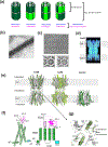
Fig. 1.
Fig. 1.. Connexin gap junction channels.
(a) Various types of connexin hemichannels can assemble to form distinct gap junction channels. Connexin can form homo- or hetero-hexameric hemichannels where they form homotypic or heterotypic gap junctions. Note that the pattern of heteromeric and heterotopic protomer arrangements are arbitrary since they have not been determined experimentally. (b) Electron microscopic observation of the gap junction in Mauthner cell synapses. (c) Isolated gap junction sheets in a 2D crystalline array viewed from the ‘top’ (top panel). Maps of the gap junction channels from image analysis in the presence (left) and absence (right) of Ca2+. (d) Maps calculated from cryo-electron crystallography showing rod-like structures representing transmembrane helices. (e) X-ray crystallographic structure of Cx26 (left, PDB code: 2ZW3) and cryo-EM structures of Cx50 (middle, PDB code: 7JJP) and Cx31.3 hemichannel (right, PDB code: 6L3T). Sticks in the Cx50 structure represent resolved lipid molecules at the extracellular leaflet of the plausible lipid bilayer. Gray belts represent the approximate location of the lipid bilayers. Note that the Cx50 model was built from the averaged cryo-EM map of native Cx46/50. (f) A monomer of Cx31.1 and the schematic presentation of the structural topology. Note that in Cx26 and Cx50, the beta strand in the TM1-2 loop and ICH are missing or not resolved. (g) The gap junction formation occurs through interactions of the extracellular domain (ECD). Shown here is the interface of the Cx26 gap junction channel. The images in panel b, c, and d have been adapted from the article by Robertson [30], Unwin and Zampighi [35], Unwin and Ennis [36], and Unger et al. [37] Copyright was obtained from the journals, J. Cell Biology, Nature, and Science.
Fig. 2.
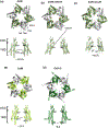
Fig. 2.. Pore sizes of connexin channels.
(a-e) The views of hemichannels from the extracellular side (upper panels) and from the side (lower panels). Only two opposing subunits are shown for clarity. The locations of the narrowest constrictions at the N-terminal regions are highlighted with dotted lines except for Cx26 calcium (panel b; calcium in red spheres) where the N-terminal ends are not well resolved. At low pH, the N-terminal region narrows down (panel c). Also note that the orientations of NTHs in Cx31.3 (panel e) are different from the rest. Cx26 low pH in the closed conformation (PDB code: 6UVT) is modelled based on a 7.5 Å cryo-EM map. Cx26 calcium (PDB code: 5ER7) is from the X-ray crystallographic data at 3.28 Å.
Fig. 3.
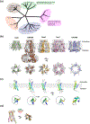
Fig. 3.. Overview of large-pore channels.
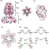
(a) Phylogenic tree all members of LRRC8, pannexin, and CALHM and some representative members of connexin and innexin families. Other family members of connexin and innexin are listed next to the tree. (b) Representative structures from different large-pore channels from the side (upper panels) and the top (lower panels) of the membrane plane. The PDB codes of Cx26, LRRC8A, Panx1, Inx6, and CALHM1 shown here are 2ZW3, 6G9O, 6VD7, 5H1Q, and 6VAM, respectively. Only one hemichannel of the Cx26 gap junction structure is shown here for clear comparison with others. The numbering in the lower panel is to show different oligomeric states. (c) The monomeric subunits of Cx26, LRRC8A, Panx1, Inx-6, and CALHM1 viewed from the side (upper panel) and from the top (lower panel) of the membrane plane where the first, second, third, and fourth transmembrane helices are colored blue, cyan, green, and yellow, respectively. (d) Superposition of the transmembrane region of Cx26, LRRC8A, Panx1, and Inx6 from the side (left) and the top (right) showing the similar helical orientation. The transmembrane helices of CALHM1 cannot be superimposed and therefore was excluded here.
Fig. 4.
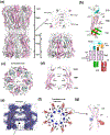
Fig. 4.. Structures of Inx-6 gap junction channel and hemichannel.
(a) The Inx-6 gap junction channel (left) and hemichannel (right) with subunits colored in alternating pink and white. The domain layers are partitioned into the transmembrane domain (TMD), the extracellular domain (ECD), and the intracellular domain (ICD). The two hemichannels interact at the ECD. Spheres in pale cyan are a conserved sequence (YYQWV) in the innexin family. (b) A monomer of Inx-6 and the schematic presentation of the structural topology. (c) Inx-6 hemichannel viewed from the extracellular side showing the octameric arrangement. The dotted line (18 Å) is drawn between Ala7 of the opposing subunits. (d) Side view showing two opposing subunits. Distances of narrow constrictions are measured between E1Hs, NTHs, and CLH1s. (e) The side view of cryo-EM density illustrating the presence of lipid-like density in Inx-6 in nanodiscs. (f) In addition to the lipid-like density, two amorphous densities, X (blue mesh) and Y (red mesh), are observed. (g) Close-up view of a single protomer and the locations of the X and Y densities. The X and Y densities are not present in the structure of the NTH deleted construct (residue 2–19 deletion).
Fig. 5.
Fig. 5.. Structure of homo-hexameric LRRC8A.
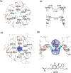
(a) Shown here is the structure of mouse LRRC8A (PDB code: 6G9O) where alternate subunits are colored brown and white. The side view shows four layers, ECD, TMD, ICL, and LRR. (b) Top views of LRRC8A in different layers. ECD, TMD, and ICL have C6 or pseudo-C6 symmetry whereas the LRR layer has C3 symmetry. (c) The structure of the LRRC8A protomer. The color code of the structure (left panel) is the same as the one for the schematic topology presentation (right panel). (d-e) The structure of human LRRC8A in detergent refined using C3 symmetry (PDB code: 5ZSU) where two distinct interfaces, ‘loose interface’ (panel d) and ‘tight interface’ (panel e) are observed. The gap in the ‘loose interface’ is more pronounced in the LRR layer than the ICL and TMD layers. (f) The structure of mouse LRRC8A in lipid nanodisc (PDB: 6NZZ) refined with C6 symmetry. In this study, the LRR layer was not modeled due to disordered density. The extent of inter-subunit packing is between those of ‘loose interface’ and ‘tight interface’ in panel d and e. (g) In this lipid nanodisc sample, the six inter-subunit interfaces in the TMD equally contain three molecules of phospholipids per interface, which strengthen the subunit-subunit interactions.
Fig. 6.
Fig. 6.. Narrow constriction at ECD of LRRC8A.
(a) The structure of human LRRC8A (PDB code: 5ZSU) looking ‘down’ the ECD. The narrow constriction is formed by six arginine residues (Arg103, cyan sticks), which are located on ECL1Hs. (b) The side view of the narrow constriction. Here only the two subunits opposite each other are shown for clarity. The Arg103 ring has a diameter of 7.6 Å whereas the second narrowest constriction with a diameter of 13.7 Å is created by Thr48 on TM1. (c and d) The narrow constriction in ECD is a binding site for a channel blocker, DCPIB (brown stick). Shown in mesh is the cryo-EM density likely representing DCPIB viewed from the top (c) and side (d). DCPIB density plugs ECD constriction around Arg103. The LRRC8A-DCPIB structure shown here is in the expanded conformation (PDB code: 6NZZ)
Fig. 7.
Fig. 7.. Cryo-EM structure of homo-hexameric LRRC8D.
(a) Shown here is the structure of human LRRC8D (PDB code: 6M04) where alternate subunits are colored magenta and gray. The side view shows four layers, ECD, TMD, ICL, and LRR as in LRRC8A. (b) Top view without the LRR layer showing pseudo-C6 symmetry. (c) Top view of LRR showing C2 symmetry. (d) Top view showing the narrow constriction at ECD formed by Phe143 (green sticks). (e) Side view of human LRRC8D (left) and human LRRC8A (right). Here only the two subunits opposite to each other are shown for clarity. The diameters of the constriction sites at the ECD and TMD are wider in LRRC8D than LRRCA.
Fig. 8.
Fig. 8.. Overall topology and extracellular gate of Panx1.
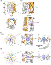
(a) Side view of the structure of human Panx1 (PDB: 6WBF). Panx1 harbors extracellular domain (ECD), transmembrane domain (TMD), and intracellular domain (ICD) layers. Sticks in the ECD and TMD represent N-linked glycosylation and bound lipids, respectively. (b) Topology of a Panx1 protomer viewed from within the plane of the membrane (left) and the schematic presentation of the structural topology (right). Disordered segments of the protein, including the intracellular loop and C-terminus, are represented by a dashed line. Caspase-7 cleaves off the last forty seven residues (scissors). (c) Close-up view of the extracellular constriction where Trp74 and Arg75 are shown as sticks (left panel). The ~9 Å diameter constriction is formed by Trp74 located on the N-terminal end of EH1. Cross-sectional view of the apo structure showing distance between Trp74 and Arg75 from neighboring protomers and potential cation-pi interactions (right panel).
Fig. 9.
Fig. 9.. Features of Panx1 structures.
(a) The human Panx1 structure (PDB: 6WBG) viewed from the side of the membrane plane (top) and the cytoplasm (bottom). The cytoplasmic face of the channel pore is wide open with the measured diameter of ~40 Å. Lipids, N-linked glycosylation, and the side tunnel residues are shown in sticks (top). The dotted square is the location of the side tunnel. (b) Close-up views of the side tunnel at the inter-protomer interfaces viewed from the outside (top) and inside (bottom) of the central pore. The side tunnel residues are located on TM1, TM2, and the loop between TM1 and NTH. The ordered lipids (purple sticks) are located at the entrance of the side tunnel. (c-d) Top-down view of the extracellular face of caspase-7 cleaved human Panx1 with CBX (‘+CBX’; left and middle panel c) (PDB: 6WBG) and without CBX (no CBX, left and middle panel d) (PDB: 6WBI). Note that densities (blue mesh) in the ECD narrow constriction are observed in both no CBX and +CBX structures. Cross-sectional view of the ‘+CBX’ structure (right panel c). Side view of the density in no CBX showing clear density around Trp74 (right panel d). Note that the density is not sufficiently resolved to determine the presence of CBX although the molecular model (green sticks in panel c) was arbitrarily placed in the published PDB coordinates.
Fig. 10.
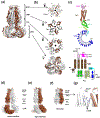
Fig. 10.. Basic architecture of CALHM.
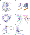
(a) Structure of CALHM1 from zebrafish (PDB ID: 6LYG) as an example. The structure is viewed from the plane of the membrane (upper panel) and the cytoplasm (lower panel). CALHM has an extracellular domain (ECD), a transmembrane domain (TMD) and an intracellular domain (ICD). Note that TMD2 and TMD4 in neighboring subunits form contacts. In the cytoplasm, the CTHs from a neighbor (n+1) and a neighbor of the neighbor (n+2) subunits interact with each other to stabilize the octameric arrangement. (b) Topology of a CALHM subunit. Each subunit contains four transmembrane helices (TM1-4), the N-terminal helix (NTH) in the middle of the central cavity, and ~40 residue long cytoplasmic helix (CTH). The remainder of the C-terminal structures are omitted for clarity. (c) Superposition of CALHM1 (slate), CALHM2 (magenta), CALHM4 (green), and CALHM5 (cyan) protomers (left panel) and of CALHM2 bound to RuR (magenta) and CALHM6 (orange) protomers (right panel) to illustrate that the arrangement of TM1 and NTH (highlighted) are different between the two groups. The NTH arrangement in CALHM1 differs from that in CALHM4 and 5 (left panel). The arrangement of TM2-4 is similar in all CALHMs. (d) CALHM1 viewed from the extracellular region to show locations of Asp121 (magenta sphere) on TM3, Arg154 (yellow sphere) in the extracellular domain, and Pro86 (cyan sphere) in the cytoplasmic loop between TM2 and 3. The residue numbers are based on human CALHM1. (e) Density likely representing cholesteryl hemisuccinate (CHS; green stick and density in slate mesh) is present around Asp121 in the structure of CALHM1 from killifish (PDB code: 6LMT).
Fig. 11.
Fig. 11.. Oligomeric assembly of CALHM proteins.
Structures of CALHM1, 2, 4, 5, and 6 and CLHM1 to illustrate variable numbers of oligomeric assembly. The PDB IDs used here are 6VAM, 6LMV, 6LOM, 6VAK, 6VAI, 6YTK, 6YTL, 6YTV, and 6YTX for CALHM1, CLHM1 (9-mer), CLHM1 (10-mer junction), CALHM2, CALHM2 (gap junction), CALHM4 (10-mer junction), CALHM4 (11-mer junction), CALHM6 (10-mer junction), and CALHM6 (11-mer junction), respectively.
Fig. 12.
Fig. 12.. Subunit interactions and CTH orientations in CALHM proteins.
(a) Structures of CALHM proteins viewed from the cytoplasm where the C-terminal domains containing CTHs are highlighted. The PDB IDs used here are 6VAM, 6LMV, 6LOM, 6VAK, 6YTK, 6YTL, 7D61, 6YTV, 6YTX, 6VAL, and 6LMX for CALHM1, CLHM1 (9-mer), CLHM1 (10-mer junction), CALHM2, CALHM4 (10-mer junction), CALHM4 (11-mer junction), CALHM5 (11-mer), CALHM6 (10-mer junction), CALHM6 (11-mer junction), CALHM1-2 chimera (11-mer), and CALHM1-2 chimera (9-mer), respectively. (b) TM4-CTH orientation for CALHM1 viewed from the cytoplasm. The critical Pro residue in the TM4-CTH loop is shown as a black sphere. TM2 from the neighboring subunit (Subunit B) is shown. (c-g) The TM4-CTH orientations of CLHM1 (c), CALHM2, 4, and 6 (d), CALHM1-2 (9-mer) chimera (e), CALH1-2 (11-mer) chimera (f), and CALHM5 (g) in comparison with CALHM1. The TM2-3 loop from the neighboring subunit in CALHM5 is shown in red. (h) Sequence alignment of the TM4-CTH linker region. For clarity, sequences from the human orthologues are shown for CALHM1-6. The critical prolines are highlighted by gray shades. Note that the C.elegans (ce) CLHM1 and CALHM5 do not have proline residues in the TM4-CTH linker. CALHM1-2 (11-mer; chicken CALHM1 and human CALHM2 chimera) and CALHM1-2 (9-merl killifish CALHM1 and human CALHM2 chimera) have distinct construct designs which place the critical proline residues at different positions.
Fig. 13.
Fig. 13.. Configurations of TMD1 and NTH and assessment of cryo-EM density.
(a-g) Structures of CALHM1 (a), CALHM2 (in EDTA) (b), CALHM2-RuR (c), CALHM2-gap (CALHM2-gap-choi) (d), CALHM4 (10-mer) (e), CALHM5 (f), and CALHM6 (10-mer) (g) viewed from the extracellular side (upper panels) and the plane of the membrane (lower panels). The orientations of TMD1 (1) and NTH can be classified as cylindrical (CALHM1, 2, 4, and 5) and conical (CALHM2-RuR, CALHM2-gap, and CALHM6). Note that CALHM2-gap from Syrjanen et al has the cylindrical conformation. In panel c, putative binding of RuR molecules are shown in blue spheres). (h) Comparison of conical conformations between CALHM2-RuR and CALHM6 (10-mer). Here, TM2-4 are superposed to highlight different orientations of TM1s. (i) Cryo-EM density (blue mesh) around the extracellular side of TM1 and 3 of CALHM2-RuR (left) and CALHM2-gap-choi (right). RuR is in stick (PDB code: 6UIW). A similar density (green mesh) in the equivalent region in CALHM2-gap-choi is surrounded by dotted lines. Note that CALHM2-gap-choi does not contain RuR.

Similar articles

Cited by

References

    1. Bevans CG, Kordel M, Rhee SK, Harris AL. Isoform composition of connexin channels determines selectivity among second messengers and uncharged molecules. J Biol Chem. 1998;273:2808–16. - PubMed
    1. Goldberg GS, Lampe PD, Nicholson BJ. Selective transfer of endogenous metabolites through gap junctions composed of different connexins. Nat Cell Biol. 1999;1:457–9. - PubMed
    1. Goodenough DA, Paul DL. Gap Junctions. 2019.
    1. Paul DL, Ebihara L, Takemoto LJ, Swenson KI, Goodenough DA. Connexin46, a novel lens gap junction protein, induces voltage-gated currents in nonjunctional plasma membrane of Xenopus oocytes. J Cell Biol. 1991;115:1077–89. - PMC - PubMed
    1. DeVries SH, Schwartz EA. Hemi-gap-junction channels in solitary horizontal cells of the catfish retina. J Physiol. 1992;445:201–30. - PMC - PubMed

Publication types

LinkOut - more resources