

Visualization of neurofibrillary tangle maturity in Alzheimer's disease: A clinicopathologic perspective for biomarker research
- PMID: 33797838
- PMCID: PMC8478697
- DOI: 10.1002/alz.12321
Visualization of neurofibrillary tangle maturity in Alzheimer's disease: A clinicopathologic perspective for biomarker research
Abstract
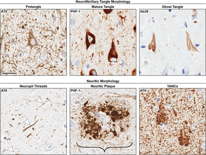
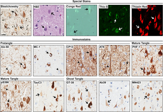
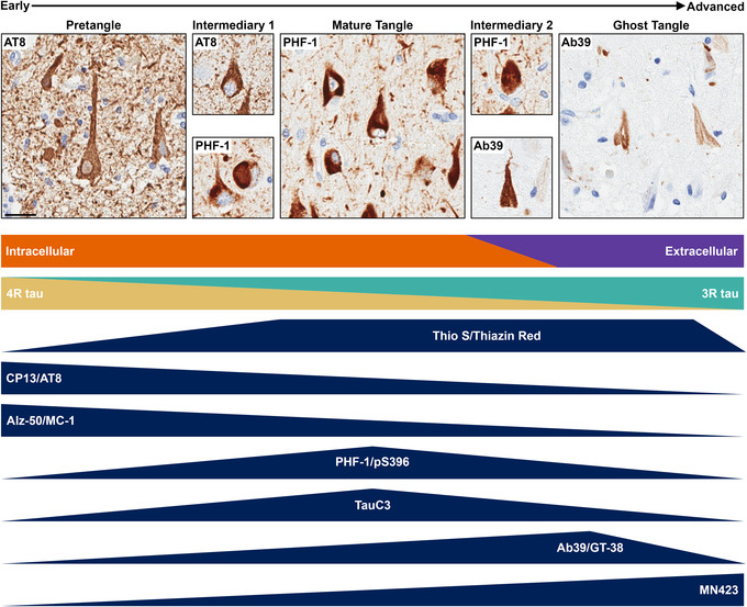
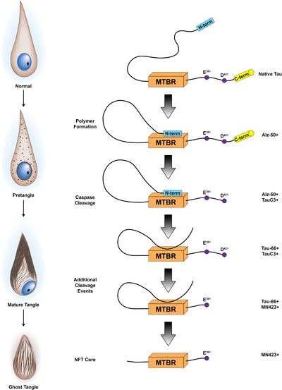
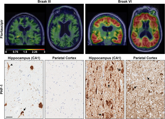
Neurofibrillary tangles, one of the neuropathologic hallmarks of Alzheimer's disease, have a dynamic lifespan of maturity that associates with progressive neuronal dysfunction and cognitive deficits. As neurofibrillary tangles mature, the biology of the neuron undergoes extensive changes that may impact biomarker recognition and therapeutic targeting. Neurofibrillary tangle maturity encompasses three levels: pretangles, mature tangles, and ghost tangles. In this review, we detail distinct and overlapping characteristics observed in the human brain regarding morphologic changes the neuron undergoes, conversion from intracellular to extracellular space, tau immunostaining patterns, and tau isoform expression changes across the lifespan of the neurofibrillary tangle. Post-translational modifications of tau such as phosphorylation, ubiquitination, conformational events, and truncations are discussed to contextualize tau immunostaining patterns. We summarize accumulated and emerging knowledge of neurofibrillary tangle maturity, discuss the current tools used to interpret the dynamic nature in the postmortem brain, and consider implications for cognitive dysfunction and tau biomarkers.
Keywords: Alzheimer's disease; neurofibrillary tangles; neuropathology; tau; tau positron emission tomography.
© 2021 The Authors. Alzheimer's & Dementia published by Wiley Periodicals LLC on behalf of Alzheimer's Association.
Conflict of interest statement
Dr. Moloney reports no disclosures. Dr. Lowe serves as a consultant for Bayer Schering Pharma, Philips Molecular Imaging, Piramal Imaging, AVID Radiopharmaceuticals, Eisai Inc., and GE Healthcare and receives research support from GE Healthcare, Siemens Molecular Imaging, AVID Radiopharmaceuticals, the NIH (NIA, NCI), and the MN Partnership for Biotechnology and Medical Genomics. Dr. Murray served as a paid consultant for AVID Radiopharmaceuticals.
Figures


Similar articles
-
Phosphorylated tau sites that are elevated in Alzheimer's disease fluid biomarkers are visualized in early neurofibrillary tangle maturity levels in the post mortem brain.Alzheimers Dement. 2023 Mar;19(3):1029-1040. doi: 10.1002/alz.12749. Epub 2022 Aug 3. Alzheimers Dement. 2023. PMID: 35920592 Free PMC article.
-
Characterization of neurofibrillary tangle immunophenotype signatures to classify tangle maturity in Alzheimer's disease.Alzheimers Dement. 2024 Jul;20(7):4803-4817. doi: 10.1002/alz.13922. Epub 2024 Jun 17. Alzheimers Dement. 2024. PMID: 38884346 Free PMC article.
-
Aluminum and Neurofibrillary Tangle Co-Localization in Familial Alzheimer's Disease and Related Neurological Disorders.J Alzheimers Dis. 2020;78(1):139-149. doi: 10.3233/JAD-200838. J Alzheimers Dis. 2020. PMID: 32925074 Free PMC article.
-
Are tangles as toxic as they look?J Mol Neurosci. 2011 Nov;45(3):438-44. doi: 10.1007/s12031-011-9566-7. Epub 2011 Jun 3. J Mol Neurosci. 2011. PMID: 21638071 Free PMC article. Review.
-
Pretangles and neurofibrillary changes: similarities and differences between AD and CBD based on molecular and morphological evolution.Neuropathology. 2014 Dec;34(6):571-7. doi: 10.1111/neup.12108. Epub 2014 Feb 26. Neuropathology. 2014. PMID: 24612177 Review.
Cited by
-
State-of-the-art imaging of neuromodulatory subcortical systems in aging and Alzheimer's disease: Challenges and opportunities.Neurosci Biobehav Rev. 2023 Jan;144:104998. doi: 10.1016/j.neubiorev.2022.104998. Epub 2022 Dec 13. Neurosci Biobehav Rev. 2023. PMID: 36526031 Free PMC article. Review.
-
Could tau-PET imaging contribute to a better understanding of the different patterns of clinical progression in Alzheimer's disease? A 2-year longitudinal study.Alzheimers Res Ther. 2023 May 3;15(1):91. doi: 10.1186/s13195-023-01237-2. Alzheimers Res Ther. 2023. PMID: 37138309 Free PMC article.
-
Novelties on Neuroinflammation in Alzheimer's Disease-Focus on Gut and Oral Microbiota Involvement.Int J Mol Sci. 2024 Oct 19;25(20):11272. doi: 10.3390/ijms252011272. Int J Mol Sci. 2024. PMID: 39457054 Free PMC article. Review.
-
Optimized method development and validation for determining donepezil in rat plasma: A liquid-liquid extraction, LC-MS/MS, and design of experiments approach.PLoS One. 2024 Sep 6;19(9):e0309802. doi: 10.1371/journal.pone.0309802. eCollection 2024. PLoS One. 2024. PMID: 39240870 Free PMC article.
-
Global neuropathologic severity of Alzheimer's disease and locus coeruleus vulnerability influences plasma phosphorylated tau levels.Mol Neurodegener. 2022 Dec 27;17(1):85. doi: 10.1186/s13024-022-00578-0. Mol Neurodegener. 2022. PMID: 36575455 Free PMC article.
References
-
- Alzheimer A. Über eine eigenartige Erkankung der Hirnrinde. Allg Z Psychiatr Ps. 1907;18:177‐179.
-
- Stelzmann RA, Schnitzlein HN, Murtagh FR. An english translation of Alzheimer's 1907 paper, “Uber eine eigenartige Erkankung der Hirnrinde”. Clin Anat. 1995;8:429‐431. - PubMed
-
- Gomez‐Isla T, Hollister R, West H, et al. Neuronal loss correlates with but exceeds neurofibrillary tangles in Alzheimer's disease. Ann Neurol. 1997;41:17‐24. - PubMed
-
- Giannakopoulos P, Herrmann FR, Bussiere T, et al. Tangle and neuron numbers, but not amyloid load, predict cognitive status in Alzheimer's disease. Neurology. 2003;60:1495‐1500. - PubMed
Publication types
MeSH terms
Substances
Grants and funding
LinkOut - more resources
Full Text Sources
Other Literature Sources
Medical
Research Materials
