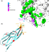
The virus-host interface: Molecular interactions of Alphacoronavirus-1 variants from wild and domestic hosts with mammalian aminopeptidase N
- PMID: 33786949
- PMCID: PMC8251223
- DOI: 10.1111/mec.15910
The virus-host interface: Molecular interactions of Alphacoronavirus-1 variants from wild and domestic hosts with mammalian aminopeptidase N
Abstract
The Alphacoronavirus-1 species include viruses that infect numerous mammalian species. To better understand the wide host range of these viruses, better knowledge on the molecular determinants of virus-host cell entry mechanisms in wildlife hosts is essential. We investigated Alphacoronavirus-1 infection in carnivores using long-term data on Serengeti spotted hyenas (Crocuta crocuta) and molecular analyses guided by the tertiary structure of the viral spike (S) attachment protein's interface with the host receptor aminopeptidase N (APN). We sequenced the complete 3'-end region of the genome of nine variants from wild African carnivores, plus the APN gene of 15 wild carnivore species. Our results revealed two outbreaks of Alphacoronavirus-1 infection in spotted hyenas associated with genetically distinct canine coronavirus type II (CCoVII) variants. Within the receptor binding domain (RBD) of the S gene the residues that directly bind to the APN receptor were conserved in all variants studied, even those infecting phylogenetically diverse host taxa. We identified a variable region within RBD located next to a region that directly interacts with the APN receptor. Two residues within this variable region were under positive selection in hyena variants, indicating that both sites were associated with adaptation of CCoVII to spotted hyena APN. Analysis of APN sequences revealed that most residues that interact with the S protein are conserved in wild carnivores, whereas some adjacent residues are highly variable. Of the variable residues, four that are critical for virus-host binding were under positive selection and may modulate the efficiency of virus attachment to carnivore APN.
Keywords: aminopeptidase N; carnivores; coronavirus; human APN; spike protein; virus-host interaction.
© 2021 The Authors. Molecular Ecology published by John Wiley & Sons Ltd.
Figures


Similar articles
-
Broad receptor engagement of an emerging global coronavirus may potentiate its diverse cross-species transmissibility.Proc Natl Acad Sci U S A. 2018 May 29;115(22):E5135-E5143. doi: 10.1073/pnas.1802879115. Epub 2018 May 14. Proc Natl Acad Sci U S A. 2018. PMID: 29760102 Free PMC article.
-
Coronavirus genotype diversity and prevalence of infection in wild carnivores in the Serengeti National Park, Tanzania.Arch Virol. 2013 Apr;158(4):729-34. doi: 10.1007/s00705-012-1562-x. Epub 2012 Dec 5. Arch Virol. 2013. PMID: 23212740 Free PMC article.
-
Characterization of CCoV-HuPn-2018 spike protein-mediated viral entry.J Virol. 2023 Sep 28;97(9):e0060123. doi: 10.1128/jvi.00601-23. Epub 2023 Sep 7. J Virol. 2023. PMID: 37676001 Free PMC article.
-
Evolution and Interspecies Transmission of Canine Distemper Virus-An Outlook of the Diverse Evolutionary Landscapes of a Multi-Host Virus.Viruses. 2019 Jun 26;11(7):582. doi: 10.3390/v11070582. Viruses. 2019. PMID: 31247987 Free PMC article. Review.
-
Bat-Origin Coronaviruses Expand Their Host Range to Pigs.Trends Microbiol. 2018 Jun;26(6):466-470. doi: 10.1016/j.tim.2018.03.001. Epub 2018 Apr 18. Trends Microbiol. 2018. PMID: 29680361 Free PMC article. Review.
Cited by
-
Natural selection differences detected in key protein domains between non-pathogenic and pathogenic feline coronavirus phenotypes.Virus Evol. 2023 Mar 15;9(1):vead019. doi: 10.1093/ve/vead019. eCollection 2023. Virus Evol. 2023. PMID: 37038392 Free PMC article.
-
Natural selection differences detected in key protein domains between non-pathogenic and pathogenic Feline Coronavirus phenotypes.bioRxiv [Preprint]. 2023 Jan 11:2023.01.11.523607. doi: 10.1101/2023.01.11.523607. bioRxiv. 2023. Update in: Virus Evol. 2023 Mar 15;9(1):vead019. doi: 10.1093/ve/vead019. PMID: 36712007 Free PMC article. Updated. Preprint.
-
Detection and characterization of novel luchacoviruses, genus Alphacoronavirus, in saliva and feces of meso-carnivores in the northeastern United States.J Virol. 2023 Nov 30;97(11):e0082923. doi: 10.1128/jvi.00829-23. Epub 2023 Oct 26. J Virol. 2023. PMID: 37882520 Free PMC article.
-
Epigenetic signatures of social status in wild female spotted hyenas (Crocuta crocuta).Commun Biol. 2024 Mar 28;7(1):313. doi: 10.1038/s42003-024-05926-y. Commun Biol. 2024. PMID: 38548860 Free PMC article.
-
Molecular detection using hybridization capture and next-generation sequencing reveals cross-species transmission of feline coronavirus type-1 between a domestic cat and a captive wild felid.Microbiol Spectr. 2024 Oct 3;12(10):e0006124. doi: 10.1128/spectrum.00061-24. Epub 2024 Aug 19. Microbiol Spectr. 2024. PMID: 39158411 Free PMC article.
References
-
- Allison, A. B. , Harbison, C. E. , Pagan, I. , Stucker, K. M. , Kaelber, J. T. , Brown, J. D. , Ruder, M. G. , Keel, M. K. , Dubovi, E. J. , Holmes, E. C. , & Parrish, C. R. (2012). The role of multiple hosts in the cross‐species transmission and emergence of a pandemic parvovirus. Journal of Virology, 86, 865–872. 10.1128/JVI.06187-11 - DOI - PMC - PubMed
-
- Bandelt, H.‐J. , Forster, P. , & Röhl, A. (1999). Median‐joining networks for inferring intraspecific phylogenies. Molecular Biology and Evolution, 16, 37–48. - PubMed
-
- Benbacer, L. , Kut, E. , Besnardeau, L. , Laude, H. , & Delmas, B. (1997). Interspecies aminopeptidase‐N chimeras reveal species‐specific receptor recognition by canine coronavirus, feline infectious peritonitis virus, and transmissible gastroenteritis virus. Journal of Virology, 71, 734–737. - PMC - PubMed
Publication types
MeSH terms
Substances
Grants and funding
LinkOut - more resources
Full Text Sources
Other Literature Sources
Miscellaneous

