Molecular Mechanisms Underlying Synaptic and Axon Degeneration in Parkinson's Disease
- PMID: 33737866
- PMCID: PMC7960781
- DOI: 10.3389/fncel.2021.626128
Molecular Mechanisms Underlying Synaptic and Axon Degeneration in Parkinson's Disease
Abstract
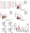
Parkinson's disease (PD) is a progressive neurodegenerative disease that impairs movement as well as causing multiple other symptoms such as autonomic dysfunction, rapid eye movement (REM) sleep behavior disorder, hyposmia, and cognitive changes. Loss of dopamine neurons in the substantia nigra pars compacta (SNc) and loss of dopamine terminals in the striatum contribute to characteristic motor features. Although therapies ease the symptoms of PD, there are no treatments to slow its progression. Accumulating evidence suggests that synaptic impairments and axonal degeneration precede neuronal cell body loss. Early synaptic changes may be a target to prevent disease onset and slow progression. Imaging of PD patients with radioligands, post-mortem pathologic studies in sporadic PD patients, and animal models of PD demonstrate abnormalities in presynaptic terminals as well as postsynaptic dendritic spines. Dopaminergic and excitatory synapses are substantially reduced in PD, and whether other neuronal subtypes show synaptic defects remains relatively unexplored. Genetic studies implicate several genes that play a role at the synapse, providing additional support for synaptic dysfunction in PD. In this review article we: (1) provide evidence for synaptic defects occurring in PD before neuron death; (2) describe the main genes implicated in PD that could contribute to synapse dysfunction; and (3) show correlations between the expression of Snca mRNA and mouse homologs of PD GWAS genes demonstrating selective enrichment of Snca and synaptic genes in dopaminergic, excitatory and cholinergic neurons. Altogether, these findings highlight the need for novel therapeutics targeting the synapse and suggest that future studies should explore the roles for PD-implicated genes across multiple neuron types and circuits.
Keywords: Dementia with Lewy Bodies; GWAS; Parkinson’s disease; degeneration; synapse; α-synuclein.
Copyright © 2021 Gcwensa, Russell, Cowell and Volpicelli-Daley.
Conflict of interest statement
The authors declare that the research was conducted in the absence of any commercial or financial relationships that could be construed as a potential conflict of interest.
Figures


Similar articles
-
α-Synuclein BAC transgenic mice exhibit RBD-like behaviour and hyposmia: a prodromal Parkinson's disease model.Brain. 2020 Jan 1;143(1):249-265. doi: 10.1093/brain/awz380. Brain. 2020. PMID: 31816026
-
Synaptic dysfunction in Parkinson's disease.Adv Exp Med Biol. 2012;970:553-72. doi: 10.1007/978-3-7091-0932-8_24. Adv Exp Med Biol. 2012. PMID: 22351072 Review.
-
Early Degeneration of Both Dopaminergic and Serotonergic Axons - A Common Mechanism in Parkinson's Disease.Front Cell Neurosci. 2016 Dec 22;10:293. doi: 10.3389/fncel.2016.00293. eCollection 2016. Front Cell Neurosci. 2016. PMID: 28066188 Free PMC article. Review.
-
Depopulation of dense α-synuclein aggregates is associated with rescue of dopamine neuron dysfunction and death in a new Parkinson's disease model.Acta Neuropathol. 2019 Oct;138(4):575-595. doi: 10.1007/s00401-019-02023-x. Epub 2019 May 31. Acta Neuropathol. 2019. PMID: 31165254 Free PMC article.
-
Behavioral defects associated with amygdala and cortical dysfunction in mice with seeded α-synuclein inclusions.Neurobiol Dis. 2020 Feb;134:104708. doi: 10.1016/j.nbd.2019.104708. Epub 2019 Dec 16. Neurobiol Dis. 2020. PMID: 31837424 Free PMC article.
Cited by
-
Brain-Region-Specific Differences in Protein Citrullination/Deimination in a Pre-Motor Parkinson's Disease Rat Model.Int J Mol Sci. 2024 Oct 17;25(20):11168. doi: 10.3390/ijms252011168. Int J Mol Sci. 2024. PMID: 39456949 Free PMC article.
-
Motor deficits and brain pathology in the Parkinson's disease mouse model hA53Ttg.Front Neurosci. 2024 Sep 20;18:1462041. doi: 10.3389/fnins.2024.1462041. eCollection 2024. Front Neurosci. 2024. PMID: 39371610 Free PMC article.
-
Druggable transcriptomic pathways revealed in Parkinson's patient-derived midbrain neurons.NPJ Parkinsons Dis. 2022 Oct 18;8(1):134. doi: 10.1038/s41531-022-00400-0. NPJ Parkinsons Dis. 2022. PMID: 36258029 Free PMC article.
-
DNA Damage and Parkinson's Disease.Int J Mol Sci. 2024 Apr 10;25(8):4187. doi: 10.3390/ijms25084187. Int J Mol Sci. 2024. PMID: 38673772 Free PMC article. Review.
-
Characterizing the Retinal Phenotype of the Thy1-h[A30P]α-syn Mouse Model of Parkinson's Disease.Front Neurosci. 2021 Sep 7;15:726476. doi: 10.3389/fnins.2021.726476. eCollection 2021. Front Neurosci. 2021. PMID: 34557068 Free PMC article.
References
Grants and funding
LinkOut - more resources
Full Text Sources
Other Literature Sources
Miscellaneous

