. 2021 Feb 11;11(1):3551.
doi: 10.1038/s41598-021-82893-y.

Dissociation of the respiratory syncytial virus F protein-specific human IgG, IgA and IgM response

Affiliations

Dissociation of the respiratory syncytial virus F protein-specific human IgG, IgA and IgM response

Kristina Borochova et al. Sci Rep. .

Abstract

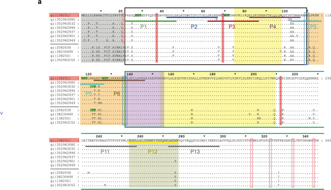
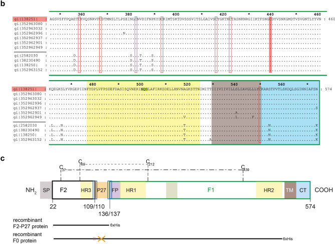
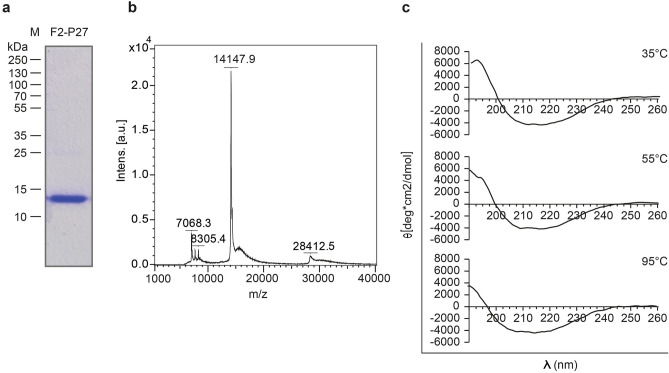
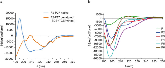
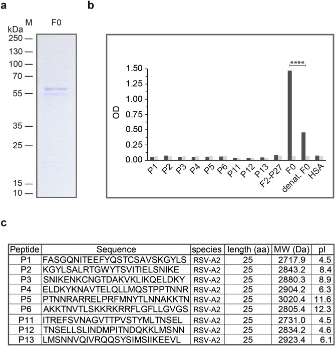
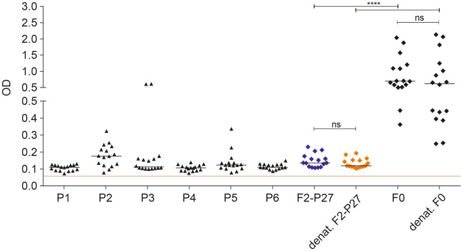
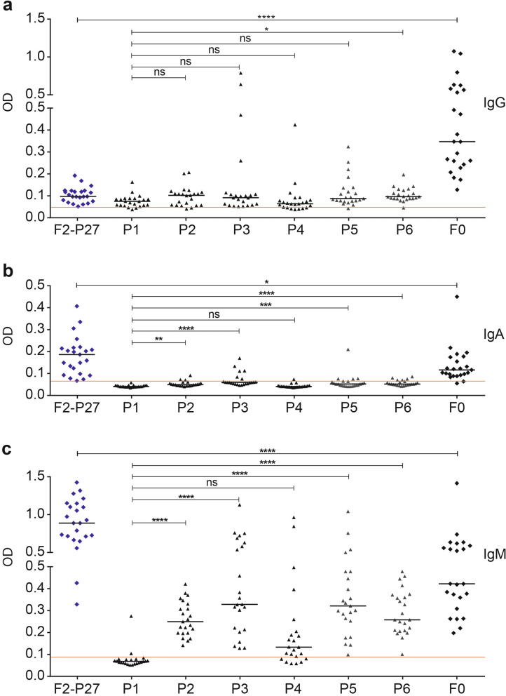
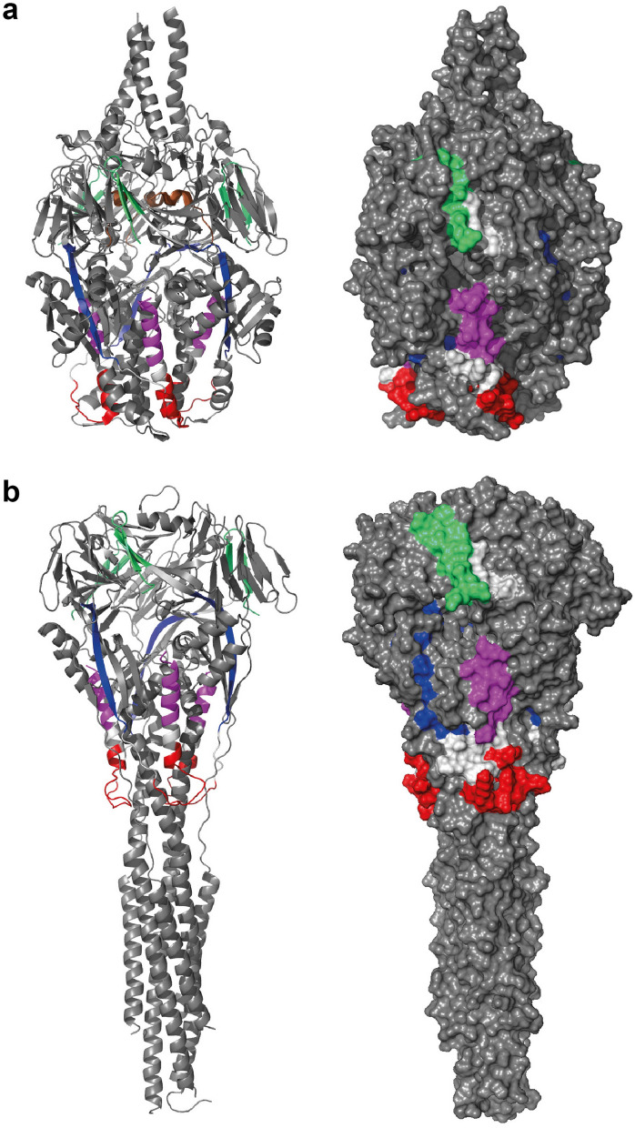
Human respiratory syncytial virus (RSV) is one of the most important causes of severe respiratory tract infections in early childhood. The only prophylactic protection is the neutralizing antibody, palivizumab, which targets a conformational epitope of the RSV fusion (F) protein. The F protein is generated as a F0 precursor containing two furin cleavage sites allowing excision of the P27 fragment and then gives rise to a fusion-competent version consisting of the N-terminal F2 subunit and the a C-terminal F1 subunits linked by two disulphide bonds. To investigate natural human F-specific antibody responses, F2 conferring the species-specificity of RSV, was expressed in Escherichia coli. Furthermore, the F0 protein, comprising both subunits F2 and F1, was expressed as palivizumab-reactive glycoprotein in baculovirus-infected insect cells. Six overlapping F2-derived peptides lacking secondary structure were synthesized. The analysis of IgG, IgA and IgM responses of adult subjects to native versions and denatured forms of F2 and F0 and to unfolded F2-derived peptides revealed that mainly non-conformational F epitopes, some of which represented cryptic epitopes which are not exposed on the proteins were recognized. Furthermore, we found a dissociation of IgG, IgA and IgM antibody responses to F epitopes with F2 being a major target for the F-specific IgM response. The scattered and dissociated immune response to F may explain why the natural RSV-specific antibody response is only partially protective underlining the need for vaccines focusing human antibody responses towards neutralizing RSV epitopes.


Conflict of interest statement

Rudolf Valenta has received research grants from Viravaxx, Vienna, Austria and serves as a consultant for this company. The other authors have no conflicts of interest to report.

Figures

Figure 1
Figure 1
Illustration of A2 strain-derived recombinant F2-, F0-proteins and synthetic peptides. Alignment of the amino acid sequence of F0 from the A2 strain, subgroup A (top line), with the most variable strains from the two antigenic subgroups, A and B. Accession numbers for subgroups A and B (https://www.ncbi.nlm.nih.gov/) on the left margin are separated by a black horizontal line, amino acid numbers are indicated on top and on the right margin (a, b). Identical amino acids are indicated by dots, conserved cysteines are indicated by red boxes. Cysteines forming disulfide bonds between the F1 fragment (green box) and F2 fragment (black box) are indicated as filled red boxes whereas other cysteines are shown by open red boxes. The two furin cleavage sites (amino acid aa 109–110; aa 136–137) are indicated by blue boxes and predicted N-linked glycosylation sites are highlighted in green. The following parts are indicated: Signal peptide (grey box), heptad repeats HR1-3 (yellow boxes), cleaved peptide P27 (orange box), fusion peptide FP (purple box), transmembrane domain (brown box), cytoplasmic tail (blue box) and palivizumab binding site (khaki box). Synthetic peptides (P1, P2, P3, P4, P5, P6, P11, P12 and P13) are shown with horizontal bars. Schematic diagram of the complete fusion protein with disulfide bonds between the F2 and F1 fragment (c). Different parts are colored as in (a, b). Recombinant F2-P27 and F0 with C-terminal hexahistidine tails are indicated below.
Figure 1
Figure 1
Illustration of A2 strain-derived recombinant F2-, F0-proteins and synthetic peptides. Alignment of the amino acid sequence of F0 from the A2 strain, subgroup A (top line), with the most variable strains from the two antigenic subgroups, A and B. Accession numbers for subgroups A and B (https://www.ncbi.nlm.nih.gov/) on the left margin are separated by a black horizontal line, amino acid numbers are indicated on top and on the right margin (a, b). Identical amino acids are indicated by dots, conserved cysteines are indicated by red boxes. Cysteines forming disulfide bonds between the F1 fragment (green box) and F2 fragment (black box) are indicated as filled red boxes whereas other cysteines are shown by open red boxes. The two furin cleavage sites (amino acid aa 109–110; aa 136–137) are indicated by blue boxes and predicted N-linked glycosylation sites are highlighted in green. The following parts are indicated: Signal peptide (grey box), heptad repeats HR1-3 (yellow boxes), cleaved peptide P27 (orange box), fusion peptide FP (purple box), transmembrane domain (brown box), cytoplasmic tail (blue box) and palivizumab binding site (khaki box). Synthetic peptides (P1, P2, P3, P4, P5, P6, P11, P12 and P13) are shown with horizontal bars. Schematic diagram of the complete fusion protein with disulfide bonds between the F2 and F1 fragment (c). Different parts are colored as in (a, b). Recombinant F2-P27 and F0 with C-terminal hexahistidine tails are indicated below.
Figure 2
Figure 2
Characterization of recombinant F2-P27 protein. (a) Coomassie brilliant blue-stained SDS-PAGE showing purified F2-P27 subunit. Molecular weights (kDa) are indicated on the left margin. (b) Mass spectrometry of recombinant F2-P27 subunit. The mass/charge (m/z) ratios are shown on the x-axes, and the intensities are displayed on the y-axes in arbitrary units (a.u.). (c) Far-UV CD spectra of F2-P27 subunit at different temperatures (35°C, 55°C and 95°C). Mean residue ellipticities (θ) (y-axes) at given wavelengths (x-axes).
Figure 3
Figure 3
Circular dichroism analysis of recombinant F2-P27 protein subunit and F2-derived synthetic peptides. (a) Circular dichroism spectra of native (blue) or denatured (orange) F2-P27 subunit. (b) Circular dichroism spectra of F2-derived synthetic peptides (P1: green; P2: dark blue; P3: red; P4: purple; P5: cyan; P6: brown). Mean residue ellipticities (θ) (y-axes) at given wavelengths (x-axes) are shown.
Figure 4
Figure 4
Characterization of recombinant F0 protein. (a) Coomassie brilliant blue-stained SDS-PAGE of recombinant F0 protein. Molecular weights (kDa) are indicated on the left margin. (b) Reactivity of palivizumab to native, denatured F0 and F0-derived peptides by ELISA. Optical density (OD) values (y-axis) correspond to bound human IgG antibodies, palivizumab (black bars) or buffer control (grey bars). Significant difference of palivizumab binding to native and denatured F0 is indicated (p < 0.0001; Mann–Whitney-U-test). (c) Characterization of synthetic peptides used (sequences, length, molecular weights, isoelectric points: pIs).
Figure 5
Figure 5
Effects of denaturation on human IgG responses to recombinant F2-P27 and F0. Shown are IgG levels (y-axis: optical density values) as scatter plots (16 adult individuals) specific for native (dark blue) and denatured (orange) recombinant F2-P27 subunit, native and denatured F0 and native F2-derived synthetic peptides (P1–P6) (x-axis). Horizontal lines within plots indicate median values. The cut-off (mean of buffer control plus three times standard deviation) is indicated by a red line. Significant differences of antibody responses are indicated: ns (not significant); *p < 0.05; **p < 0.001; ***p < 0.0001.
Figure 6
Figure 6
Human antibody responses to recombinant F2-P27, F0 and F2-derived synthetic peptides. Shown are IgG (a), IgA (b) and IgM (c) levels (y-axis: optical density values) in sera from 23 adult individuals to recombinant F2-P27, F0 and F2-derived synthetic peptides (P1–P6), (x-axis). Horizontal lines within scatter plots indicate median values. The cut-off (mean of buffer control plus three times standard deviation) is indicated by a red line. Significant differences of antibody responses are indicated: ns (not significant); *p < 0.05; **p < 0.001; ***p < 0.0001.
Figure 7
Figure 7
The surface exposure of the peptides in the folded forms of the RSV-F protein. (a) the pre-fusion F protein (PDB 3RRR) and (b) the post-fusion F protein (PDB 6APD) are shown in a cartoon-presentation (left panel) and as a surface presentation (right panel). Both forms exist as trimers with the three-fold axis aligned with the vertical axis. The peptides have been colored (P1 green, P2 blue, P3 red, P4 violet and P6 brown; the atomic coordinates for P5 and P6 are missing completely in the post-fusion F protein structure and only 10 residues of P6 have been determined in the pre-fusion F protein structure (brown α-helix in a). P1, P3 and P4 display several surface exposed regions, whereas P2 is mainly buried.

Similar articles

Cited by

References

    1. Nair H, et al. Global burden of acute lower respiratory infections due to respiratory syncytial virus in young children: A systematic review and meta-analysis. Lancet. 2010;375:1545–1555. doi: 10.1016/S0140-6736(10)60206-1. - DOI - PMC - PubMed
    1. Lozano R, et al. Global and regional mortality from 235 causes of death for 20 age groups in 1990 and 2010: A systematic analysis for the Global Burden of Disease Study 2010. Lancet. 2012;380:2095–2128. doi: 10.1016/S0140-6736(12)61728-0. - DOI - PMC - PubMed
    1. Jartti T, et al. Bronchiolitis needs a revisit: Distinguishing between virus entities and their treatments. Allergy Eur. J. Allergy Clin. Immunol. 2019;74:40–52. doi: 10.1111/all.13624. - DOI - PMC - PubMed
    1. Hall CB, Long CE, Schnabel KC. Respiratory syncytial virus infections in previously healthy working adults. Clin. Infect. Dis. 2001;33:792–796. doi: 10.1086/322657. - DOI - PubMed
    1. Falsey AR, Hennessey PA, Formica MA, Cox C, Walsh EE. Respiratory syncytial virus infection in elderly and high-risk adults. N. Engl. J. Med. 2005;352:1749–1759. doi: 10.1056/NEJMoa043951. - DOI - PubMed

Publication types

MeSH terms