. 2021 Feb;41(2):734-754.
doi: 10.1161/ATVBAHA.120.314655. Epub 2020 Dec 10.

Single Mutation in the NFU1 Gene Metabolically Reprograms Pulmonary Artery Smooth Muscle Cells

Affiliations

Single Mutation in the NFU1 Gene Metabolically Reprograms Pulmonary Artery Smooth Muscle Cells

Joel James et al. Arterioscler Thromb Vasc Biol. 2021 Feb.

Abstract

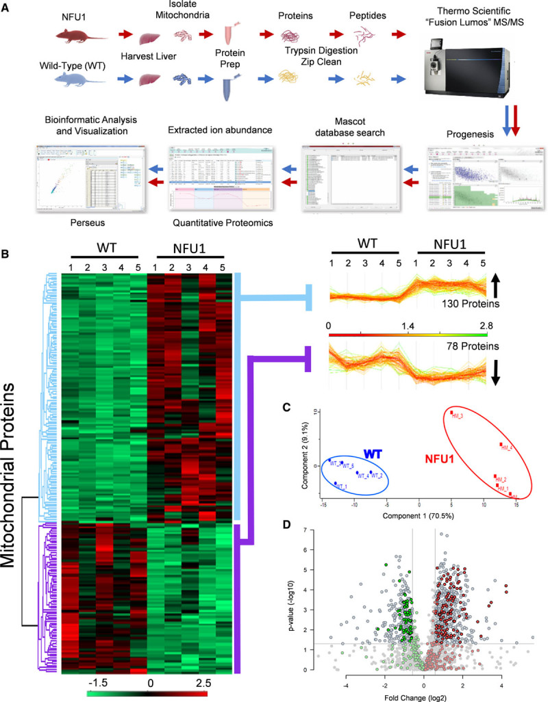
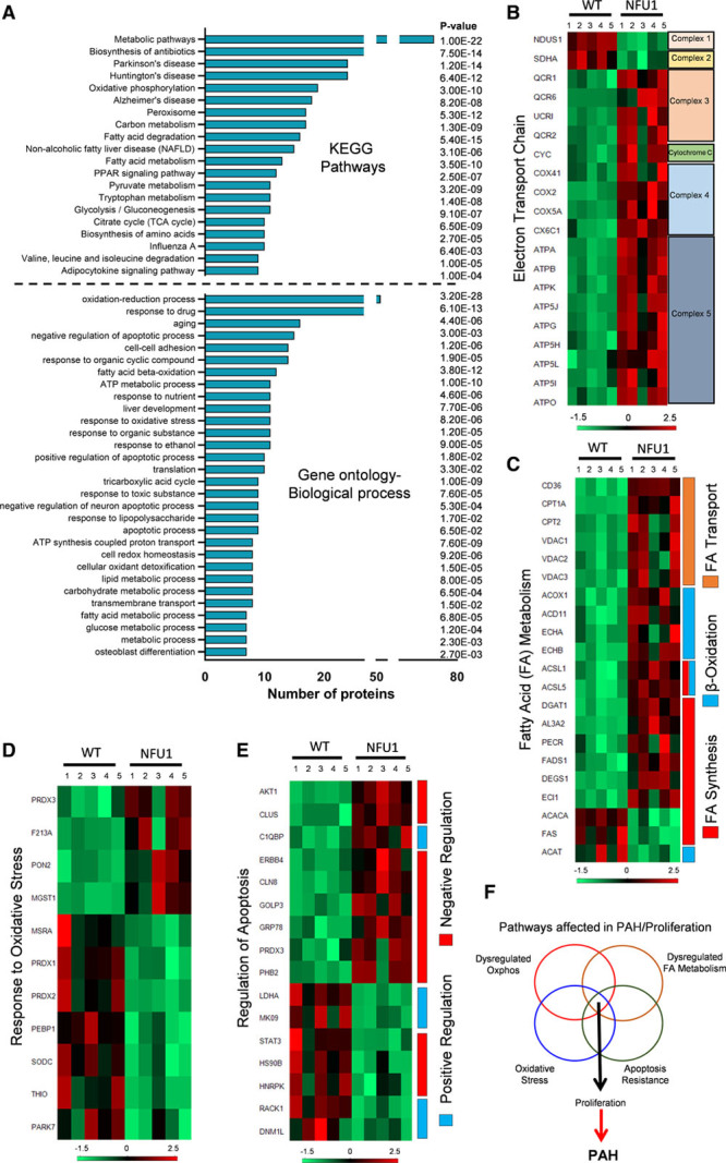
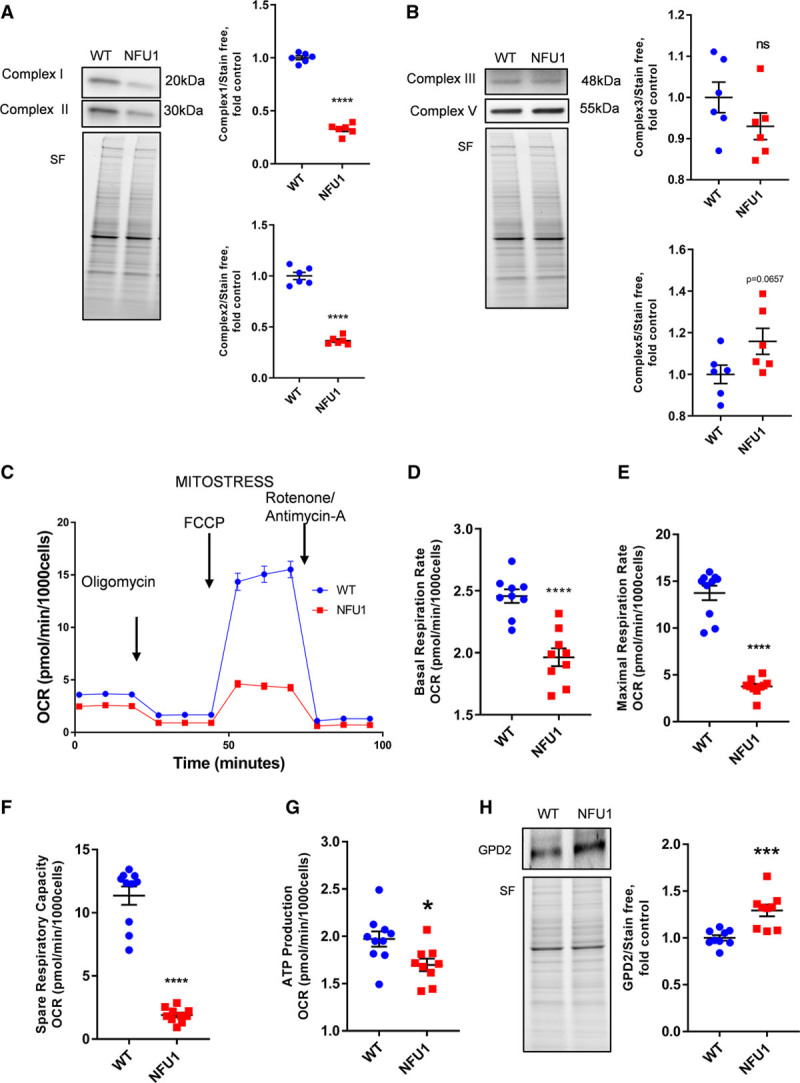
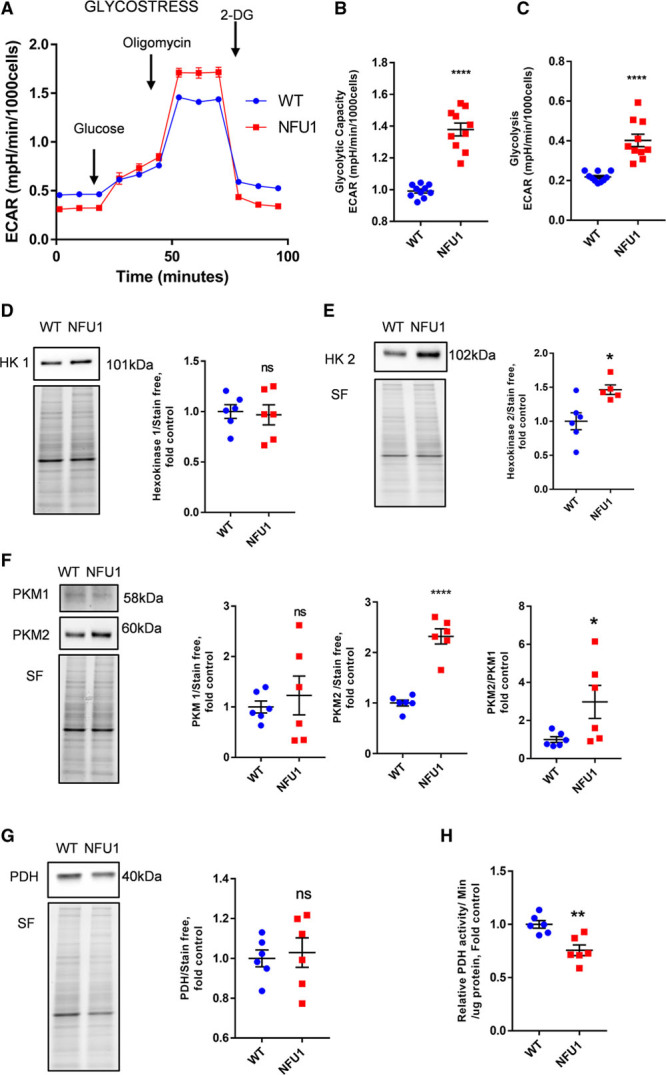
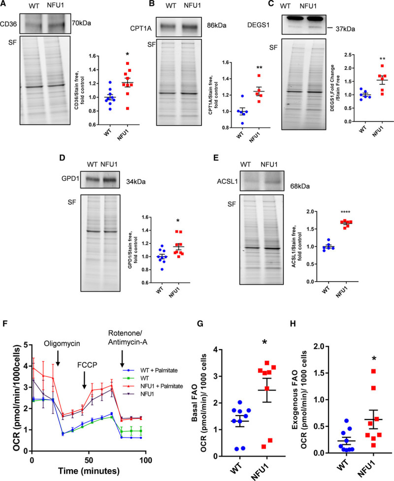
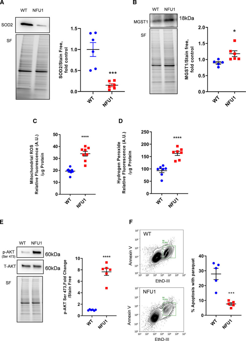
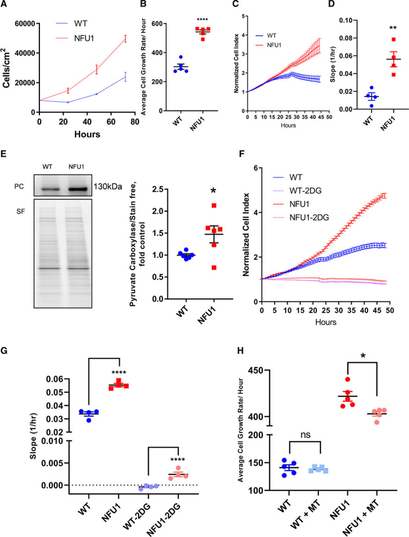
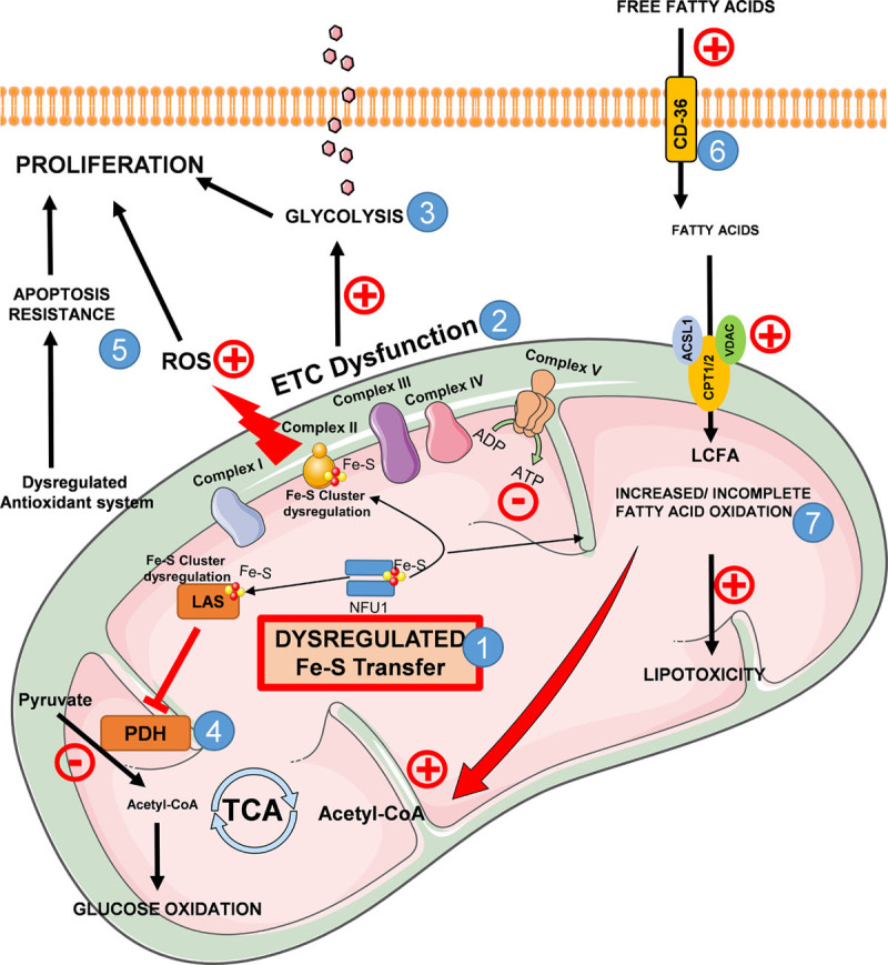
Objective: NFU1 is a mitochondrial iron-sulfur scaffold protein, involved in iron-sulfur assembly and transfer to complex II and LAS (lipoic acid synthase). Patients with the point mutation NFU1G208C and CRISPR/CAS9 (clustered regularly interspaced short palindromic repeats/clustered regularly interspaced short palindromic repeat-associated 9)-generated rats develop mitochondrial dysfunction leading to pulmonary arterial hypertension. However, the mechanistic understanding of pulmonary vascular proliferation due to a single mutation in NFU1 remains unresolved. Approach and Results: Quantitative proteomics of isolated mitochondria showed the entire phenotypic transformation of NFU1G206C rats with a disturbed mitochondrial proteomic landscape, involving significant changes in the expression of 208 mitochondrial proteins. The NFU1 mutation deranged the expression pattern of electron transport proteins, resulting in a significant decrease in mitochondrial respiration. Reduced reliance on mitochondrial respiration amplified glycolysis in pulmonary artery smooth muscle cell (PASMC) and activated GPD (glycerol-3-phosphate dehydrogenase), linking glycolysis to oxidative phosphorylation and lipid metabolism. Decreased PDH (pyruvate dehydrogenase) activity due to the lipoic acid shortage is compensated by increased fatty acid metabolism and oxidation. PASMC became dependent on extracellular fatty acid sources due to upregulated transporters such as CD36 (cluster of differentiation 36) and CPT (carnitine palmitoyltransferase)-1. Finally, the NFU1 mutation produced a dysregulated antioxidant system in the mitochondria, leading to increased reactive oxygen species levels. PASMC from NFU1 rats showed apoptosis resistance, increased anaplerosis, and attained a highly proliferative phenotype. Attenuation of mitochondrial reactive oxygen species by mitochondrial-targeted antioxidant significantly decreased PASMC proliferation.

Conclusions: The alteration in iron-sulfur metabolism completely transforms the proteomic landscape of the mitochondria, leading toward metabolic plasticity and redistribution of energy sources to the acquisition of a proliferative phenotype by the PASMC.

Keywords: glycolysis; metabolism; mitochondria; myocytes; pulmonary artery; smooth muscle.


Conflict of interest statement

None.

Figures

Figure 1.
Figure 1.
Mitochondrial proteome is markedly altered in NFU1 mutants. A, To identify differences in proteomic expression between WT (wild type) and NFU1G206C homozygous mutant rats, mitochondria were isolated, proteins prepared, subjected to trypsin digestion, and analyzed by tandem mass spectrometry. Quantification of the data was executed in Progenesis QI for proteomics, and peptide/protein identification was performed by database searching with Mascot. Quantification of changes in peptide/protein abundance was performed via extracted ion abundance. The resulting quantitative proteomics data were further processed by Perseus for visual representation of the findings. B, Unbiased hierarchical clustering of the 208 significantly affected mitochondrial proteins in the NFU1 mutant vs WT groups confirmed that the expression patterns across the different individual biological samples cluster together accordingly as WT or NFU1 mutant. A heat map and linked dendrogram of the hierarchical clustering results provide a visual representation of the clustered matrix, and the associated profile plots further reveal consistency within groups of the corresponding protein expression patterns (2 boxes to the right of the heat map). C, Unbiased principal component analysis of the 208 significantly affected proteins revealed that the protein expression differences of the individual biological samples within each group were consistent and no outliers were detected (n=5). D, Volcano plot representing proteins that are over- or underexpressed in WT rats vs NFU1 G206C homozygous mutant rats with the mitochondrial proteins highlighted in green and red.
Figure 2.
Figure 2.
Functional annotation of significant proteins from the proteomic data using the Database for Annotation, Visualization and Integrated Discovery. A, Representation of top 20 pathways in KEGG and top 30 processes in the gene ontology-biological process. Pathways were sorted based on the abundance of the number of proteins associated. B, Heat map indicating abundance of proteins associated with the mitochondrial electron transport chain shows decreased levels of complex I and II and increased complexes III to V in the NFU1 group. C, Heat map of fatty acid metabolism proteins within the mitochondria shows dysregulated fatty acid (FA) oxidation and synthesis in NFU1 rats. D, Heat map indicating an abundance of mitochondrial proteins associated with regulating oxidative stress shows a misbalance in NFU1 mitochondria compared with WT (wild type). E, Heat map of mitochondrial proteins regulating apoptosis shows a shift favoring NFU1 survival. F, Possible dysregulated pathways that lead to proliferation and pulmonary arterial hypertension (PAH) in the NFU1 pulmonary artery smooth muscle cell (PASMC). KEGG indicates Kyoto Encyclopedia of Genes and Genomes; PPAR, peroxisome proliferator-activated receptor; and TCA, citrate cycle.
Figure 3.
Figure 3.
NFU1 mutant pulmonary artery smooth muscle cells show altered electron transport chain proteins and decreased mitochondrial respiration. A, Complex 1 and complex 2 protein expressions are significantly decreased in the NFU1 group. B, Complex 3 expression was unchanged, but complex 5 expression was slightly elevated in the NFU1 group. C, Representation of overall mitochondrial oxygen consumption rates. Basal (D), maximal (E), and spare respiration rates (F) are significantly decreased in the NFU1 group. G, Mitochondrial ATP production is significantly decreased in the NFU1 group. H, GPD (glycerol-3-phosphate dehydrogenase)-2 protein expression is significantly increased in the NFU1 group (mean±SEM; n=6 to 10; *, ***, **** vs WT P<0.05, 0.001, 0.0001, Student t test). FCCP indicates carbonyl cyanide 4-(trifluoromethoxy)phenylhydrazone; ns, nonsignificant; OCR, oxygen consumption rate; SF, stain-free normalization; and WT, wild type.
Figure 4.
Figure 4.
NFU1 mutant pulmonary artery smooth muscle cells (PASMCs) are highly glycolytic. A, The glyco-stress assay shows overall extracellular acidification rates (ECAR). NFU1 PASMCs show (B) higher glycolytic capacity and (C) higher basal glycolysis than the WT (wild type) group. Expression levels of glycolytic proteins: (D) HK (hexokinase) 1 expression was unaltered between the groups, but (E) HK2 was increased in the NFU1 groups; (F) PKM (pyruvate kinase)-1 was unaltered between the groups, but PKM2 and the PKM2/PKM1 ratio were increased in the NFU1 group. G, PDH (pyruvate dehydrogenase) expression was unchanged, but (H) PDH activity was decreased in the NFU1 group. PDH and HK2 have been probed from the same membrane after stripping (mean±SEM; n=5 to 10; *, **, **** vs WT P<0.05, 0.01, 0.0001, Student t test). 2-DG indicates 2-deoxyglucose; ns, nonsignificant; and SF, stain-free normalization.
Figure 5.
Figure 5.
Alterations in fatty acid (FA) metabolism. A–E, CD36 (cluster of differentiation 36), CPT1A (carnitine palmitoyltransferase 1A), DEGS1 (delta 4-desaturase, sphingolipid 1), GPD (glycerol-3-phosphate dehydrogenase)-1, and ACSL1 (acyl-Co-A synthetase-1) protein expression is significantly increased in the NFU1 group. F, Seahorse-based analysis showing the overall levels of FA oxidation (FAO) the WT (wild type) and NFU1 group. G, Basal FAO is higher in the NFU1 group. Basal FAO is calculated from Figure 6B, measured as the difference in basal oxygen consumption rate (OCR) and OCR after rotenone/antimycin-A addition. H, Exogenously supplemented (palmitic acid) FAO is higher in the NFU1 group. Exogenous FAO, calculated from Figure 6B, is measured as the maximal OCR (FCCP [carbonyl cyanide 4-(trifluoromethoxy)phenylhydrazone]) difference between the respective palmitate groups and the groups without palmitate; OCR [(WT+palmitate)−(WT)], OCR [(NFU1+palmitate)−NFU1] (mean±SEM; n=5 to 10; *, **, **** vs WT P<0.05, 0.01, 0.0001, Student t test). SF indicates stain-free normalization.
Figure 6.
Figure 6.
Oxidative status and apoptosis resistance. SOD (superoxide dismutase)-2 protein expression is significantly decreased (A) and MGST1 (microsomal glutathione S-transferase 1) protein expression is increased (B) in the NFU1 group. C, Mitochondrial reactive oxygen species (mROS) determined by MitoSox Red shows increased mROS in the NFU1 group. D, Hydrogen peroxide production is increased in the NFU1 group. E, p-AKT (phospho-protein kinase B) is significantly upregulated in the NFU1 group. F, Apoptosis assay (annexin V–ethidium bromide) shows that the NFU1 group has decreased apoptosis during paraquat-induced stress. Graph represented as the percentage of total pulmonary artery smooth muscle cells undergoing apoptosis (mean±SEM; n=5 to 8; *, ***, **** vs WT P<0.05, 0.001, 0.0001, Student t test). ROS indicates reactive oxygen species; SF, stain-free normalization; and WT, wild type.
Figure 7.
Figure 7.
Cell proliferation is increased in pulmonary artery smooth muscle cells isolated from the NFU1 and WT (wild type) rats. A, Growth curves of WT and NFU1 pulmonary artery smooth muscle cell (PASMC) identified by cell counting. B, NFU1 PASMCs have higher proliferation rates in comparison with the WT PASMC (mean±SEM; n=5; ** vs WT P<0.01, Student t test). C, Proliferation rates as determined by the iCELLigence System (ACEA Biosciences), indicating real-time variations in electrical impedance, representing the cell growth index. D, Slope shows significantly increased growth rates in the NFU1 PASMC (mean±SEM; n=4; ** vs WT P<0.01, Student t test). E, PC (pyruvate carboxylase) expression is increased in the NFU1 PASMC (mean±SEM; n=5; * vs WT P<0.05, Student t test). F, Proliferation rate of both WT and NFU1 PASMC is inhibited by 2-deoxyglucose (2DG); however, (G) NFU1 PASMCs show significantly increased growth rate than WT PASMCs when treated with 2DG (25 mmol/L; mean±SEM; n=4; **** vs WT P<0.0001, Student t test). NFU1 mutant PASMC proliferation is decreased with the mitochondrial antioxidant mito-tempo (MT). H, NFU1 PASMC treated with 500 nM MT for 48 h shows decreased growth rates while in WT PASMC, the growth rate is unchanged with MT treatment (mean±SEM; n=5; * vs NFU1 P<0.05, Student t test). ns indicates nonsignificant; and SF, stain-free normalization.
Figure 8.
Figure 8.
Consequences of NFU1 mutation on metabolic reprogramming of smooth muscle cells. (1) Mutation in the NFU1 protein causes defective iron-sulfur (Fe-S) cluster transfer to mitochondrial complex II and LAS (lipoic acid synthase). (2) Fe-S cluster transfer defects cause dysfunctional electron transport chain (ETC), leading to decreased ATP production in NFU1 pulmonary artery smooth muscle cell (PASMC). (3) Dysfunctional ETC and decreased mitochondrial ATP cause a compensatory metabolic switch, increasing glycolysis, thus leading to proliferation. (4) PDH (pyruvate dehydrogenase) activity is decreased as a result of impaired LAS, causing decreased glucose oxidation. (5) Impaired mitochondrial complexes lead to increased reactive oxygen species (ROS) production. In conjunction with dysregulated antioxidant system and apoptosis resistance, ROS production increases proliferation. (6) Fatty acid (FA) import is elevated in NFU1 PASMC via elevated CD36 (cluster of differentiation 36) expression. (7) Increased fatty acid import in conjunction with increased FA oxidation could balance decreased glucose oxidation and further supplement proliferation. Incomplete FA oxidation leads to lipotoxicity. ACSL1 indicates acyl-Co-A synthetase; AU, arbitrary units; CPT, carnitine palmitoyltransferase; EthDIII, ethidium homodimer III; LCFA, long-chain fatty acids; p-AKT, phospho-protein kinase B; T-AKT, total-protein kinase B; TCA, citrate cycle; and VDAC, voltage-dependent anion-selective channel protein.

Similar articles

Cited by

References

    1. Chan SY, Loscalzo J. Pathogenic mechanisms of pulmonary arterial hypertension. J Mol Cell Cardiol. 2008;44:14–30. doi: 10.1016/j.yjmcc.2007.09.006 - PMC - PubMed
    1. Stenmark KR, Fagan KA, Frid MG. Hypoxia-induced pulmonary vascular remodeling: cellular and molecular mechanisms. Circ Res. 2006;99:675–691. doi: 10.1161/01.RES.0000243584.45145.3f - PubMed
    1. Humbert M, Guignabert C, Bonnet S, Dorfmüller P, Klinger JR, Nicolls MR, Olschewski AJ, Pullamsetti SS, Schermuly RT, Stenmark KR, et al. Pathology and pathobiology of pulmonary hypertension: state of the art and research perspectives. Eur Respir J. 2019;53:1801887. - PMC - PubMed
    1. Marshall JD, Bazan I, Zhang Y, Fares WH, Lee PJ. Mitochondrial dysfunction and pulmonary hypertension: cause, effect, or both. Am J Physiol Lung Cell Mol Physiol. 2018;314:L782–L796. doi: 10.1152/ajplung.00331.2017 - PMC - PubMed
    1. Xu S, Xu X, Zhang J, Ying K, Shao Y, Zhang R. Pulmonary hypertension as a manifestation of mitochondrial disease: a case report and review of the literature. Medicine (Baltimore). 2017;96:e8716 doi: 10.1097/MD.0000000000008716 - PMC - PubMed

Publication types

MeSH terms