

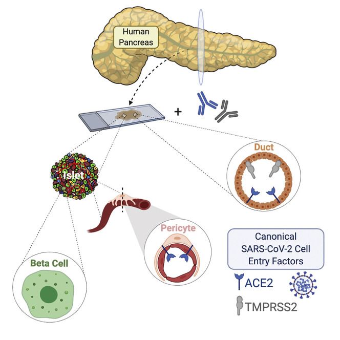
SARS-CoV-2 Cell Entry Factors ACE2 and TMPRSS2 Are Expressed in the Microvasculature and Ducts of Human Pancreas but Are Not Enriched in β Cells
- PMID: 33207245
- PMCID: PMC7664344
- DOI: 10.1016/j.cmet.2020.11.006
SARS-CoV-2 Cell Entry Factors ACE2 and TMPRSS2 Are Expressed in the Microvasculature and Ducts of Human Pancreas but Are Not Enriched in β Cells
Abstract
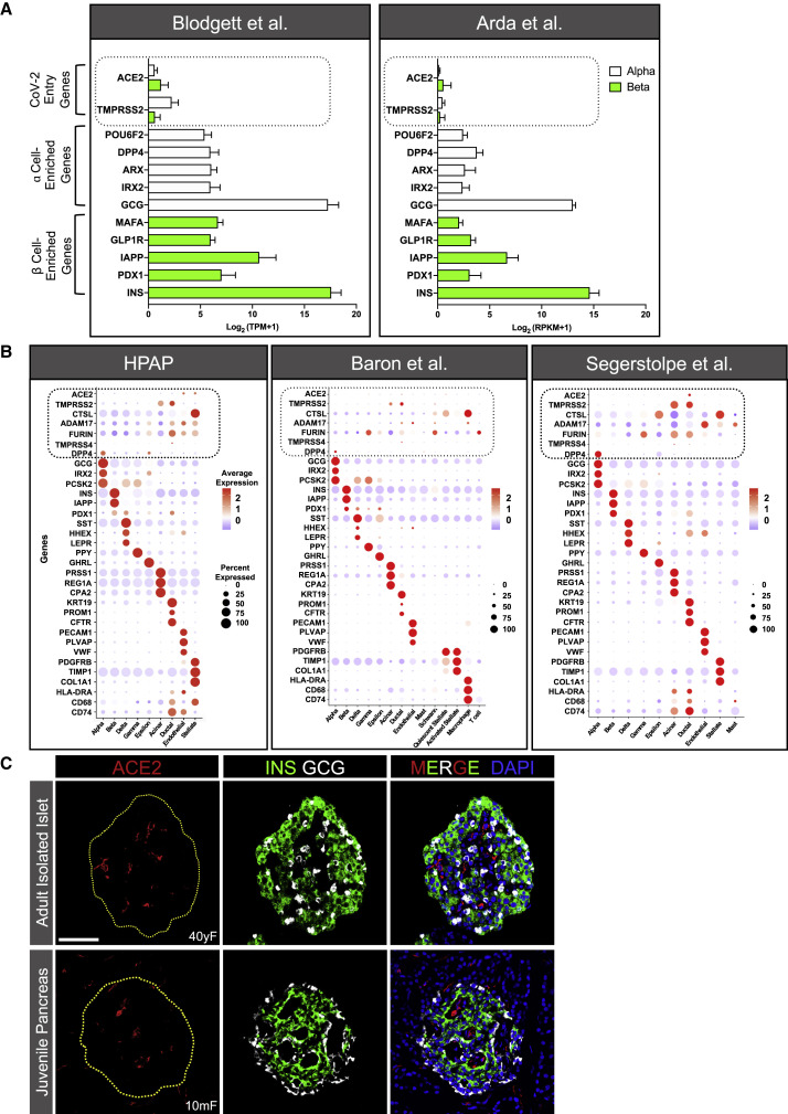
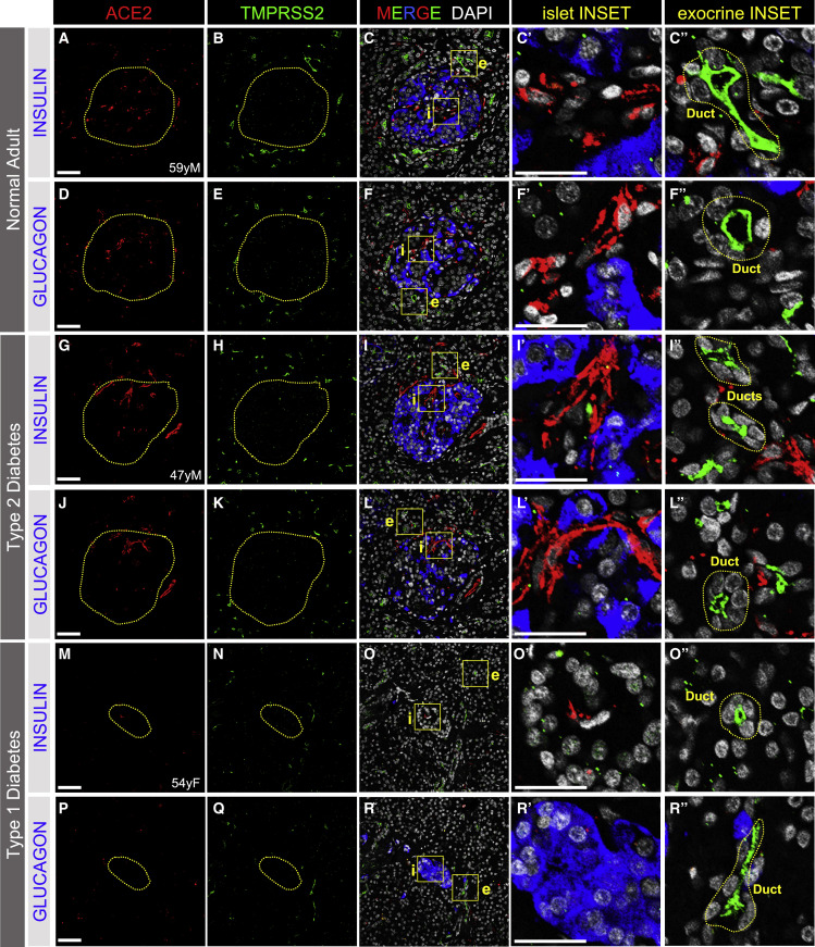
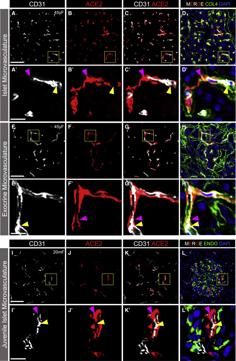
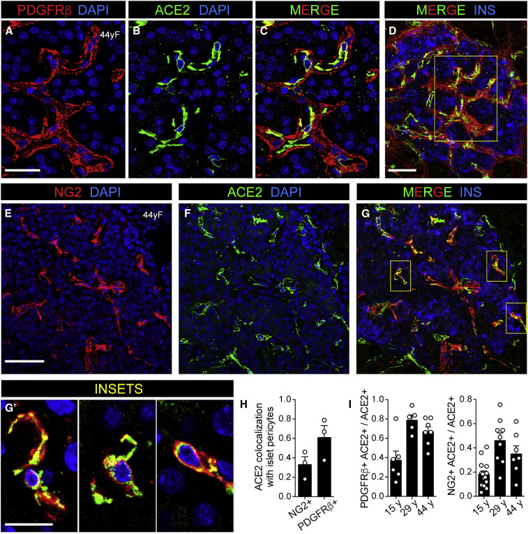
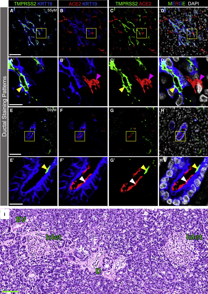
Isolated reports of new-onset diabetes in individuals with COVID-19 have led to the hypothesis that SARS-CoV-2 is directly cytotoxic to pancreatic islet β cells. This would require binding and entry of SARS-CoV-2 into β cells via co-expression of its canonical cell entry factors, angiotensin-converting enzyme 2 (ACE2) and transmembrane serine protease 2 (TMPRSS2); however, their expression in human pancreas has not been clearly defined. We analyzed six transcriptional datasets of primary human islet cells and found that ACE2 and TMPRSS2 were not co-expressed in single β cells. In pancreatic sections, ACE2 and TMPRSS2 protein was not detected in β cells from donors with and without diabetes. Instead, ACE2 protein was expressed in islet and exocrine tissue microvasculature and in a subset of pancreatic ducts, whereas TMPRSS2 protein was restricted to ductal cells. These findings reduce the likelihood that SARS-CoV-2 directly infects β cells in vivo through ACE2 and TMPRSS2.
Keywords: ACE2; COVID-19; SARS-CoV-2; TMPRSS2; beta cell; duct; islet; microvasculature; pancreas; pericyte.
Copyright © 2020 Elsevier Inc. All rights reserved.
Conflict of interest statement
Declaration of Interests The authors declare no competing interests.
Figures
Update of
-
SARS-CoV-2 Cell Entry Factors ACE2 and TMPRSS2 are Expressed in the Pancreas but are Not Enriched in Islet Endocrine Cells.bioRxiv [Preprint]. 2020 Oct 20:2020.08.31.275719. doi: 10.1101/2020.08.31.275719. bioRxiv. 2020. Update in: Cell Metab. 2020 Dec 1;32(6):1028-1040.e4. doi: 10.1016/j.cmet.2020.11.006. PMID: 33106804 Free PMC article. Updated. Preprint.
Similar articles
-
Expression of SARS-CoV-2 Entry Factors in the Pancreas of Normal Organ Donors and Individuals with COVID-19.Cell Metab. 2020 Dec 1;32(6):1041-1051.e6. doi: 10.1016/j.cmet.2020.11.005. Epub 2020 Nov 13. Cell Metab. 2020. PMID: 33207244 Free PMC article.
-
SARS-CoV-2 Cell Entry Factors ACE2 and TMPRSS2 are Expressed in the Pancreas but are Not Enriched in Islet Endocrine Cells.bioRxiv [Preprint]. 2020 Oct 20:2020.08.31.275719. doi: 10.1101/2020.08.31.275719. bioRxiv. 2020. Update in: Cell Metab. 2020 Dec 1;32(6):1028-1040.e4. doi: 10.1016/j.cmet.2020.11.006. PMID: 33106804 Free PMC article. Updated. Preprint.
-
Lymphocytes regulate expression of the SARS-CoV-2 cell entry factor ACE2 in the pancreas of T2DM patients.Diabet Med. 2023 Oct;40(10):e15106. doi: 10.1111/dme.15106. Epub 2023 Apr 11. Diabet Med. 2023. PMID: 37014274
-
Contributions of human ACE2 and TMPRSS2 in determining host-pathogen interaction of COVID-19.J Genet. 2021;100(1):12. doi: 10.1007/s12041-021-01262-w. J Genet. 2021. PMID: 33707363 Free PMC article. Review.
-
SARS-CoV-2 and pancreas: a potential pathological interaction?Trends Endocrinol Metab. 2021 Nov;32(11):842-845. doi: 10.1016/j.tem.2021.07.004. Epub 2021 Jul 24. Trends Endocrinol Metab. 2021. PMID: 34373155 Free PMC article. Review.
Cited by
-
Transient Hyperglycemia in a Patient With Type 2 Diabetes After COVID-19 Messenger RNA Vaccination: A Case Report.Cureus. 2024 Jul 6;16(7):e63983. doi: 10.7759/cureus.63983. eCollection 2024 Jul. Cureus. 2024. PMID: 39105031 Free PMC article.
-
Digestive system infection by SARS‑CoV‑2: Entry mechanism, clinical symptoms and expression of major receptors (Review).Int J Mol Med. 2023 Mar;51(3):19. doi: 10.3892/ijmm.2023.5222. Epub 2023 Jan 20. Int J Mol Med. 2023. PMID: 36660939 Free PMC article. Review.
-
Diabetic Ketoacidosis and COVID-19: A Case Series From an Inner-City Community Teaching Hospital in New York.Cureus. 2022 Jul 5;14(7):e26580. doi: 10.7759/cureus.26580. eCollection 2022 Jul. Cureus. 2022. PMID: 35936183 Free PMC article.
-
COVID-19 and Endocrine Disorders - Emerging Links in this Puzzle.Indian J Endocrinol Metab. 2021 Jan-Feb;25(1):1-3. doi: 10.4103/2230-8210.322027. Indian J Endocrinol Metab. 2021. PMID: 34386385 Free PMC article. No abstract available.
-
SARS-CoV2 infects pancreatic beta cells in vivo and induces cellular and subcellular disruptions that reflect beta cell dysfunction.Res Sq [Preprint]. 2021 Jul 20:rs.3.rs-592374. doi: 10.21203/rs.3.rs-592374/v1. Res Sq. 2021. PMID: 34312617 Free PMC article. Preprint.
References
Publication types
MeSH terms
Substances
Grants and funding
- UC4 DK104211/DK/NIDDK NIH HHS/United States
- U01 DK112217/DK/NIDDK NIH HHS/United States
- U24 DK104162/DK/NIDDK NIH HHS/United States
- UC4 DK112232/DK/NIDDK NIH HHS/United States
- U01 DK089572/DK/NIDDK NIH HHS/United States
- UC4 DK112217/DK/NIDDK NIH HHS/United States
- U01 DK123743/DK/NIDDK NIH HHS/United States
- R24 DK106755/DK/NIDDK NIH HHS/United States
- U01 DK104162/DK/NIDDK NIH HHS/United States
- P30 DK019525/DK/NIDDK NIH HHS/United States
- R01 DK117147/DK/NIDDK NIH HHS/United States
- UC4 DK098085/DK/NIDDK NIH HHS/United States
- UC4 DK108120/DK/NIDDK NIH HHS/United States
- U2C DK059637/DK/NIDDK NIH HHS/United States
- U24 DK098085/DK/NIDDK NIH HHS/United States
- 2020063/DDCF/Doris Duke Charitable Foundation/United States
- I01 BX000666/BX/BLRD VA/United States
- P30 DK020593/DK/NIDDK NIH HHS/United States
- U01 DK123594/DK/NIDDK NIH HHS/United States
- K01 DK111757/DK/NIDDK NIH HHS/United States
- P01 HL075462/HL/NHLBI NIH HHS/United States
- P30 CA068485/CA/NCI NIH HHS/United States
- U24 DK059637/DK/NIDDK NIH HHS/United States
- U01 DK123716/DK/NIDDK NIH HHS/United States
LinkOut - more resources
Full Text Sources
Medical
Miscellaneous
