

Recent advances in 3D bioprinting of musculoskeletal tissues
- PMID: 33166949
- PMCID: PMC8312688
- DOI: 10.1088/1758-5090/abc8de
Recent advances in 3D bioprinting of musculoskeletal tissues
Abstract
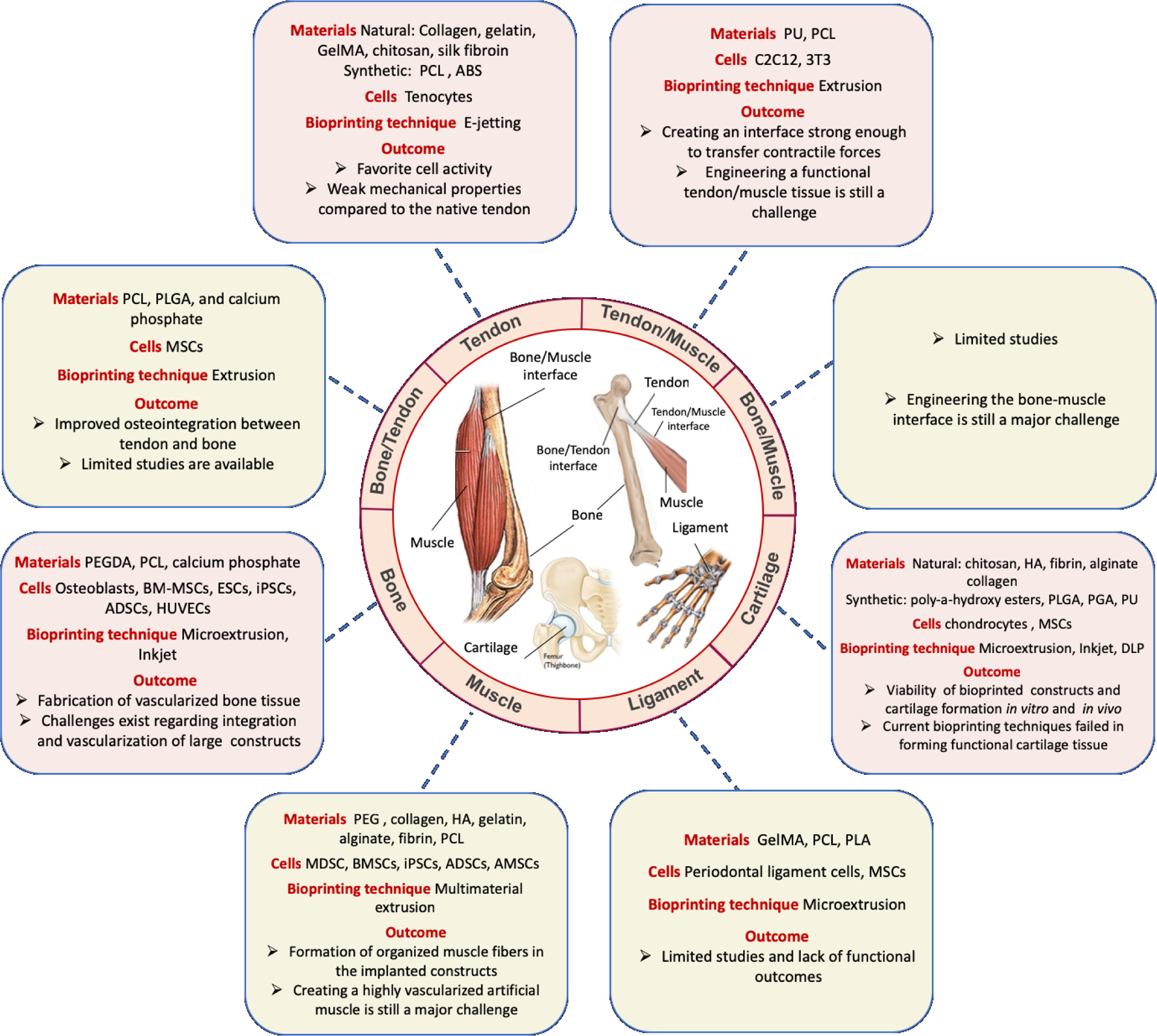
The musculoskeletal system is essential for maintaining posture, protecting organs, facilitating locomotion, and regulating various cellular and metabolic functions. Injury to this system due to trauma or wear is common, and severe damage may require surgery to restore function and prevent further harm. Autografts are the current gold standard for the replacement of lost or damaged tissues. However, these grafts are constrained by limited supply and donor site morbidity. Allografts, xenografts, and alloplastic materials represent viable alternatives, but each of these methods also has its own problems and limitations. Technological advances in three-dimensional (3D) printing and its biomedical adaptation, 3D bioprinting, have the potential to provide viable, autologous tissue-like constructs that can be used to repair musculoskeletal defects. Though bioprinting is currently unable to develop mature, implantable tissues, it can pattern cells in 3D constructs with features facilitating maturation and vascularization. Further advances in the field may enable the manufacture of constructs that can mimic native tissues in complexity, spatial heterogeneity, and ultimately, clinical utility. This review studies the use of 3D bioprinting for engineering bone, cartilage, muscle, tendon, ligament, and their interface tissues. Additionally, the current limitations and challenges in the field are discussed and the prospects for future progress are highlighted.
Keywords: 3D bioprinting; bone; graft; musculoskeletal; tissue defects; tissue engineering.
© 2021 IOP Publishing Ltd.
Figures
Similar articles
-
Tissue-Specific Decellularized Extracellular Matrix Bioinks for Musculoskeletal Tissue Regeneration and Modeling Using 3D Bioprinting Technology.Int J Mol Sci. 2021 Jul 22;22(15):7837. doi: 10.3390/ijms22157837. Int J Mol Sci. 2021. PMID: 34360604 Free PMC article. Review.
-
3D Bioprinting for Cartilage and Osteochondral Tissue Engineering.Adv Healthc Mater. 2017 Nov;6(22). doi: 10.1002/adhm.201700298. Epub 2017 Aug 14. Adv Healthc Mater. 2017. PMID: 28804984 Review.
-
Use of electroconductive biomaterials for engineering tissues by 3D printing and 3D bioprinting.Essays Biochem. 2021 Aug 10;65(3):441-466. doi: 10.1042/EBC20210003. Essays Biochem. 2021. PMID: 34296738 Review.
-
3D Bioprinting:principles, fantasies and prospects.J Stomatol Oral Maxillofac Surg. 2019 Apr;120(2):128-132. doi: 10.1016/j.jormas.2018.12.014. Epub 2019 Jan 1. J Stomatol Oral Maxillofac Surg. 2019. PMID: 30609384
-
Application and development of 3D bioprinting in cartilage tissue engineering.Biomater Sci. 2022 Sep 27;10(19):5430-5458. doi: 10.1039/d2bm00709f. Biomater Sci. 2022. PMID: 35972308 Review.
Cited by
-
MicroRNA-29c-tetrahedral framework nucleic acids: Towards osteogenic differentiation of mesenchymal stem cells and bone regeneration in critical-sized calvarial defects.Cell Prolif. 2024 Jul;57(7):e13624. doi: 10.1111/cpr.13624. Epub 2024 Feb 27. Cell Prolif. 2024. PMID: 38414296 Free PMC article.
-
Decellularized Extracellular Matrix-Based Bioinks for Tendon Regeneration in Three-Dimensional Bioprinting.Int J Mol Sci. 2022 Oct 26;23(21):12930. doi: 10.3390/ijms232112930. Int J Mol Sci. 2022. PMID: 36361719 Free PMC article. Review.
-
High-throughput bioprinting of spheroids for scalable tissue fabrication.Nat Commun. 2024 Nov 21;15(1):10083. doi: 10.1038/s41467-024-54504-7. Nat Commun. 2024. PMID: 39572584 Free PMC article.
-
Applications of functionally-adapted hydrogels in tendon repair.Front Bioeng Biotechnol. 2023 Feb 2;11:1135090. doi: 10.3389/fbioe.2023.1135090. eCollection 2023. Front Bioeng Biotechnol. 2023. PMID: 36815891 Free PMC article. Review.
-
Tissue-Specific Decellularized Extracellular Matrix Bioinks for Musculoskeletal Tissue Regeneration and Modeling Using 3D Bioprinting Technology.Int J Mol Sci. 2021 Jul 22;22(15):7837. doi: 10.3390/ijms22157837. Int J Mol Sci. 2021. PMID: 34360604 Free PMC article. Review.
References
-
- Christensen LV, Physiology and pathophysiology of skeletal muscle contractions. Part II. Static activity, Journal of oral rehabilitation, 13 (1986) 463–477. - PubMed
-
- Christensen LV, Physiology and pathophysiology of skeletal muscle contractions. Part I. Dynamic activity, Journal of oral rehabilitation, 13 (1986) 451–461. - PubMed
-
- Gobbi A, Francisco RA, Lubowitz JH, Allegra F, Canata G, Osteochondral Lesions of the Talus: Randomized Controlled Trial Comparing Chondroplasty, Microfracture, and Osteochondral Autograft Transplantation, Arthroscopy: The Journal of Arthroscopic & Related Surgery, 22 (2006) 1085–1092. - PubMed
-
- Jackson DW, Grood ES, Goldstein JD, Rosen MA, Kurzweil PR, Cummings JF, Simon TM, A comparison of patellar tendon autograft and allograft used for anterior cruciate ligament reconstruction in the goat model, The American Journal of Sports Medicine, 21 (1993) 176–185. - PubMed
Publication types
MeSH terms
Grants and funding
LinkOut - more resources
Full Text Sources
Other Literature Sources