From macro to micro: a combined bioluminescence-fluorescence approach to monitor bacterial localization
- PMID: 33103833
- PMCID: PMC8614114
- DOI: 10.1111/1462-2920.15296
From macro to micro: a combined bioluminescence-fluorescence approach to monitor bacterial localization
Abstract
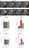
Bacterial bioluminescence is widely used to study the spatiotemporal dynamics of bacterial populations and gene expression in vivo at a population level but cannot easily be used to study bacterial activity at the level of individual cells. In this study, we describe the development of a new library of mini-Tn7-lux and lux::eyfp reporter constructs that provide a wide range of lux expression levels, and which combine the advantages of both bacterial bioluminescence and fluorescent proteins to bridge the gap between macro- and micro-scale imaging techniques. We demonstrate that a dual bioluminescence-fluorescence approach using the lux operon and eYFP can be used to monitor bacterial movement in plants both macro- and microscopically and demonstrate that Pseudomonas syringae pv phaseolicola can colonize the leaf vascular system and systemically infect leaves of common bean (Phaseolus vulgaris). We also show that bacterial bioluminescence can be used to study the impact of plant immune responses on bacterial multiplication, viability and spread within plant tissues. The constructs and approach described in this study can be used to study the spatiotemporal dynamics of bacterial colonization and to link population dynamics and cellular interactions in a wide range of biological contexts.
© 2020 The Authors. Environmental Microbiology published by Society for Applied Microbiology and John Wiley & Sons Ltd.
Figures


Similar articles
-
Pseudomonas syringae pv. syringae B728a Regulates Multiple Stages of Plant Colonization via the Bacteriophytochrome BphP1.mBio. 2017 Oct 24;8(5):e01178-17. doi: 10.1128/mBio.01178-17. mBio. 2017. PMID: 29066541 Free PMC article.
-
Confocal imaging of Pseudomonas syringae pv. phaseolicola colony development in bean reveals reduced multiplication of strains containing the genomic island PPHGI-1.Mol Plant Microbe Interact. 2010 Oct;23(10):1294-302. doi: 10.1094/MPMI-05-10-0114. Mol Plant Microbe Interact. 2010. PMID: 20672876
-
Spatial and temporal dynamics of primary and secondary metabolism in Phaseolus vulgaris challenged by Pseudomonas syringae.Physiol Plant. 2015 Jan;153(1):161-74. doi: 10.1111/ppl.12237. Epub 2014 Jul 4. Physiol Plant. 2015. PMID: 24871330
-
The pH of the leaf apoplast is critical for the formation of Pseudomonas syringae-induced lesions on leaves of the common bean (Phaseolus vulgaris).Plant Sci. 2020 Jan;290:110328. doi: 10.1016/j.plantsci.2019.110328. Epub 2019 Nov 5. Plant Sci. 2020. PMID: 31779895
-
Early detection of bean infection by Pseudomonas syringae in asymptomatic leaf areas using chlorophyll fluorescence imaging.Photosynth Res. 2008 Apr;96(1):27-35. doi: 10.1007/s11120-007-9278-6. Epub 2007 Nov 14. Photosynth Res. 2008. PMID: 18000760
Cited by
-
ScAnalyzer: an image processing tool to monitor plant disease symptoms and pathogen spread in Arabidopsis thaliana leaves.Plant Methods. 2024 May 31;20(1):80. doi: 10.1186/s13007-024-01213-3. Plant Methods. 2024. PMID: 38822355 Free PMC article.
-
Single-cell level LasR-mediated quorum sensing response of Pseudomonas aeruginosa to pulses of signal molecules.Sci Rep. 2024 Jul 13;14(1):16181. doi: 10.1038/s41598-024-66706-6. Sci Rep. 2024. PMID: 39003361 Free PMC article.
-
Microbiome diversity protects against pathogens by nutrient blocking.Science. 2023 Dec 15;382(6676):eadj3502. doi: 10.1126/science.adj3502. Epub 2023 Dec 15. Science. 2023. PMID: 38096285 Free PMC article.
-
Sensor-based phenotyping of above-ground plant-pathogen interactions.Plant Methods. 2022 Mar 21;18(1):35. doi: 10.1186/s13007-022-00853-7. Plant Methods. 2022. PMID: 35313920 Free PMC article. Review.
-
A versatile Tn7 transposon-based bioluminescence tagging tool for quantitative and spatial detection of bacteria in plants.Plant Commun. 2021 Jul 20;3(1):100227. doi: 10.1016/j.xplc.2021.100227. eCollection 2022 Jan 10. Plant Commun. 2021. PMID: 35059625 Free PMC article.
References
-
- Asai, S. , Takamura, K. , Suzuki, H. , and Setou, M. (2008) Single‐cell imaging of c‐fos expression in rat primary hippocampal cells using a luminescence microscope. Neurosci Lett 434: 289–292. - PubMed
-
- Bae, C. , Han, S.W. , Song, Y.‐R. , Kim, B.‐Y. , Lee, H.‐J. , Lee, J.‐M. , et al. (2015) Infection processes of xylem‐colonizing pathogenic bacteria: possible explanations for the scarcity of qualitative disease resistance genes against them in crops. Theor Appl Genet 128: 1219–1229. - PubMed
-
- Bennett, M. , Mehta, M. , and Grant, M. (2005) Biophoton imaging: a nondestructive method for assaying R gene responses. MPMI 18: 95–102. - PubMed
-
- Bogs, J. , Bruchmüller, I. , Erbar, C. , and Geider, K. (1998) Colonization of host plants by the fire blight pathogen Erwinia amylovora marked with genes for bioluminescence and fluorescence. Phytopathology™ 88: 416–421. - PubMed
Publication types
MeSH terms
Grants and funding
LinkOut - more resources
Full Text Sources
Other Literature Sources
Miscellaneous

