Review
. 2021 Jan;17(1):115-124.
doi: 10.1002/alz.12192. Epub 2020 Oct 19.

Hypothesis: Tau pathology is an initiating factor in sporadic Alzheimer's disease

Affiliations
Review

Hypothesis: Tau pathology is an initiating factor in sporadic Alzheimer's disease

Amy F T Arnsten et al. Alzheimers Dement. 2021 Jan.

Abstract

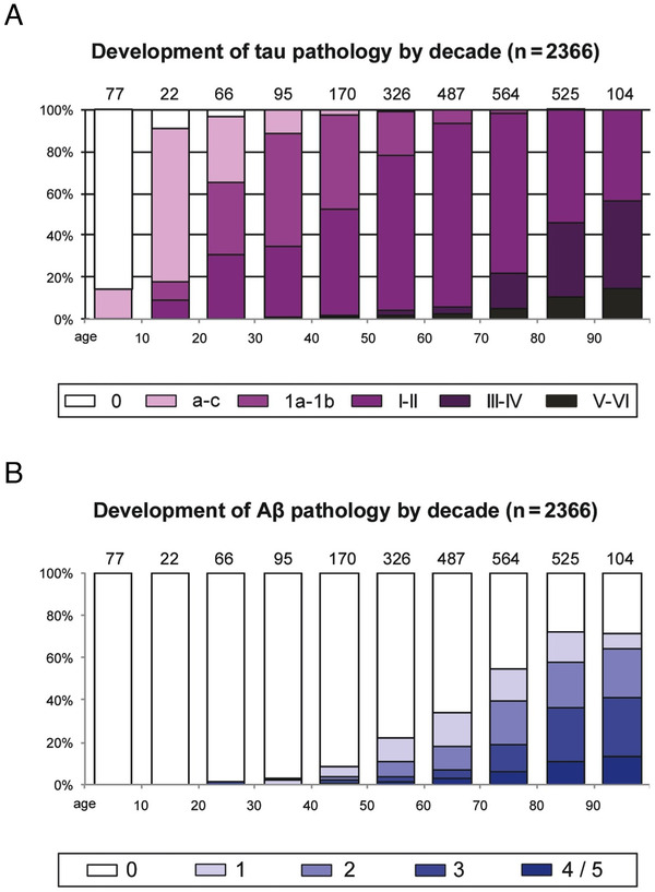
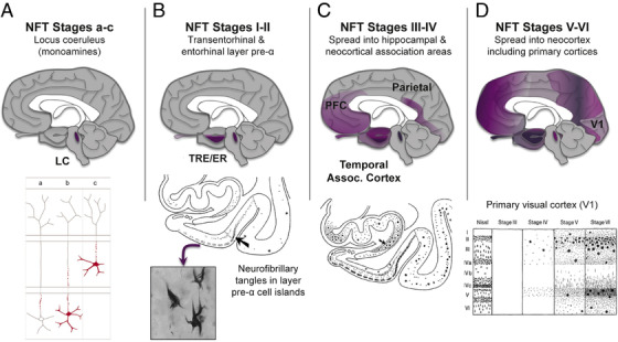
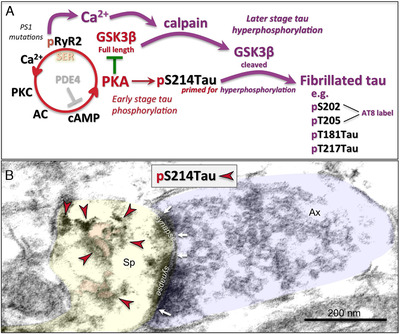
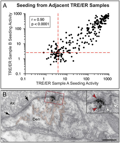
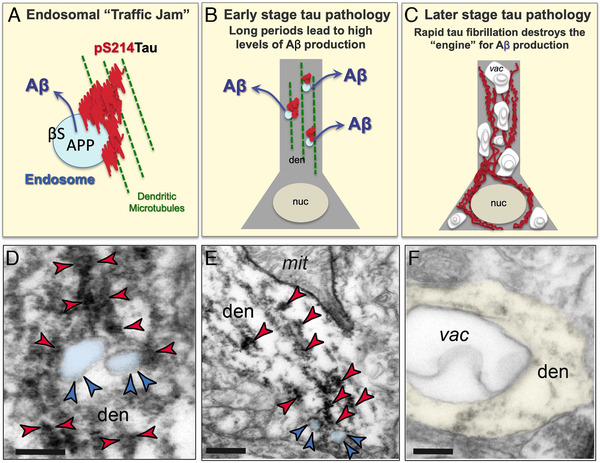
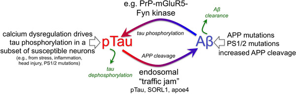
The etiology of the common, sporadic form of Alzheimer's disease (sAD) is unknown. We hypothesize that tau pathology within select projection neurons with susceptible microenvironments can initiate sAD. This postulate rests on extensive data demonstrating that in human brains tau pathology appears about a decade before the formation of Aβ plaques (Aβps), especially targeting glutamate projection neurons in the association cortex. Data from aging rhesus monkeys show abnormal tau phosphorylation within vulnerable neurons, associated with calcium dysregulation. Abnormally phosphorylated tau (pTau) on microtubules traps APP-containing endosomes, which can increase Aβ production. As Aβ oligomers increase abnormal phosphorylation of tau, this would drive vicious cycles leading to sAD pathology over a long lifespan, with genetic and environmental factors that may accelerate pathological events. This hypothesis could be testable in the aged monkey association cortex that naturally expresses characteristics capable of promoting and sustaining abnormal tau phosphorylation and Aβ production.

Keywords: abnormally phosphorylated tau; association cortex; calcium; rhesus monkey; sporadic Alzheimer's disease; tau seeding; β-amyloid.


Conflict of interest statement

Amy F.T. Arnsten and Yale receive royalties from the USA sales of Intuniv (extended release guanfacine). They do not receive royalties from non‐USA or generic sales of Intuniv. The other authors have no actual or potential conflicts of interest to declare.

Figures

FIGURE 1
FIGURE 1
(A) The frequency of fibrillar tau pathology in the human brain across the lifespan. White columns indicate an absence of AT8‐positive abnormal tau. Color‐coded key and columns in violet shading show the relevant frequency of cases at all stages of AT8‐positive tau lesions. The number of cases in each decade appears directly above the columns. (B) The frequency of fibrillar amyloid beta (Aβ) pathology in the human brain across the lifespan. Columns in blue shading indicate the relevant frequency of individuals at various Aβ plaque phases. The number of cases in each decade also appears above each column. Note that tau pathology arises much earlier than Aβ plaques. Adapted with permission from 3
FIGURE 2
FIGURE 2
The sequence of tau pathology in the human brain. (A) Tau pathology is first seen in the subcortical nuclei that project widely to the cerebral cortex, such as the noradrenergic locus coeruleus (LC, subcortical stages a‐c). However, these cells do not degenerate until later stages (eg, neurofibrillary tangle [NFT] stages III/IV). Abnormal tau in the LC is shown in red in the bottom portion of panel A. Notably, tau pathology (stage a) begins in the proximal axon and is followed by lesions filling the somatodendritic domain of coeruleus neurons (stage b). Next, tau‐positive axons and/or nerve cells develop in other non‐thalamic subcortical nuclei with diffuse cortical projections (stage c). (B) Tau pathology is initially seen in the cerebral cortex in glutamatergic neurons of the transentorhinal region (TRE) and then entorhinal region (ER) cortices (NFT stages I‐II, arrow in the lower portion of panel B points to the border between the two). Initially, the tau pathology is confined predominantly to layer pre‐α (inset shows neurofibrillary tangles in pre‐α). (C) Tau pathology then develops in the deeper layers of TRE and ER, in the CA1 sector of the Ammon's horn (arrow in the lower portion of panel C points to the prosubiculum) and in the adjoining temporal neocortex (NFT stages III‐IV). (D) Tau pathology finally spreads to the secondary and then primary visual, auditory, somatosensory, and somatomotor cortices at the latest stages (NFT stages V‐VI). The lower portion of panel D shows, for example, the progression of pathology in the primary visual cortex, V1, in greater detail. Based on 3 and 9 with permission
FIGURE 3
FIGURE 3
The possible role of calcium dysregulation in tau pathology. (A) Schematic illustration of potential molecular signaling events contributing to early‐ and later‐stage abnormal tau phosphorylation. Many glutamate synapses on spines in the dorsolateral prefrontal cortex (dlPFC) express the molecular machinery to magnify calcium signaling near the synapse, where cyclic adeonosine monophosphate (cAMP)‐ protein kinase A (PKA) signaling increases calcium release from the smooth endoplasmic reticulum (SER) through calcium channel ryanodine receptors (RyR2), a process held in check by phosphodiesterases (PDE4), which catabolizes cAMP. 17 However, PDE4 is lost with advancing age, 17 leading to increased PKA activity. This includes increased PKA phosphorylation of ryanodine receptors (pS2808RyR2), which causes calcium leakage into the cytosol, 16 similar to presenilin mutations in autosomal dominant AD. 28 Increased PKA activity also phosphorylates tau at S214, 17 priming tau for hyperphosphorylation by the kinase, GSK3β, 38 a key factor in driving sporadic Altzheimer's disease (sAD) tau pathology. Full length GSK3β is inhibited by PKA, which may hold this process in check when cytosolic calcium levels are normal. However, when there are sufficiently high levels of calcium to activate the protease calpain, this may “switch” the system into later stage, tau hyperphosphorylation, as calpain can cleave GSK3β at the N‐terminus, removing its inhibition by PKA. 48 Disinhibition of GSK3β would initiate hyperphosphorylation of pS214Tau at key sites for fibrillation, 38 including those labeled by the AT8 antibody currently used to diagnose AD, and pT181Tau and pT217Tau, which have potential as novel, in vivo biomarkers. 49 , 50 Disinhibited GSK3β signaling can also degrade PDE4s, 51 which would further dysregulate calcium and PKA signaling and tau phosphorylation. Thus, the activation of calpain may be a major step in activating advanced pathological events, consistent with it heralding the rise of fibrillated tau pathology in the sAD brain. 30 , 31 However, even early‐stage tau phosphorylation at S214 may have pathological consequences, as its aggregation on microtubules interferes with endosomal trafficking, which may contribute to the production of Aβ (see Figure 5 below). (B) An example of a glutamatergic‐like synapse on a spine in the aged dlPFC, where immunolabeling for PKA‐phosphorylated tau (pS214Tau; indicated by the red arrowheads) is expressed on and near the SER (pseudocolored in pink). The spine (Sp) is pseudocolored in yellow; the axon terminal (Ax) is pseudocolored in blue; the synapse is perforated and is delineated by white arrows; the scale bar indicates 200 nm. Note that pS214Tau labeling in aged dlPFC was first reported in 17
FIGURE 4
FIGURE 4
Propagation of tau pathology in vulnerable cortical circuits. (A) Figure reproduced with permission from (42) on network propagation (“seeding”) that starts in the transentorhinal region (TRE) and entorhinal region (ER). Seeding of abnormal tau was most effective at earliest time points from these cortical regions. The generation of large amounts of pTau in TRE/ER early in the aging process may arise from early calcium dysregulation in these circuits, as seen in aging monkey. 16 (B) A Paspalas 16 immunoEM image capturing pS214Tau trafficking (red arrowheads in inset) between excitatory neurons in layer pre‐α of the middle‐aged rhesus monkey ER (from 16 with permission). Black arrows point to the synaptic membrane; white arrowheads indicate an omega‐shaped profile on the plasma membrane. The smooth endoplasmic reticulum (SER) is pseudocolored in pink. Abbreviations: Ax, axon; Den, dendrite. Scale bar = 200 nm
FIGURE 5
FIGURE 5
Endosomal “traffic jams” may drive amyloid beta (Aβ) pathology. (A) Schematic of an endosomal “traffic jam” similar to that described for genetic insults to retromer signaling, 45 showing an endosome containing amyloid precursor protein (APP) and β secretase trapped in aggregated pS214Tau on a microtubule. (B) A schematic diagram of early stage tau pathology in a dendrite (den), where aggregations of phosphorylated tau (red) on microtubules (green) trap endosomes (blue) and exacerbate the production of Aβ, which is then released into the extracellular space, e.g. after axonal transport. Real examples of aggregated pS214Tau on microtubules trapping endosomes are shown in panels D and E. nuc = nucleus. (C) A schematic diagram of later stage tau pathology, with fibrillated tau in the dendrite and soma now accompanied by extensive autophagic vacuoles (vac), where normal neuronal organelles are now lost, and the cellular engine for Aβ production has deteriorated. A real example of an autophagic vacuole in a dendrite is shown in panel F. (D, E) ImmunoEM of the aged monkey layer III dorsolateral prefrontal cortex (dlPFC) showing early‐stage, soluble pS214Tau (indicated by red arrowheads) aggregating on microtubules in dendrites (den) where it traps enlarged endosomes (pseudocolored in blue, and indicated by blue arrowheads). Aggregations of pS214Tau are often seen near dysmorphic mitochondria (mit), consistent with local calcium dysregulation. Note that the dlPFC is a site of extensive Aβ pathology in sporadic Altzheimer's disease. Interestingly, rhesus monkeys are apoE4 genotype, 52 which may increase the numbers of endosomes in this species. 53 Scale bar in B: 200 nm; scale bar in C: 200 nm. (F) With advanced age, pTau‐afflicted dendrites fill with autophagic vacuoles (vac), such as the one seen here in dlPFC, and lose normal organelles, including those needed to generate Aβ. Scale bar in D: 200 nm. Scale bars in D‐F: 200 nm
FIGURE 6
FIGURE 6
Potentially interacting vicious cycles in sporadic Altzheimer's disease (sAD). Cartoon showing pTau driving Aβ production and Aβ driving tau phosphorylation, setting up a vicious cycle. In sAD, cell biological changes in the aging association cortex (eg, calcium dysregulation) can increase early stage tau pathology that subsequently generates Aβ cleavage via trapping of endosomes in aggregated pTau. Genetic alterations in retromer signaling (eg, SORL1) may exacerbate the risk of sAD by further exacerbating these “endosomal traffic jams” which increase amyloid precursor protein (APP) cleavage to Aβ in endosomes. In contrast, in familial Altzheimer's, primary genetic perturbations in amyloid signaling (eg, duplications in APP) can initiate the degenerative process, which subsequently increases tau hyperphosphorylation. In both cases, vicious cycles would be set in motion that ultimately lead to amyloid plaques and neurofibrillary tangles. Thus, AD pathology could have multiple initiating factors that can lead to a similar phenotype

Similar articles

Cited by

References

    1. Hardy J, Selkoe DJ. The amyloid hypothesis of Alzheimer's disease: progress and problems on the road to therapeutics. Science. 2002;297:353‐356. - PubMed
    1. Morris GP, Clark IA, Vissel B. Questions concerning the role of amyloid‐β in the definition, aetiology and diagnosis of Alzheimer's disease. Acta Neuropathol. 2018;136:663‐689. - PMC - PubMed
    1. Braak H, Del Tredici K. Neuroanatomy and pathology of sporadic Alzheimer's disease. Adv Anat Embryol Cell Biol. 2015;215:1‐162. - PubMed
    1. Huang LK, Chao SP, Hu CJ. Clinical trials of new drugs for Alzheimer disease. J Biomed Sci. 2020;27:18. - PMC - PubMed
    1. Arboleda‐Velasquez JF, Lopera F, O'Hare M, et al. Resistance to autosomal dominant Alzheimer's disease in an APOE3 Christchurch homozygote: a case report. Nat Med. 2019;25:1680‐1683. - PMC - PubMed

Publication types