. 2020 Oct 14;6(42):eabb8941.
doi: 10.1126/sciadv.abb8941. Print 2020 Oct.

cGAS suppresses genomic instability as a decelerator of replication forks

Affiliations

cGAS suppresses genomic instability as a decelerator of replication forks

Hao Chen et al. Sci Adv. .

Abstract

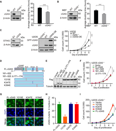
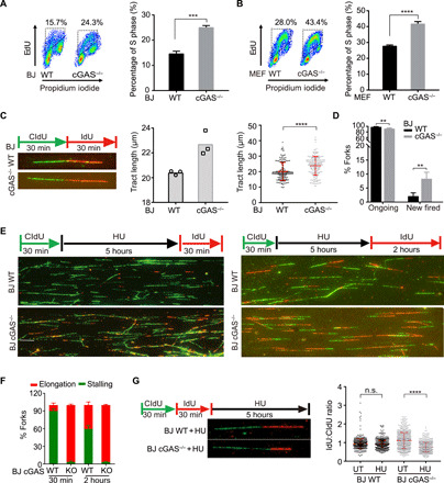
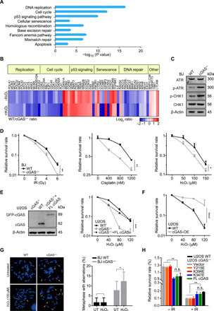
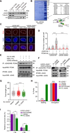
The cyclic GMP-AMP synthase (cGAS), a sensor of cytosolic DNA, is critical for the innate immune response. Here, we show that loss of cGAS in untransformed and cancer cells results in uncontrolled DNA replication, hyperproliferation, and genomic instability. While the majority of cGAS is cytoplasmic, a fraction of cGAS associates with chromatin. cGAS interacts with replication fork proteins in a DNA binding-dependent manner, suggesting that cGAS encounters replication forks in DNA. Independent of cGAMP and STING, cGAS slows replication forks by binding to DNA in the nucleus. In the absence of cGAS, replication forks are accelerated, but fork stability is compromised. Consequently, cGAS-deficient cells are exposed to replication stress and become increasingly sensitive to radiation and chemotherapy. Thus, by acting as a decelerator of DNA replication forks, cGAS controls replication dynamics and suppresses replication-associated DNA damage, suggesting that cGAS is an attractive target for exploiting the genomic instability of cancer cells.


Figures

Fig. 1
Fig. 1. Nuclear localization and DNA binding activity of cGAS are required to suppress cell proliferation, independently of STING and cGAMP.
(A and B) Western blot (WB) of cGAS in WT and cGAS−/− BJ and MEF cells. Thirty-thousand cells were seeded into each well of a six-well plate. The number of BJ WT/cGAS−/− (A) and MEF WT/cGAS−/− (B) cells was counted on the indicated day to determine the average population doubling time. (C) WB of STING in BJ and U2OS WT cells and cGAS in WT, cGAS−/−, and green fluorescent protein (GFP)–cGAS stably overexpressed WT U2OS cells (cGAS-OE). Proliferation rates of WT, cGAS−/−, and cGAS-OE U2OS cells were determined as described in (A) and (B). (D and E) Schematic structure of cGAS truncated proteins. WB of flag-tagged cGAS deletions and mutants in U2OS cGAS−/− cells is shown. (F) Thirty-thousand cells were seeded into each well of a six-well plate. The number of U2OS cGAS−/− cells transfected with flag-tagged deletion and flag-vector plasmids was counted every day until reaching high density. (G) U2OS cGAS−/− cells were transfected with flag-tagged cGAS point mutant plasmids and tested by immunostaining (scale bar, 10 μm). (H) Quantification of percentage of cells with nuclear cGAS (n = 50). Three independent experiments were done. (I) Thirty-thousand cells were seeded into each well of a six-well plate. The number of U2OS cGAS−/− cells transfected with flag-tagged full length (FL), Y215E, K347E, K394E, and flag-vector plasmids was counted every day until reaching high density. Note that the same vector control is used in (F) and (I). ***P < 0.001, Mann-Whitney test.
Fig. 2
Fig. 2. cGAS deficiency endows a hyper-replicative cell state associated with replication defects.
(A and B) The staining of EdU and propidium iodide (PI) in BJ WT (A), MEF WT (B), and corresponding cGAS−/− cells was analyzed by flow cytometry. The percentage of S phase from flow cytometry analysis is shown. (C) Representative images of nascent DNA tract length in BJ WT and cGAS−/− cells (left), quantification of DNA tract length in either BJ WT or cGAS−/− cells from three independent experiments (middle), and nascent DNA tract length of one representative experiment (right) (n = 200, mean ± SD in each experiment). Statistical analysis was done with the two-sided Mann-Whitney test; ****P < 0.0001. (D) DNA fiber analysis of ongoing and new fired forks (n = 600) in BJ WT and cGAS−/− cells. Mean ± SD is shown. (E) The replication fork status of BJ WT and cGAS−/− cells treated with 4 mM HU for 5 hours and released for 30 min or 2 hours. Representative images of fiber assay are shown (scale bar, 10 μm). (F) DNA fiber analysis of replication fork stalling and elongation (n = 1800) in BJ WT and cGAS−/− cells treated with HU. Means and SD of three independent experiments are shown. (G) Ratios of IdU and CldU length (each group, n > 660) are shown in BJ WT and cGAS−/− cells treated with or without [untreated (UT)] 4 mM HU for 5 hours. Median ratio is indicated in red. **P < 0.01, ***P < 0.001, Mann-Whitney test. n.s., not significant.
Fig. 3
Fig. 3. Fast replication renders cGAS-deficient cells sensitive to radiation and chemo-drugs.
(A) RNA-seq expression data of BJ WT and cGAS−/− cell were analyzed. The enrichment KEGG (Kyoto Encyclopedia of Genes and Genomes) pathway comparison between gene expression from RNA-seq in BJ WT and cGAS−/− cells is shown. (B) Heat map of RNA expression in BJ WT and cGAS−/− cells. A gene list was generated using genes showing a 1.5-fold change cutoff, 10 reads or more, and P value of <0.05. (C) Level of phospho-ATR and CHK1 in BJ WT and cGAS−/− cells by WB. (D) Colony formation assay for BJ WT and cGAS−/− cells with treatment of the indicated dose of ionizing radiation (IR), cisplatin, or H2O2. (E) Colony formation assay for U2OS WT, cGAS−/−, and GFP-cGAS stably expressed cGAS−/− (cGAS−/− + FL cGAS) cells with the indicated dose of H2O2. (F) Colony formation assay for U2OS WT and cGAS-OE with treatment of indicated dose of H2O2. (G) Representative metaphases of BJ WT and cGAS−/− cells treated with 100 μM H2O2 for 24 hours. Quantification of incidence of total chromosomal aberrations (n > 500 in one experiment). Three independent experiments were done. (H) Colony formation assay for U2OS WT and cGAS−/− cells transfected with mutant plasmids treated without and with 4-Gy IR. **P < 0.01, ****P < 0.0001. Mann-Whitney test. n.s., not significant.
Fig. 4
Fig. 4. DNA binding of cGAS is required for fork stalling after damage and drug resistance.
(A) Cytosolic, soluble nuclear, and chromatin fractions from U2OS cells were immunoblotted for cGAS and indicated proteins. (B) U2OS cells expressing GFP-cGAS were pulled down for interactome analysis. The results of mass spectrometry identified several major replication fork components. The schematic structure of cGAS pulled down with a replication fork complex via DNA or based on the proximity at replication forks. (C) Representative PLA of U2OS WT and cGAS−/− cells (scale bar, 10 μm). (D) Quantification of colocalization between cGAS and PCNA. Three independent experiments were done, mean ± SD is shown, and statistical analysis was done with the two-sided Mann-Whitney test; ****P < 0.001 and **** P < 0.0001. (E) Interaction of PCNA and cGAS detected with coimmunoprecipitation (Co-IP). U2OS cells were treated with or without 4 mM HU or 1% EtBr before Co-IP analysis with anti-cGAS antibody. (F) Interaction of cGAS mutants and PCNA detected with Co-IP. U2OS cGAS−/− cells were transfected with cGAS flag-tagged FL, K394E mutant, or NLS-K394E mutant for 36 hours before Co-IP analysis with anti-cGAS antibody. (G) Analysis of nascent DNA tract length (n = 600) in BJ cGAS−/− cells transfected with K394E mutant and corresponding control plasmids. For all experiments in this study, three independent experiments were done, mean ± SD is shown, and statistical analysis was done with the two-sided Mann-Whitney test; **** P < 0.0001. (H) DNA fiber analysis of replication fork stalling and elongation (n = 1800) in BJ cGAS−/− cells transfected with DNA binding defective mutants. Means and SD of three independent experiments are shown. (I) Colony formation assay for U2OS cGAS−/− cells expressing NLS-K394E mutants without and with 4-Gy IR treatment.

Similar articles

Cited by

References

    1. Jackson S. P., Bartek J., The DNA-damage response in human biology and disease. Nature 461, 1071–1078 (2009). - PMC - PubMed
    1. Sun L., Wu J., Du F., Chen X., Chen Z. J., Cyclic GMP-AMP synthase is a cytosolic DNA sensor that activates the type I interferon pathway. Science 339, 786–791 (2013). - PMC - PubMed
    1. Li T., Chen Z. J., The cGAS-cGAMP-STING pathway connects DNA damage to inflammation, senescence, and cancer. J. Exp. Med. 215, 1287–1299 (2018). - PMC - PubMed
    1. Wang H., Hu S., Chen X., Shi H., Chen C., Sun L., Chen Z. J., cGAS is essential for the antitumor effect of immune checkpoint blockade. Proc. Natl. Acad. Sci. U.S.A. 114, 1637–1642 (2017). - PMC - PubMed
    1. Pépin G., Gantier M. P., cGAS-STING activation in the tumor microenvironment and its role in cancer immunity. Adv. Exp. Med. Biol. 1024, 175–194 (2017). - PubMed

Publication types