Review
. 2020 Sep 29;9(10):2204.
doi: 10.3390/cells9102204.

Notch Signaling in Breast Cancer: A Role in Drug Resistance

Affiliations
Review

Notch Signaling in Breast Cancer: A Role in Drug Resistance

McKenna BeLow et al. Cells. .

Abstract

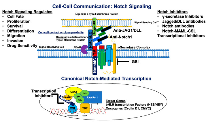
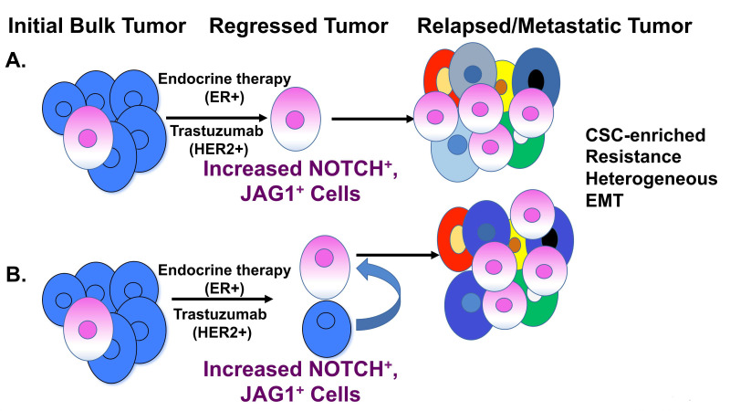
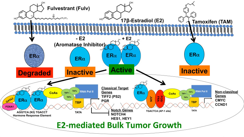
Breast cancer is a heterogeneous disease that can be subdivided into unique molecular subtypes based on protein expression of the Estrogen Receptor, Progesterone Receptor, and/or the Human Epidermal Growth Factor Receptor 2. Therapeutic approaches are designed to inhibit these overexpressed receptors either by endocrine therapy, targeted therapies, or combinations with cytotoxic chemotherapy. However, a significant percentage of breast cancers are inherently resistant or acquire resistance to therapies, and mechanisms that promote resistance remain poorly understood. Notch signaling is an evolutionarily conserved signaling pathway that regulates cell fate, including survival and self-renewal of stem cells, proliferation, or differentiation. Deregulation of Notch signaling promotes resistance to targeted or cytotoxic therapies by enriching of a small population of resistant cells, referred to as breast cancer stem cells, within the bulk tumor; enhancing stem-like features during the process of de-differentiation of tumor cells; or promoting epithelial to mesenchymal transition. Preclinical studies have shown that targeting the Notch pathway can prevent or reverse resistance through reduction or elimination of breast cancer stem cells. However, Notch inhibitors have yet to be clinically approved for the treatment of breast cancer, mainly due to dose-limiting gastrointestinal toxicity. In this review, we discuss potential mechanisms of Notch-mediated resistance in breast cancer cells and breast cancer stem cells, and various methods of targeting Notch through γ-secretase inhibitors, Notch signaling biologics, or transcriptional inhibitors. We also discuss future plans for identification of novel Notch-targeted therapies, in order to reduce toxicity and improve outcomes for women with resistant breast cancer.

Keywords: Notch; breast cancer; cancer stem cells; resistance.


Conflict of interest statement

The authors declare no conflict of interest.

Figures

Figure 1
Figure 1
Notch ligands and receptors. The Signal-sending cell expresses Notch ligands, type I transmembrane proteins. These include Jagged1, Jagged2, DLL1, DLL3, and DLL4. Each ligand contains a short cytoplasmic tail, followed by the transmembrane domain, EGF-like repeats, a DOS domain, DLS binding domain, and MNNL domain. The signal-receiving cells express the Notch receptors, including Notch1, Notch2, Notch3, and Notch4. Each receptor contains EGF-like domains, LNR (Lin-Notch repeats), HD (heterodimerization domain), transmembrane domain, RAM (RBPJ-associated molecule), ANK (Ankyrin repeats), TAD (transactivation domain), and PEST (Pro-Glu-Ser-Thr) domains.
Figure 2
Figure 2
Canonical Notch signaling and inhibitors. The signal-sending cell expressing a Notch ligand (Jagged/DLL) is in close proximity to the signal-receiving cell expressing a Notch receptor. The ligand binds the Notch receptor through EGF-like repeats, and this interaction dislodges the extracellular portion of Notch (NotchEC) from the transmembrane portion (NotchIC-TM). NotchEC is endocytosed with the ligand into the signal sending cell. The removal of NotchEC exposes the ADAM cleavage site on the ecto-domain of the NotchIC-TM. ADAM-mediated cleavage creates a substrate (NEXT: Notch extracellular truncation) for the γ-secretase complex. This subsequent cleavage produces a product (NotchIC) that translocates to the nucleus. NotchIC binds to CSL and recruits MAML. MAML recruits other transcriptional co-activators and the RNA POL II initiation machinery to initiate transcription of Notch target genes including basic helix loop helix (bHLH) proteins of the HES and HEY family of transcriptional repressors. Other genes include the oncogenes, CMYC and CCND1 (Cyclin D1) for initiation of the cell cycle. Notch signaling is inhibited by a variety of molecules that include γ-secretase inhibitors (GSIs), antibodies directed against Notch ligands and receptors, and transcriptional inhibitors that target the NotchIC-MAML-CSL ternary complex. Notch regulates cell fate, proliferation, survival, differentiation, migration, invasion, and sensitivity to cancer drugs.
Figure 3
Figure 3
Role of breast cancer stem cells. (A) The initial cancer stem-cell hypothesis proposes that a small population of cells exist within the bulk tumor and these cells have inherent stem-like properties of drug resistance, self-renewal, and tumor-initiating potential. Upon treatment with endocrine or HER2-targeted therapy, such as trastuzumab, the majority of the bulk tumor regresses, leaving a small population of cells. These cells have high Notch activity due to increased Notch and/or Jagged1 (JAG1) expression. The Notch-enriched cells are cancer stem cells (CSC) with properties of drug resistance and give rise to a heterogeneous tumor that has undergone epithelial-to-mesenchymal transition (EMT). These CSCs are responsible for tumor relapse and metastatic spread. (B) An alternative hypothesis proposes that, upon regression of the bulk tumor, Notch signaling is elevated in both CSCs and non-CSCs. The Notch/Jagged-high-expressing CSCs have properties of self-renewal, drug resistance, and expand into a heterogeneous tumor with more aggressive features. Additionally, non-CSCs may de-differentiate to CSCs under prolonged treatment conditions due to increased Notch and/or Jagged expression. The converted CSCs may give rise to a heterogeneous tumor with drug resistance and metastatic properties.
Figure 4
Figure 4
Estrogen signaling and regulation of Notch. The ERα ligand 17β-estradiol (E2) crosses the plasma membrane and binds with high affinity to the ERα monomer. Upon binding its ligand, ERα dimerizes and is in active conformation to enter the nucleus and activate transcription. Classical ER signaling requires the homodimers to bind hormone response elements. These hormone-response elements are frequently near FOXA1 and GATA3 binding elements. It is thought that ER homodimers are recruited to hormone-response elements through interaction with FOXA1 and/or GATA3. Once bound to the hormone-response elements, ER dimers recruit co-activator proteins, including p300 and other epigenetic modifying proteins, to unwind DNA and bring in the transcription initiation complex. Classical ER target genes, PGR, and TIFF2 are expressed, while other genes, such as NOTCH4, HES1, and HEY1, are repressed. Non-classical ER signaling requires interaction of an ER monomer or dimer with other transcription factors (i.e., AP-1) to promote activation of AP-1-driven genes including CMYC and CCND1 (Cyclin D1). In this fashion, E2 promotes tumor growth. Endocrine therapy includes treatment with estrogen-depleting (-E2) agents, such as aromatase inhibitors, and direct competitive inhibition, using tamoxifen (TAM) or fulvestrant (Fulv). TAM is an inhibitor of ER activity in breast cancer cells. Fulv is an ER degrader, and this results in complete downregulation of ER and ER signaling. Targeting ER expression and activity inhibits tumor growth. However, the consequence of ER inhibition is Notch activation and subsequent enrichment of Notch-high CSCs and resistance.
Figure 5
Figure 5
HER2 signaling and regulation of Notch. Gene amplification of the ERBB2 gene results in overproduction of the HER2 protein. High expression of HER2 on the cell surface may promote homodimerization or heterodimerization with HER1, HER3, or HER4. Dimerization results in activation of the HER2 kinase domain and phosphorylation (P) of specific tyrosine (Y) residues within the cytoplasmic tail. The PY residues are docking sites for adaptor proteins including Grb2, Shc, the p85 subunit of PI-3Kinase, and others. HER2 signaling activates many kinase cascades, including the Ras-Raf-MEK-ERK, PKC, JAK-STAT, PI3Kinase-AKT-mTOR, and NF-κB pathways. Through a diverse transcriptional program, genes are expressed that promote cell-cycle progression, migration, and invasion. Other genes, including NOTCH1, JAG1, and HEY1, are repressed. Upon treatment with HER2-targeted therapy, using humanized monoclonal antibodies such as trastuzumab and pertuzumab or a small molecule tyrosine kinase inhibitor (lapatinib or neratinib), Notch genes are de-repressed. High Notch signaling promotes CSC enrichment, resistance, and increased tumor-initiating potential.

Similar articles

Cited by

References

    1. Siegel R.L., Mph K.D.M., Jemal A. Cancer statistics, 2020. CA Cancer J. Clin. 2020;70:7–30. doi: 10.3322/caac.21590. - DOI - PubMed
    1. Dai X., Xiang L., Li T., Bai Z. Cancer hallmarks, biomarkers and breast cancer molecular subtypes. J. Cancer. 2016;7:1281–1294. doi: 10.7150/jca.13141. - DOI - PMC - PubMed
    1. Hussain S.A., Williams S., Stevens A., Rea D.W. Endocrine therapy for early breast cancer. Expert Rev. Anticancer Ther. 2004;4:877–888. doi: 10.1586/14737140.4.5.877. - DOI - PubMed
    1. Tremont A., Lu J., Cole J.T. Endocrine therapy for early breast cancer: Updated review. Ochsner J. 2017;17:405–411. - PMC - PubMed
    1. Boccardo F., Rubagotti A., Amoroso D., Mesiti M., Pacini P., Gallo L., Sismondi P., Giai M., Genta F., Mustacchi G., et al. Italian breast cancer adjuvant chemo-hormone therapy cooperative group trials. Adv. Struct. Saf. Stud. 1998;152:453–470. doi: 10.1007/978-3-642-45769-2_44. - DOI - PubMed

Publication types

MeSH terms

Substances