. 2020 Sep 22:9:e56829.
doi: 10.7554/eLife.56829.

BiteOscope, an open platform to study mosquito biting behavior

Affiliations

BiteOscope, an open platform to study mosquito biting behavior

Felix Jh Hol et al. Elife. .

Abstract

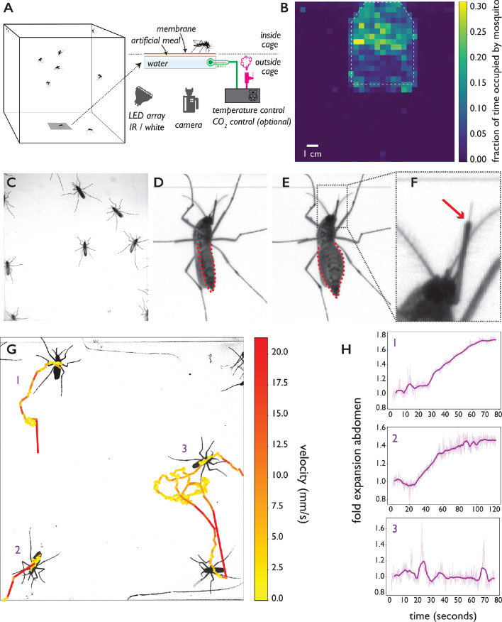
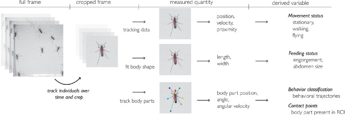
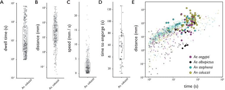
Female mosquitoes need a blood meal to reproduce, and in obtaining this essential nutrient they transmit deadly pathogens. Although crucial for the spread of mosquito-borne diseases, blood feeding remains poorly understood due to technological limitations. Indeed, studies often expose human subjects to assess biting behavior. Here, we present the biteOscope, a device that attracts mosquitoes to a host mimic which they bite to obtain an artificial blood meal. The host mimic is transparent, allowing high-resolution imaging of the feeding mosquito. Using machine learning, we extract detailed behavioral statistics describing the locomotion, pose, biting, and feeding dynamics of Aedes aegypti, Aedes albopictus, Anopheles stephensi, and Anopheles coluzzii. In addition to characterizing behavioral patterns, we discover that the common insect repellent DEET repels Anopheles coluzzii upon contact with their legs. The biteOscope provides a new perspective on mosquito blood feeding, enabling the high-throughput quantitative characterization of this lethal behavior.

Keywords: Mosquito; behavior; blood feeding; contact-dependent sensing; ecology; ethology; imaging; neuroscience.

Plain language summary

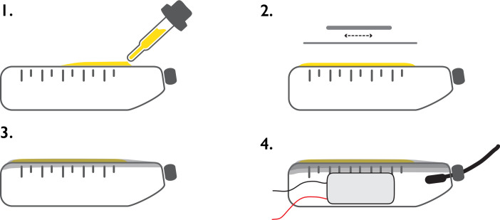
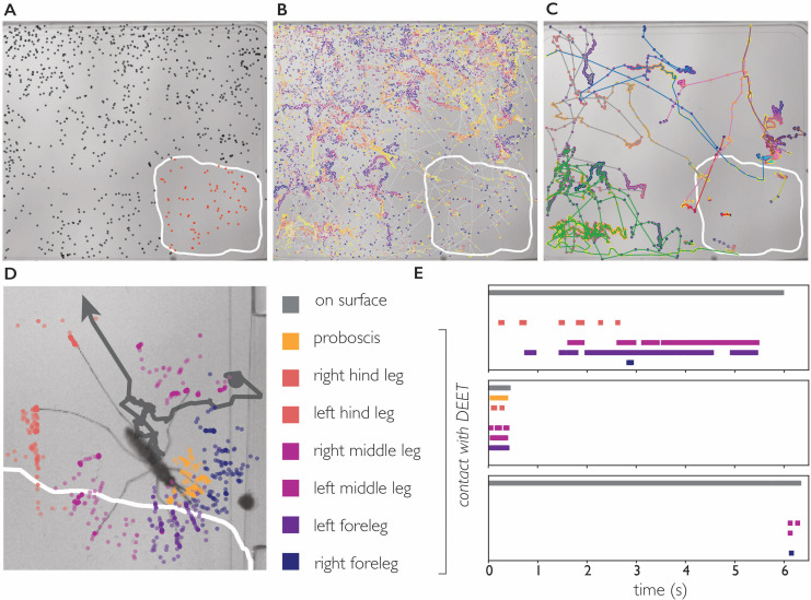
Scientists often sacrifice their own skin to study how mosquitos drink blood. They allow mosquitos to bite them in laboratory settings so they can observe the insects’ feeding behavior. By observing blood feeding, scientists hope to find ways to prevent deadly diseases like malaria, which is transmitted by bites from mosquitos carrying the malaria parasite. These studies are not only unpleasant for the volunteers, they also have important limitations. For example, it is too risky to use pathogen-infected mosquitos that could make the volunteers sick. A device called the biteOscope developed by Hol et al. may give scientists and their skin a reprieve. The device has a transparent skin-like covering that attracts mosquitos and supplies them an artificial blood meal when they bite. The device captures high-resolution images of the insects’ behavior. It is small enough to fit in a backpack when disassembled, costs about $900 to $3,500 US dollars, and is suitable for use in the laboratory or in the field. Using machine-learning techniques, Hol et al. also developed an automated system for analyzing the images. The researchers tested the device on four types of disease-transmitting mosquitos. In one set of experiments, Anopheles mosquitos were recorded interacting with a biteOscope partially coated with an insect repellent called DEET. The images captured by the biteOscope showed that the mosquitos are attracted to the warm surface and land on the part coated with DEET. But when their legs come in contact with the repellent, they leave. The biteOscope provides scientists a new way to study blood feeding, even in mosquitos infected with dangerous pathogens. It might also be used to test new ways to prevent mosquitos from biting and spreading disease. Because the device is portable and relatively inexpensive, it may enable larger studies in a variety of settings.


Conflict of interest statement

FH, LL, MP No competing interests declared

Figures

Figure 1.
Figure 1.. The biteOscope.
(A) Schematic of the set up. The bite substrate consists of a water bath (cell culture flask) that is mounted in the floor or wall of a cage, allowing freely flying mosquitoes access. An artificial meal is applied on the outside surface of the culture flask and covered using a Parafilm membrane, water in the flask is temperature controlled using a Raspberry Pi reading a temperature probe, and a Peltier element for heating (0.1 accuracy). The Raspberry Pi optionally controls the inflow of gas. Illumination is provided by an array of white or IR LEDs. A camera and lens situated outside the cage images mosquitoes (abdominal view) through the bite substrate. (B) Two-dimensional histogram (heatmap) showing mosquito presence on the bite substrate (indicated with a dashed line) and on the surrounding wall. Mosquitoes spend more time on the bite surface. (C) Raw image of Ae. aegypti on the bite substrate. (D-F) Images of an Ae. aegypti mosquito that has pierced the membrane and inserted its stylet into the meal. After imbibing, the abdomen dilates. The red arrow in (F) indicates the tip of the labium where the stylets (visible as a thin needle-like structure) pierce the surface and enter the artificial meal. (G) Tracks showing movement of Ae. aegypti on the bite substrate, color of tracks indicates velocity. (H) Fold expansion of the abdomen over time, indicating full engorgement in mosquitoes 1 and 2, and no feeding in mosquito 3 of panel (G).
Figure 1—figure supplement 1.
Figure 1—figure supplement 1.. Schematic of bite substrate assembly.
Assembly of the bite substrate. Step 1: The artificial meal is applied to the top surface of a flask containing degassed water (for the top surface of a 70 mL falcon tissue culture flask, a volume of approximately 1.6 mL is used). Step 2: The liquid is spread with a pipette tip to cover the entire top surface of the flask, and a rectangular section of Parafilm is stretched to approximately twice its original size (in x and y direction). Step 3: The Parafilm is stretched over the top surface of the flask to create a liquid cell and secured on the sides of the flask (by gently pressing). Step 4: A 5 mm wide ribbon of Parafilm is wrapped around the edge of the flask to provide additional support against leaking, the Peltier element (heating) is attached to the side of the flask using tape, and the temperature probe is inserted in the flask.
Figure 1—figure supplement 2.
Figure 1—figure supplement 2.. Overview of the computational pipeline.
Figure 2.
Figure 2.. Behavioral statistics of An. coluzzii (A–D) and all four species (E).
Each datapoint is derived from an individual trajectory, boxes indicate quartiles. (A) The time spent on the bite surface (n=349). (B) The total distance covered walking on the surface during a trajectory (n=349). (C) The mean velocity during a trajectory (n=349). (D) The time from landing to full engorgement (for trajectories leading to full engorgement, n=48). (E) The duration of a trajectory (total time for trajectories not leading to engorgement (transparent dots), time to full engorgement for trajectories that led to full engorgement (opaque circles)) versus the distance covered during that trajectory. The different colors denote different species, Ae. aegypti: magenta, Ae. albopictus: black, An. stephensi: cyan, An. coluzzii: yellow.
Figure 2—figure supplement 1.
Figure 2—figure supplement 1.. Behavioral statistics of Ae. aegypti, Ae. albopictus, An. stephensi, and An. coluzzii.
Each datapoint is derived from an individual trajectory. (A) The time spent on the bite surface. (B) The total distance covered walking on the surface during this time. (C) The mean velocity during a trajectory. (D) The time from landing to full engorgement (for trajectories leading to full engorgement).
Figure 3.
Figure 3.. Feeding behavior of individual Ae. albopictus.
(A, B) Ethograms of individual Ae. albopictus interacting with a bite substrate offering a PBS only meal, n=10 (A), and a meal consisting of PBS + 1 mM ATP, n=9 (B). Distinct exploratory bouts appear as continuous blocks in the ethogram and are labeled according to the behavior being displayed: flight (yellow), walking (purple), and stationary (dark blue), the time of engorgement to full repletion is marked by a black box and a white asterix. (C) Behavioral statistics of the data displayed in A and B showing the total time spent on the bite substrate (left, no significant difference p=0.39, Wilcoxon rank-sum test), the number of exploratory bouts undertaken (middle, significantly different p=0.020, Wilcoxon rank-sum test), and the length of individual bouts (right, significantly different p=9×10⁻⁴, Wilcoxon rank-sum test), of Ae. albopictus exploring the PBS only substrate (labeled 0) and those that engorged to full repletion on the PBS + ATP substrate (labeled 1). Individual data points are shown in purple, the mean and associated 95% confidence interval are depicted by a black dot and bar, respectively. Individuals that were offered the PBS + ATP substrate but did not feed to full repletion were excluded from this analysis.
Figure 3—figure supplement 1.
Figure 3—figure supplement 1.. The accuracy of automatic classification of locomotion behaviors (stationary, walking, flight) is 89% and exceeds 80% for a range of parameter values.
The accuracy of automatic classification of locomotion behaviors (stationary, walking, flight) presented in Figure 3A,B is 89% and exceeds 80% for a range of parameter values. (A) Threshold used to distinguish stationary from walking. (B) Threshold used to distinguish walking from flight.
Figure 4.
Figure 4.. Body part tracking reveals movement patterns of specific behaviors.
Color coding of plots in panels A-F are displayed at the bottom of the figure. (A–C) Trajectories of the tips of the six legs and proboscis of an Ae. albopictus female grooming her antennae (A), walking (B), and probing (C). (D–F) Time traces showing egocentric x (full lines) and y (dashed lines) coordinates of the body parts of mosquitoes shown in A-C. Anterior grooming is characterized by smooth periodic movement in the x and y planes. During walking the x-coordinate shows a swing that alternates between fore, middle, and hind leg; probing shows rapid pulling of the fore and middle legs towards the body. (G–I) Continuous wavelet transforms of the body part coordinates highlight the periodicity of movements. The amplitude of the spectrogram is indicated by the color, going from low (purple) to high (yellow). Yellow bands indicate periodic movement of a body part. Spectrograms of the seven body parts are stacked and separated by white lines (color coding on the right shows stacking order, with the x-coordinate of the body part on top, and y-coordinate on the bottom (x, and y coordinates are separated by a dashed line)).
Figure 4—figure supplement 1.
Figure 4—figure supplement 1.. Two-dimensional embedding of data shown in A-I.
Two-dimensional embedding of the body part coordinates and their wavelet transforms (data from Figure 4) using t-SNE. Data points are color coded according to the behavior displayed (manually labelled) and show three distinct clusters for grooming, walking, and probing. The clustering of the data points demonstrates the richness of the data obtained from body part tracking and indicates that clustering body part coordinates and derived features may be a feasible method to classify behaviors. Robust unsupervised classification using such methods requires a larger dataset than the one presented here.
Figure 5.
Figure 5.. DEET repels An. coluzzii on contact with legs.
(A) Landings on a substrate partly coated with 50% DEET (white line indicates DEET-coated surface). Black dots indicate landings outside the DEET area, red dots indicate landings inside the DEET area. The landing rate in the DEET area is approximately 1.9 times lower compared to the non-treated surface. (B) Trajectories of mosquito movement on the surface. Dots of individual tracks are colored from purple (start of the track) to yellow (end of the track). An. coluzzii on average spend seven times longer on the non-coated surface compared to the DEET-coated surface. (C) Example tracks of mosquitoes landing on the non-treated area and subsequently entering the DEET-coated area. (D) Body part tracking of a mosquito near the edge of the DEET-coated surface. The grey line indicates the movement of the center of mass of the mosquito (a dot indicates the start of the track, arrowhead departure). Colored dots indicate the position of the legs and proboscis during the section of the trajectory where the mosquito is within reach of the DEET-coated area (indicated by the white line). (E) Ethogram showing typical behavioral patterns when a mosquito comes in contact with DEET. The grey bar (top) indicates that a mosquito is anywhere on the surface (including the uncoated area), the colored bars indicate contact of a specific appendage with DEET. The top panel corresponds to the mosquito shown in (D) illustrating a mosquito that walks toward the DEET area, contacts it with several legs, and flies away. The middle panel is an example of ‘touch and go’ contact in which a mosquito lands on the DEET area, contacts it with several legs and proboscis, and takes off. The bottom panel shows a mosquito that after a long exploratory bout outside the DEET area, takes off as soon as the right foreleg and both middle legs contact the DEET area.

Similar articles

Cited by

References

    1. Abboubakar H, Buonomo B, Chitnis N. Modelling the effects of malaria infection on mosquito biting behaviour and attractiveness of humans. Ricerche Di Matematica. 2016;65:329–346. doi: 10.1007/s11587-016-0293-9. - DOI
    1. Afify A, Betz JF, Riabinina O, Lahondère C, Potter CJ. Commonly used insect repellents hide human odors from anopheles mosquitoes. Current Biology. 2019;29:3669–3680. doi: 10.1016/j.cub.2019.09.007. - DOI - PMC - PubMed
    1. Afify A, Potter CJ. Insect repellents mediate species-specific olfactory behaviours in mosquitoes. Malaria Journal. 2020;19:1–10. doi: 10.1186/s12936-020-03206-8. - DOI - PMC - PubMed
    1. Allan D, Caswell T, Keim N, van der Wel C. trackpy: Trackpy . v0. 3.2Zenodo. 2016 doi: 10.5281/zenodo.3492186. - DOI
    1. Benton R. The neurobiology of gustation in insect disease vectors: progress and potential. Current Opinion in Insect Science. 2017;20:19–27. doi: 10.1016/j.cois.2017.02.003. - DOI - PubMed

Publication types

Substances