. 2020 Sep 22;117(38):23695-23706.
doi: 10.1073/pnas.2003932117. Epub 2020 Sep 9.

LncRNA Malat1 inhibition of TDP43 cleavage suppresses IRF3-initiated antiviral innate immunity

Affiliations

LncRNA Malat1 inhibition of TDP43 cleavage suppresses IRF3-initiated antiviral innate immunity

Wei Liu et al. Proc Natl Acad Sci U S A. .

Abstract

Long noncoding RNAs (lncRNAs) involved in the regulation of antiviral innate immune responses need to be further identified. By functionally screening the lncRNAs in macrophages, here we identified lncRNA Malat1, abundant in the nucleus but significantly down-regulated after viral infection, as a negative regulator of antiviral type I IFN (IFN-I) production. Malat1 directly bound to the transactive response DNA-binding protein (TDP43) in the nucleus and prevented activation of TDP43 by blocking the activated caspase-3-mediated TDP43 cleavage to TDP35. The cleaved TDP35 increased the nuclear IRF3 protein level by binding and degrading Rbck1 pre-mRNA to prevent IRF3 proteasomal degradation upon viral infection, thus selectively promoting antiviral IFN-I production. Deficiency of Malat1 enhanced antiviral innate responses in vivo, accompanying the increased IFN-I production and reduced viral burden. Importantly, the reduced MALAT1, augmented IRF3, and increased IFNA mRNA were found in peripheral blood mononuclear cells (PBMCs) from systemic lupus erythematosus (SLE) patients. Therefore, the down-regulation of MALAT1 in virus-infected cells or in human cells from autoimmune diseases will increase host resistance against viral infection or lead to autoinflammatory interferonopathies via the increased type I IFN production. Our results demonstrate that the nuclear Malat1 suppresses antiviral innate responses by targeting TDP43 activation via RNA-RBP interactive network, adding insight to the molecular regulation of innate responses and autoimmune pathogenesis.

Keywords: Malat1; TDP43; innate immunity; long noncoding RNA; type I interferon.


Conflict of interest statement

The authors declare no competing interest.

Figures

Fig. 1.
Fig. 1.
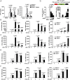
The expression of nuclear lncRNA Malat1 decreases upon viral infection. (A) Heat map of top 20 abundant nuclear lncRNAs in RAW264.7 cells at resting state which are changed more than twofold after VSV infection for 8 h. ENSMUST00000172812 (Malat1) is pointed out by black arrow. (B) Northern blot assay of Malat1 level in RAW264.7 cells along with VSV infection for indicated hours. Relative intensity of Malat1 is calculated by the ImageJ program. (C) Confocal microscope images from FISH assay of Malat1 level in RAW264.7 cells along with VSV infection for indicated hours. Relative fluorescence intensity of Malat1 is calculated by the ImageJ program (Right). Red, Malat1; blue, DAPI. (Scale bar, 5 μm.) (D) qRT-PCR analysis of relative Malat1 level in RAW264.7 cells along with VSV, EMCV, and HSV-1 infection for indicated hours, respectively. Data are representative of three independent experiments (B and C) or shown as mean ± SD of n = 3 biological replicates (C and D), two-tailed unpaired Student’s t test (C and D).
Fig. 2.
Fig. 2.
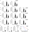
Malat1 selectively inhibits the production of type I IFNs in macrophages upon viral infection. (A) ELISA analysis of IFN-α and IFN-β in culture supernatants of Malat1+/+, Malat1−/ , and Malat1−/− rescued by Malat1 RAW264.7 cells infected with VSV or HSV-1 for 12 h, respectively. (B) qRT-PCR analysis of relative Ifna4, Ifnb1, and Isg15 mRNA level in Malat1+/+ and Malat1−/− RAW264.7 cells along with VSV infection for indicated hours. (C) qRT-PCR analysis of relative Ifna4, Ifnb1, and Isg15 mRNA level in Malat1+/+ and Malat1−/− RAW264.7 cells along with HSV-1 infection for indicated hours. (D) Luciferase assay of IFN-β promoter activity influenced by gradually enhanced Malat1 in HEK293T cells transfected with RIG-I and Malat1 vectors upon VSV infection for indicated hours (Left) and qRT-PCR analysis of Malat1 relative level in HEK293T cells above transfected with 200, 400, and 800 ng Malat1 vector, respectively (Right). (E) qRT-PCR analysis of relative Mx1, Ifit1, and Isg15 mRNA level in Malat1+/+ and Malat1−/− RAW264.7 cells treated with IFN-α for indicated hours. (F) qRT-PCR analysis of relative Mx1, Ifit1, and Isg15 mRNA level in Malat1+/+ and Malat1−/− RAW264.7 cells treated with IFN-β for indicated hours. All data are shown as mean ± SD of n = 3 biological replicates, two-tailed unpaired Student’s t test.
Fig. 3.
Fig. 3.
Deficiency of Malat1 enhances antiviral response in vivo. (A) ELISA of IFN-α and IFN-β in serum from Malat1+/+ and Malat1−/− mice infected with VSV (5 × 10⁷ pfu/g) for 18 h or HSV-1 (1 × 10⁷ pfu/g) for 24 h, respectively, via tail i.v. injection. (B) qRT-PCR analysis of relative Ifna4, Ifnb1, and Isg15 mRNA level in liver from Malat1+/+ and Malat1−/− mice corresponding to A. (C) qRT-PCR analysis of relative Ifna4, Ifnb1, and Isg15 mRNA level in spleen from Malat1+/+ and Malat1−/− mice corresponding to A. (D) qRT-PCR analysis of relative Ifna4, Ifnb1, and Isg15 mRNA level in lung from Malat1+/+ and Malat1−/− mice corresponding to A. (E) qRT-PCR analysis of relative VSV RNA replication in liver, spleen, and lung from Malat1+/+ and Malat1−/− mice corresponding to A. (F) Survival curves of 6- to 8-wk-old mice of Malat1+/+ and Malat1−/− infected with VSV (1 × 10⁸ pfu/g) via tail i.v. injection. Data are shown as mean ± SD of n = 3 biological replicates (AE) or demonstrated as Kaplan–Meier survival curve (F, n = 7), two-tailed unpaired Student’s t test (AE), or Log-rank (Mantel–Cox) test (F).
Fig. 4.
Fig. 4.
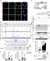
Identification of Malat1-bound TDP43 in selectively promoting IFN-I production. (A) Confocal microscope images from sequential FISH assay of Malat1 and TDP43 in RAW264.7 cells along with VSV infection for indicated hours. Red, Malat1; green, TDP43; and blue, DAPI. (Scale bar, 5 μm.) (B) iCLIP assay of V5-TDP43 overexpressed in RAW264.7 cells and the likely interaction sites of Malat1 that bind with TDP43. (C) RNA pull-down assay of different truncations of Malat1 with overexpressed V5-TDP43 in RAW264.7 cells. (D) RNA pull-down assay of Malat1 (3,006 to 4,020 nt) and Malat1 (4,569 to 5,609 nt) fragments with HEK293T cell lysates overexpressed different truncations of TDP43, respectively. (E) ELISA analysis of IFN-α and IFN-β in culture supernatants of Tardbp+/+ and Tardbp−/− RAW264.7 cells infected with VSV or HSV-1 for 12 h, respectively. (F) Luciferase assay of IFN-β promoter activity influenced by gradually enhanced TDP43 in HEK293T cells transfected with RIG-I and TDP43 vectors upon VSV infection for indicated hours (Upper) and Western blot analysis of TDP43/TDP35 level in HEK293T cells above transfected with 200, 400, and 800 ng TDP43 vector, respectively (Lower). Data are representative of three independent experiments (A, C, D, and F) or shown as mean ± SD of n = 3 biological replicates (E and F), two-tailed unpaired Student’s t test (E and F).
Fig. 5.
Fig. 5.
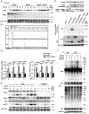
Malat1 inhibits IFN-I production by blocking TDP43 cleavage to its active form TDP35. (A) Western blot analysis of Malat1’s effect on TDP43 along with VSV infection for the indicated hours in RAW264.7 cells. (B) qRT-PCR analysis of relative Ifna4 and Ifnb1 mRNA level in Tardbp+/+, Tardbp−/−, and Tardbp−/− rescued by TDP35 RAW264.7 cells upon VSV infection for indicated hours. (C) Luciferase analysis of IFN-β promoter activity in Tardbp+/+ and Tardbp−/− L929 cells transfected with Malat1, RIG-I, as well as wild-type, truncation, or mutant of TDP43 vectors, respectively. (D) Coimmunoprecipitation analysis of interaction between TDP43 and cleaved caspase-3 affected by Malat1 in RAW264.7 cells upon virus infection for indicated hours. Data are representative of three independent experiments (A and D) or shown as mean ± SD of n = 3 biological replicates (B and C), two-tailed unpaired Student’s t test (B and C).
Fig. 6.
Fig. 6.
The cleaved TDP35 increases nuclear IRF3 protein level by binding and degrading Rbck1 pre-mRNA to prevent IRF3 proteasomal degradation upon viral infection. (A) Western blot analysis of TDP35’s effect on protein level of IRF3 in the nucleus of Tardbp+/+, Tardbp−/−, and Tardbp−/− rescued by TDP35 RAW264.7 cells upon VSV infection for indicated hours. (B) iCLIP assay of V5-TDP43 overexpressed RAW264.7 cells and the likely interaction sites of Rbck1 pre-mRNA that binding with TDP43/TDP35. (C) RNA pull-down assay of Rbck1 pre-mRNA and the different truncations or mutant of TDP43 overexpressed in HEK293T cells, respectively. (D) qRT-PCR analysis of relative pre-mRNA and mRNA level of Rbck1 in Tardbp+/+, Tardbp−/−, and Tardbp−/− rescued by TDP35 RAW264.7 cells upon virus infection for indicated hours. (E) Western blot analysis of the necessity of TDP35 for Malat1 regulating the expression of Rbck1 and phosphorylation of IRF3 in nucleus of Malat1+/+ and Malat1−/− RAW264.7 cells transfected with siRNA targeting negative control (siNC) or Tardbp (siTardbp), respectively, upon VSV infection for indicated hours. (F) Coimmunoprecipitation analysis of the K48-linked polyubiquitination on IRF3 in Malat1+/+ and Malat1−/− RAW264.7 cells infected with VSV for indicated hours. Data are representative of three independent experiments (A, C, E, and F) or shown as mean ± SD of n = 3 biological replicates (D), two-tailed unpaired Student’s t test (D).
Fig. 7.
Fig. 7.
The decreased expression of MALAT1 is associated with increased TDP43 cleavage, IRF3 protein, and IFNA expression in PBMCs of SLE patients. (A) qRT-PCR analysis of relative MALAT1 and IFNA in PBMCs from healthy donors and SLE patients. (B) Western blot analysis of key molecules in MALAT1-regulated antiviral response in PBMCs from healthy donors and SLE patients. (C) qRT-PCR analysis of relative MALAT1 and IFNA in PBMCs from SLE patients before and after effective treatment. (D) Western blot analysis of key molecules in MALAT1-regulated antiviral response in PBMCs from SLE patients before and after effective treatment. (E) Proposed working model for Malat1 as a negative regulator of antiviral innate response by targeting TDP43 activation via an RNA-RBP interactive network. Data are shown as mean ± SD of n = 12 (A) or n = 6 (C) biological replicates, two-tailed unpaired Student’s t test (A and C).

Similar articles

Cited by

References

    1. Yan N., Chen Z. J., Intrinsic antiviral immunity. Nat. Immunol. 13, 214–222 (2012). - PMC - PubMed
    1. Kretschmer S., Lee-Kirsch M. A., Type I interferon-mediated autoinflammation and autoimmunity. Curr. Opin. Immunol. 49, 96–102 (2017). - PubMed
    1. Lee-Kirsch M. A., The type I interferonopathies. Annu. Rev. Med. 68, 297–315 (2017). - PubMed
    1. McNab F., Mayer-Barber K., Sher A., Wack A., O’Garra A., Type I interferons in infectious disease. Nat. Rev. Immunol. 15, 87–103 (2015). - PMC - PubMed
    1. Liu S. et al. ., Phosphorylation of innate immune adaptor proteins MAVS, STING, and TRIF induces IRF3 activation. Science 347, aaa2630 (2015). - PubMed

Publication types

MeSH terms