Existence of SARS-CoV-2 Entry Molecules in the Oral Cavity
- PMID: 32825469
- PMCID: PMC7503451
- DOI: 10.3390/ijms21176000
Existence of SARS-CoV-2 Entry Molecules in the Oral Cavity
Abstract
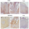
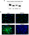
The severe acute respiratory syndrome coronavirus 2 (SARS-CoV-2) receptor, angiotensin-converting enzyme 2 (ACE2), transmembrane protease serine 2 (TMPRSS2), and furin, which promote entry of the virus into the host cell, have been identified as determinants of SARS-CoV-2 infection. Dorsal tongue and gingiva, saliva, and tongue coating samples were examined to determine the presence of these molecules in the oral cavity. Immunohistochemical analyses showed that ACE2 was expressed in the stratified squamous epithelium of the dorsal tongue and gingiva. TMPRSS2 was strongly expressed in stratified squamous epithelium in the keratinized surface layer and detected in the saliva and tongue coating samples via Western blot. Furin was localized mainly in the lower layer of stratified squamous epithelium and detected in the saliva but not tongue coating. ACE2, TMPRSS2, and furin mRNA expression was observed in taste bud-derived cultured cells, which was similar to the immunofluorescence observations. These data showed that essential molecules for SARS-CoV-2 infection were abundant in the oral cavity. However, the database analysis showed that saliva also contains many protease inhibitors. Therefore, although the oral cavity may be the entry route for SARS-CoV-2, other factors including protease inhibitors in the saliva that inhibit viral entry should be considered.
Keywords: ACE2; SARS-CoV-2; TMPRSS2; furin; oral cavity; saliva; taste cell; tongue coating.
Conflict of interest statement
The authors declare no conflict of interest.
Figures


Similar articles
-
Single-cell analysis of SARS-CoV-2 receptor ACE2 and spike protein priming expression of proteases in the human heart.Cardiovasc Res. 2020 Aug 1;116(10):1733-1741. doi: 10.1093/cvr/cvaa191. Cardiovasc Res. 2020. PMID: 32638018 Free PMC article.
-
Optimized Pseudotyping Conditions for the SARS-COV-2 Spike Glycoprotein.J Virol. 2020 Oct 14;94(21):e01062-20. doi: 10.1128/JVI.01062-20. Print 2020 Oct 14. J Virol. 2020. PMID: 32788194 Free PMC article.
-
Structural and functional modelling of SARS-CoV-2 entry in animal models.Sci Rep. 2020 Sep 28;10(1):15917. doi: 10.1038/s41598-020-72528-z. Sci Rep. 2020. PMID: 32985513 Free PMC article.
-
COVID-19 pandemic: Insights into structure, function, and hACE2 receptor recognition by SARS-CoV-2.PLoS Pathog. 2020 Aug 21;16(8):e1008762. doi: 10.1371/journal.ppat.1008762. eCollection 2020 Aug. PLoS Pathog. 2020. PMID: 32822426 Free PMC article. Review.
-
Oral Pathology in COVID-19 and SARS-CoV-2 Infection-Molecular Aspects.Int J Mol Sci. 2022 Jan 27;23(3):1431. doi: 10.3390/ijms23031431. Int J Mol Sci. 2022. PMID: 35163355 Free PMC article. Review.
Cited by
-
Prevalence of Oral Manifestations in COVID-19-Diagnosed Patients at a Tertiary Care Hospital in Kerala.J Maxillofac Oral Surg. 2024 Apr;23(2):296-300. doi: 10.1007/s12663-023-02049-5. Epub 2023 Dec 1. J Maxillofac Oral Surg. 2024. PMID: 38601253
-
Pathological changes in oral epithelium and the expression of SARS-CoV-2 entry receptors, ACE2 and furin.PLoS One. 2024 Mar 15;19(3):e0300269. doi: 10.1371/journal.pone.0300269. eCollection 2024. PLoS One. 2024. PMID: 38489333 Free PMC article.
-
SARS-CoV-2 mechanisms of cell tropism in various organs considering host factors.Heliyon. 2024 Feb 20;10(4):e26577. doi: 10.1016/j.heliyon.2024.e26577. eCollection 2024 Feb 29. Heliyon. 2024. PMID: 38420467 Free PMC article. Review.
-
Association between dysbiotic perio-pathogens and inflammatory initiators and mediators in COVID-19 patients with diabetes.Heliyon. 2024 Jan 6;10(2):e24089. doi: 10.1016/j.heliyon.2024.e24089. eCollection 2024 Jan 30. Heliyon. 2024. PMID: 38293542 Free PMC article.
-
The utility of salivary CRP and IL-6 as a non-invasive measurement evaluated in patients with COVID-19 with and without diabetes.F1000Res. 2024 Jan 11;12:419. doi: 10.12688/f1000research.130995.3. eCollection 2023. F1000Res. 2024. PMID: 38269064 Free PMC article.
References
MeSH terms
Substances
LinkOut - more resources
Full Text Sources
Miscellaneous

