. 2020 Sep;2(9):873-881.
doi: 10.1038/s42255-020-0245-2. Epub 2020 Jul 27.

Long-chain fatty acyl-CoA esters regulate metabolism via allosteric control of AMPK β1 isoforms

Affiliations

Long-chain fatty acyl-CoA esters regulate metabolism via allosteric control of AMPK β1 isoforms

Stephen L Pinkosky et al. Nat Metab. 2020 Sep.

Abstract

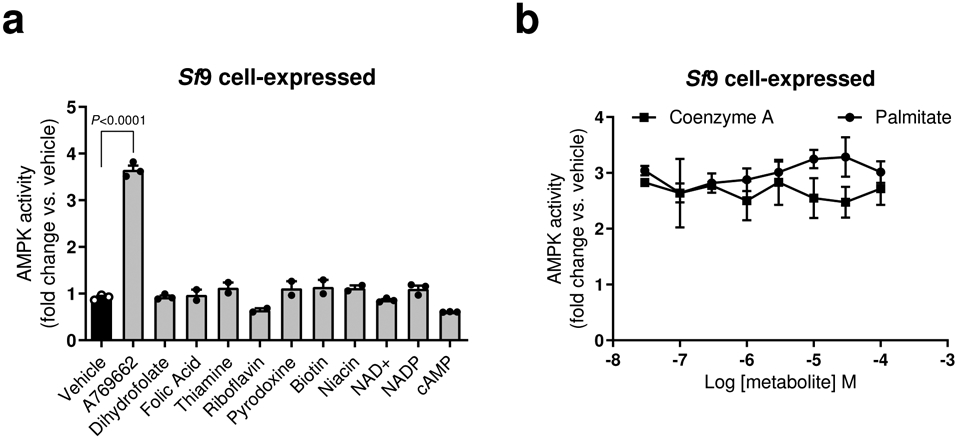
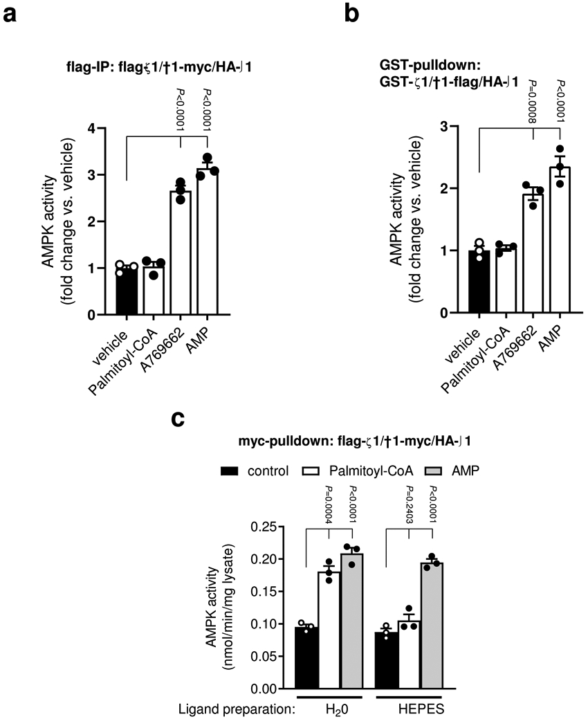
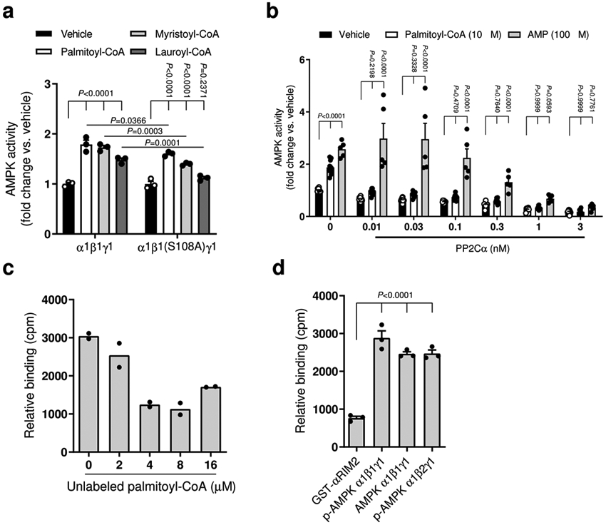
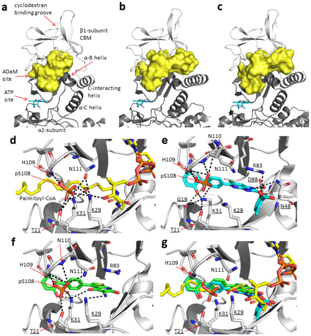
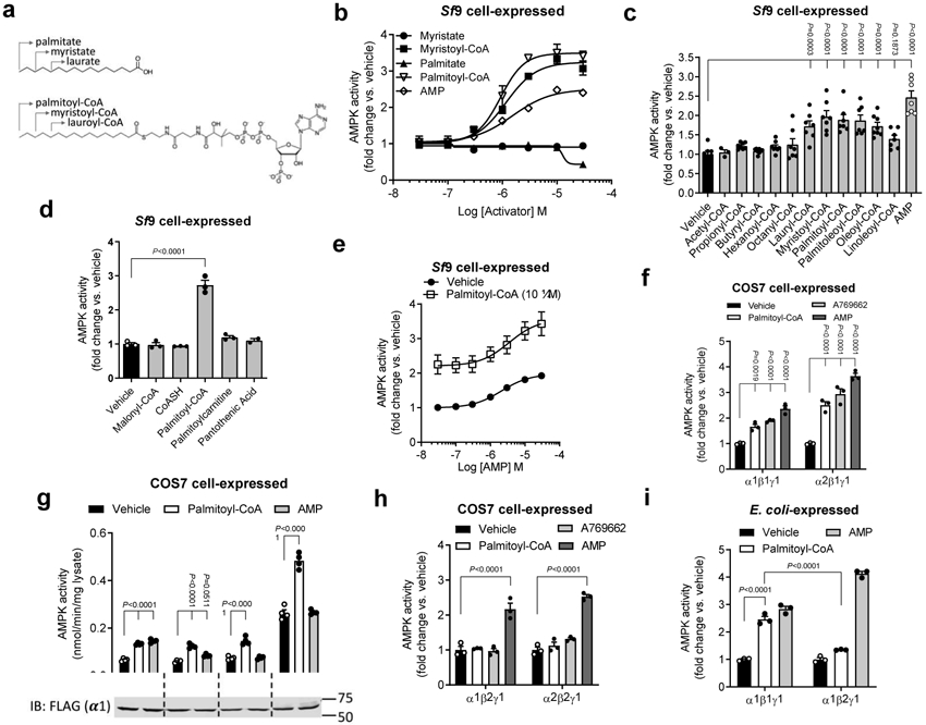
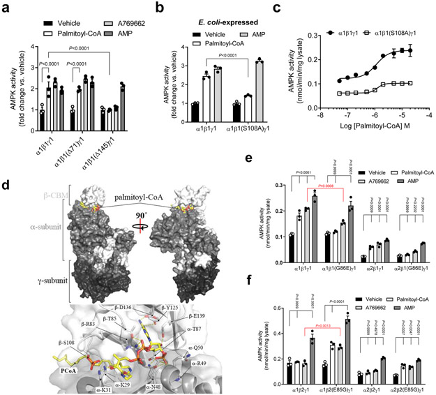
Long-chain fatty acids (LCFAs) play important roles in cellular energy metabolism, acting as both an important energy source and signalling molecules1. LCFA-CoA esters promote their own oxidation by acting as allosteric inhibitors of acetyl-CoA carboxylase, which reduces the production of malonyl-CoA and relieves inhibition of carnitine palmitoyl-transferase 1, thereby promoting LCFA-CoA transport into the mitochondria for β-oxidation2-6. Here we report a new level of regulation wherein LCFA-CoA esters per se allosterically activate AMP-activated protein kinase (AMPK) β1-containing isoforms to increase fatty acid oxidation through phosphorylation of acetyl-CoA carboxylase. Activation of AMPK by LCFA-CoA esters requires the allosteric drug and metabolite site formed between the α-subunit kinase domain and the β-subunit. β1 subunit mutations that inhibit AMPK activation by the small-molecule activator A769662, which binds to the allosteric drug and metabolite site, also inhibit activation by LCFA-CoAs. Thus, LCFA-CoA metabolites act as direct endogenous AMPK β1-selective activators and promote LCFA oxidation.


Figures

Extended Data Fig. 1
Extended Data Fig. 1. Specificity of AMPK activation by palmitoyl-CoA and effects of co-incubations with free palmitate or coenzyme A.
a, b, Activities of AMPKα1β1γ1 (Sf9 insect cell-expressed), determined by TR-FRET SAMS assay, in the presence of coenzymes (100 μM), cofactors (100 μM) and vitamins (100 μM) (a), or following 15 min pre-incubation in the presence of palmitoyl-CoA (10 μM) ± indicated concentrations of free palmitate or coenzyme A (b). Data are shown as mean fold change in AMPK activity vs. vehicle ± s.e.m. For a, n = 3 except for folic acid, thiamine, riboflavin, pyridoxine, biotin and niacin (n = 2); for b, n = 5 (palmitate incubation) or n = 3 (coenzyme A incubation). Statistical significance was calculated using one-way ANOVA with Bonferroni’s multiple comparisons test. n represent biological independent experiments.
Extended Data Fig. 2
Extended Data Fig. 2. Palmitoyl-CoA activation of purified AMPK is sensitive to the method of protein immobilization
a, b, Activities of AMPKα1β1γ1 (COS7 cell-expressed; fusion tags as indicated) were determined by 32P SAMS peptide assay ± palmitoyl-CoA (10 μM), A769662 (10 μM) or AMP (100 μM), following immobilization on anti-flag agarose (a) or glutathione-Sepharose (b). Data are shown as mean fold change in AMPK activity vs. vehicle ± s.e.m., n = 3. c, Activities of AMPKα1β1γ1 (COS7 cell-expressed; fusion tags as indicated) were determined by 32P SAMS peptide assay ± palmitoyl-CoA (10 μM), prepared in either H2O or 50 mM HEPES, following immobilization on anti-myc agarose. Data are shown as mean specific activity ± s.e.m., n = 3. Statistical significance was calculated using one-way ANOVA with Bonferroni’s multiple comparisons test. n represent biological independent experiments.
Extended Data Fig. 3
Extended Data Fig. 3. Characterization of LCFA-CoA activation of AMPK
a, Activities of AMPKα1β1γ1 (COS7 cell-expressed, WT and β1S108A mutant) were determined by 32P SAMS assay, following immobilization on anti-myc agarose, ± palmitoyl-CoA, myristoyl-CoA or lauroyl-CoA (10 μM). Data are shown as mean fold change in AMPK activity vs. vehicle ± s.e.m., n=3. Statistical significance was calculated using two-way ANOVA with Bonferroni’s multiple comparisons test. n represent biological independent experiments. b, Activities of AMPKα1β1γ1 (Sf9 insect cell-expressed) were determined by TR-FRET in the presence of the indicated concentration of phosphatase PP2Cα ± AMP (30 μM) or palmitoyl-CoA (10 μM). Data are shown as mean fold change in AMPK activity vs. vehicle ± s.e.m., n=10 except for AMP incubated (n = 5). Statistical significance was calculated using two-way ANOVA with Bonferroni’s multiple comparisons test. n represent biological independent experiments. c, d, Binding of [3H]-palmitoyl-CoA to AMPKα1β1γ1 (E. coli-expressed) ± increasing concentrations of unlabeled palmitoyl-CoA (c), or to various AMPK preparations (d). GST-αRIM2: His6-GST-LVPRGS(thrombin cleavage site)-α1(282–374). Data are shown as mean relative binding ± s.e.m. For c, n = 2; for d, n = 3. Statistical significance was calculated using one-way ANOVA with Bonferroni’s multiple comparisons test. n represent biological independent experiments.
Extended Data Fig. 4
Extended Data Fig. 4. The channel at the interface between AMPK α2- and β1-subunits used for docking palmitoyl-CoA, interactions made by pSer108 and comparison with ADaM site activators.
(a) In the PDB ID:4CFF AMPK structure (shown as cartoon), the channel (i.e. the docking protomol, yellow molecular surface) encompasses the ADaM site (located directly above the ATP binding site of the α2-subunit kinase domain) and continues into a pocket located beneath the cyclodextran binding groove of the β1-CBM. In this AMPK structure, the channel is blocked at approximately the α-B helix of the α2-subunit by residues Arg49, Arg53, Pro86 and Thr87 of the α2-subunit and Pro140, Gln154, Lys156, Asp159 and Lys172 of the β1-subunit. Whereas in the (b) 5ISO and (c) 6B1U AMPK structures, the channel runs the full width across the α2- and β1-subunit interface (i.e. from the start of the ADaM site to the C-interacting helix of the β1-subunit, including the pocket beneath the cyclodextran binding groove of the β1-CBM). Polar interactions made by pSer108 in the (d) palmitoyl-CoA:AMPKα2β1γ1 model, (e) SC4:AMPKα2β1γ1 crystal structure (PDB ID: 6B1U) and (f) A-769662:AMPKα2β1γ1 crystal structure (PDB ID: 4CFF). (g) Overlay of the palmitoyl-CoA:AMPKα2β1γ1 model with the A-769662:AMPKα2β1γ1 and SC4:AMPKα2β1γ1 crystal structures (only A-769962 and SC4 shown for clarity). The same view is shown in panels a-c and d-g, the structures have been aligned via their β1-subunit CBM. In panels a-c, the location of the ATP binding site in the α2-subunit kinase domain is indicated by staurosporine (cyan sticks). Residues from the α2-catalytic subunit are underlined. Polar interactions are indicated by black dashed lines.
Extended Data Fig. 5
Extended Data Fig. 5. Unique palmitoyl-CoA conformation clusters consistent with our experimental data.
Docking into the (a) 4CFF, (b) 5ISO and (c) 6B1U active AMPKα2β1γ1 structures (grey cartoon). Palmitoyl-CoA shown as sticks, with the carbon atoms coloured either yellow or green. (d) Overlay of all docked palmitoyl-CoA poses except for those in Clusters 2, 4 and 5 for the 5ISO AMPK structure. The overlay shows that the different conformational clusters for the 4CFF, 5ISO and 6B1U AMPK structures fall under a general binding mode. Carbon, sulphur, nitrogen, oxygen and phosphorous atoms are coloured grey, yellow, blue, red and orange respectively.
Extended Data Fig. 6
Extended Data Fig. 6. Alignment of AMPK β1 and β2 sequences from diverse species.
Alignments to human AMPKβ1Gly86 and β2Glu85 residues are in bold.
Extended Data Fig. 7
Extended Data Fig. 7. AMPKβ1N111 does not alter sensitivity to palmitoyl-CoA or AMP
AMPK activity of AMPKα1β1γ1 (WT, β1N111A or β1N111D) was determined by 32P SAMS assay, following immobilization on anti-myc agarose, ± palmitoyl-CoA (10 μM) or AMP (100 μM). Data are shown as mean fold change in activity vs. vehicle, ± s.e.m.; n = 3. Statistical significance was calculated using two-way ANOVA with Bonferroni’s multiple comparisons test. n represent biological independent experiments.
Figure 1.
Figure 1.. LCFA-CoAs are direct AMPK activators.
a, Structures of LCFAs (upper) and LCFA-CoAs (lower). b-e, Activities of AMPKα1β1γ1 (Sf9 insect cell-expressed), determined by TR-FRET SAMS assay, in the presence of increasing concentrations of myristate, palmitate, their respective CoA thioester conjugates and AMP (b), acyl-CoA esters (10 μM) with chain lengths ranging from C2 to C18 (c), metabolites or biosynthetic precursors important for palmitoyl-CoA synthesis or catabolism (d) or ± 10 μM palmitoyl-CoA and the indicated concentrations of AMP (e). Data are shown as mean fold change in activity vs. vehicle ± s.e.m. For b, n = 2; for c, n = 7 except for propionyl-, butyryl-, hexanoyl- and oleoyl-CoAs (n =8) and acetyl-CoA (n = 3); for d, n = 3 except for pantothenic acid (n = 2); for e, n = 6. Statistical significance was calculated using one-way ANOVA with Bonferroni’s multiple comparisons test. n represent biological independent experiments. f-h, Activities of AMPK complexes (COS7 cell-expressed; basal activities detailed in Extended Data Table 1), determined by 32P SAMS peptide assay following anti-myc agarose immobilization, of α1 and α2 AMPK ± palmitoyl-CoA (10 μM), A769662 (10 μM) or AMP (100 μM) (f), γ1 mutants ± AMP (100 μM) or palmitoyl-CoA (10 μM) (g) and β1 and β2 AMPK ± palmitoyl-CoA (10 μM), A769662 (10 μM) or AMP (100 μM) (h). For f, h, data are shown as mean fold change in activity vs. vehicle ± s.e.m.; n = 3. For g, data are shown as mean specific activity ± s.e.m.; n = 4. Statistical significance was calculated using two-way ANOVA with Bonferroni’s multiple comparisons test. n represent biological independent experiments. Immunoblot shows relative expression of flag-AMPK in COS7 cells. (i) Activities of purified AMPK complexes α1β1γ1 and α1β2γ1 (E. coli-expressed; basal activities detailed in Extended Data Table 2), determined by 32P SAMS peptide assay, ± palmitoyl-CoA (10 μM) or AMP (100 μM). Data are shown as mean fold change in activity vs. vehicle ± s.e.m.; n = 3. Statistical significance was calculated using two-way ANOVA with Bonferroni’s multiple comparisons test. n represent biological independent experiments.
Figure 2.
Figure 2.. LCFA-CoA activation is mediated through the AMPK ADaM site.
a, Activities of AMPKα1β1γ1 (WT or N-terminal deletions of β1 residues 1-71 (Δ71) or 1-145 (Δ145)) ± palmitoyl-CoA (10 μM) or AMP (100 μM). b, c, Activities of AMPKα1β1γ1 (WT or β1S108A) ± palmitoyl-CoA (10 μM) or AMP (100 μM) (b) or increasing concentration of palmitoyl-CoA (c). For a, b, data are shown as mean fold change in activity vs. vehicle ± s.e.m.; n = 3. For c, data are shown as mean specific activity ± s.e.m.; n = 3. Statistical significance was calculated using two-way ANOVA with Bonferroni’s multiple comparisons test. n represent biological independent experiments. d, In silico modelling of palmitoyl-CoA bound to AMPKα2β1γ1. e, f, Activities of AMPKα1β1γ1 and α2β1γ1 (WT or β1G86E) (e), or AMPK α1β2γ1 and α2β2γ1 (WT or β2E85G) (f) ± palmitoyl-CoA (10 μM), A769662 (10 μM) or AMP (100 μM). Data are shown as mean specific activity ± s.e.m.; n = 3. Statistical significance was calculated using two-way ANOVA with Bonferroni’s multiple comparisons test, or unpaired, 2-tailed Student’s t test (red P values). n represent biological independent experiments. Activities determined by 32P SAMS peptide assay. COS7 cell-expressed AMPK immobilized on anti-myc agarose was used in all experiments unless indicated.
Figure 3.
Figure 3.. FA-CoAs increase fatty acid oxidation through AMPK phosphorylation of ACC.
a, Primary mouse hepatocytes from C57Bl6J mice were treated with vehicle or palmitate (100, 250, or 500 μM) followed by western blotting for total AMPKα, phosphorylated-AMPK (p-AMPK, α-Thr172), total ACC, phosphorylated-ACC (p-ACC, Ser79/212) and β-actin. Data are shown as mean fold change in phosphorylation of AMPK (n = 5) or ACC (n = 6) vs. vehicle ± s.e.m. b, Primary mouse hepatocytes from C57Bl6J mice were treated with vehicle or 500 μM palmitate, bromo-palmitate (Br-Palmitate) or methyl-palmitate (Me-Palmitate) followed by western blotting for total ACC, phosphorylated-ACC (p-ACC, Ser79/212) and β-actin. Data are shown as mean fold change in phosphorylation of ACC (n = 6) vs. vehicle ± s.e.m. Statistical significance was calculated using one-way ANOVA with Bonferroni’s multiple comparisons test. n represent biological independent experiments. Representative immunoblots are shown. c, Respiratory Exchange Ratios (R.E.R.) of WT and ACC DKI mice following oral administration of saline (Veh), or Intralipid® (10 ml/kg). Data are shown as mean R.E.R. ± s.e.m. (WT, n = 7; ACC DKI, n = 8). Statistical significance was calculated using two-way ANOVA with Bonferroni’s multiple comparisons test. n represent biological independent experiments. d-f, WT and ACC DKI mice, following oral administration of saline (Vehicle) or Intralipid® (10 ml/kg), were measured for lipid oxidation rates calculated from R.E.R. over 4 h, starting 1 h post-gavage (d), food intake during 2 h re-feed (e) and activity levels 6 h post-gavage (f). Data are shown as mean fold change in lipid oxidation rate vs. vehicle (d), mean food intake (e) and mean total beam breaks (f) ± s.e.m. (WT, n = 7; ACC DKI, n = 8). Statistical significance was calculated using unpaired, 2-tailed Student’s t test with Bonferroni’s multiple comparisons test. n represent biological independent experiments.

Similar articles

Cited by

References

    1. Faergeman NJ & Knudsen J Role of long-chain fatty acyl-CoA esters in the regulation of metabolism and in cell signalling. Biochem J 323 ( Pt 1), 1–12 (1997). - PMC - PubMed
    1. Bortz WM & Lynen F THE INHIBITION OF ACETYL COA CARBOXYLASE BY LONG CHAIN ACYL COA DERIVATIVES. Biochem Z 337, 505–509 (1963). - PubMed
    1. McGarry JD & Foster DW In support of the roles of malonyl-CoA and carnitine acyltransferase I in the regulation of hepatic fatty acid oxidation and ketogenesis. J Biol Chem 254, 8163–8168 (1979). - PubMed
    1. McGarry JD, Takabayashi Y & Foster DW The role of malonyl-coa in the coordination of fatty acid synthesis and oxidation in isolated rat hepatocytes. J Biol Chem 253, 8294–8300 (1978). - PubMed
    1. Trumble GE, Smith MA & Winder WW Purification and characterization of rat skeletal muscle acetyl-CoA carboxylase. Eur J Biochem 231, 192–198, doi:10.1111/j.1432-1033.1995.tb20686.x (1995). - DOI - PubMed

Publication types

MeSH terms

LinkOut - more resources