Review
. 2020 Sep;21(9):485-498.
doi: 10.1038/s41583-020-0333-z. Epub 2020 Jul 22.

Emerging importance of satellite glia in nervous system function and dysfunction

Affiliations
Review

Emerging importance of satellite glia in nervous system function and dysfunction

Menachem Hanani et al. Nat Rev Neurosci. 2020 Sep.

Erratum in

Abstract

Satellite glial cells (SGCs) closely envelop cell bodies of neurons in sensory, sympathetic and parasympathetic ganglia. This unique organization is not found elsewhere in the nervous system. SGCs in sensory ganglia are activated by numerous types of nerve injury and inflammation. The activation includes upregulation of glial fibrillary acidic protein, stronger gap junction-mediated SGC-SGC and neuron-SGC coupling, increased sensitivity to ATP, downregulation of Kir4.1 potassium channels and increased cytokine synthesis and release. There is evidence that these changes in SGCs contribute to chronic pain by augmenting neuronal activity and that these changes are consistent in various rodent pain models and likely also in human pain. Therefore, understanding these changes and the resulting abnormal interactions of SGCs with sensory neurons could provide a mechanistic approach that might be exploited therapeutically in alleviation and prevention of pain. We describe how SGCs are altered in rodent models of four common types of pain: systemic inflammation (sickness behaviour), post-surgical pain, diabetic neuropathic pain and post-herpetic pain.


Conflict of interest statement

The authors declare no competing interests.

Figures

Fig. 1
Fig. 1. Location and morphology of SGCs.
a | Position of the dorsal root ganglia (DRG) in the sensory pathways leading from the skin to the brain. A paravertebral sympathetic ganglion (SG) is also indicated. These ganglia innervate most organs, including blood vessels. b | Location of the trigeminal ganglion (asterisk) which innervates the face and teeth. The three divisions of the trigeminal ganglion are indicated as V1–V3. c | Low-power electron micrograph showing the neuron–satellite glial cell (SGC) units in a DRG. Neurons are labelled N1–N6, SGCs are coloured blue. The widened area in the SGC surrounding N3 contains the cell’s nucleus. ct, connective tissue space; v, blood vessels. Scale bar, 10 µm. d | Schematic of three patterns of grouping of sensory neurons. Top: neurons are separated by a connective tissue space (indicated by arrow), and each has its own SGC sheath. Middle: a cluster of two neurons that share a common SGC sheath and are separated by a SGC process. Bottom: a cluster where the neurons share a common SGC sheath, but without an intervening SGC process. e | Schematic of a sympathetic neuron covered with an SGC envelope. The SGCs (arrows) cover the synapses. SGC processes extend beyond the neuronal soma and ensheath an axon and a dendrite. Part c adapted with permission from ref., Elsevier. Part e adapted with permission from ref., Elsevier.
Fig. 2
Fig. 2. Mechanisms of signal spread in sensory ganglia and their possible contribution to chronic pain.
a | Spread of calcium waves in sensory ganglia. The axon of neuron 1 (N1) is injured, which induces the firing of action potentials in the cell body. This causes release of ATP from the neuron, which acts on satellite glial cells (SGCs) by elevating intracellular Ca2+ in them, which in turn induces them to release ATP (and other factors, including cytokines). The increased gap junctions between SGCs facilitate the spread of IP3 within the coupled SGCs, which increases the intracellular calcium in these cells. The combination of the increased sensitivity to ATP and the increased coupling by gap junctions will promote the propagation of calcium waves, resulting in the excitation of N2. This excitation may relay nociceptive signals to the spinal cord. b | Example of calcium wave propagation based on experiments on cultured neurons and SGCs from mouse trigeminal ganglion. Electrical or mechanical stimulation of the neuron at time 0 resulted in higher intracellular Ca2+ in it. After a delay, the adjacent SGCs also responded with elevated intracellular Ca2+, with the amplitude and delay determined by proximity to the neuron. Similar results were obtained when an SGC was stimulated. Inset: schematic showing a neuron and three SGCs (labelled 1–3). c,d | Cross depolarization in the DRG. The method for demonstrating cross depolarization: stimulating the axon of a neuron (coloured red) evokes firing in it, and intracellular recording (blue electrode) from an unstimulated neuron reveals slow depolarization due to this firing (part c). Brief subthreshold depolarizing currents (indicated by downward-pointing arrows) were applied to the recorded cell before (1), during (2) and after (3) the stimulation of an axon of a nearby neuron. When applied while the neuron was depolarized by the excitation of the nearby neuron (marked by the bar below the lower trace), the brief electrical stimulus summed with the cross depolarization to reach the threshold and fire an action potential (red trace) (part d). The conclusion from this experiment is that electrical activity in one neuron releases a messenger (likely ATP), causing cross depolarization, which increases the excitability of other neurons. Part a adapted with permission from ref., Elsevier. Part b adapted with permission from ref., Cambridge University Press. Part d adapted from ref. copyright 1996, Society for Neuroscience.
Fig. 3
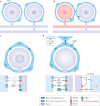
Fig. 3. Injury-induced changes in neuron–SGC bidirectional communications.
a,b | Proposed mechanism of coupled activation between neurons. Synchronous activity of adjacent neurons could arise by the spread of depolarization from neuron 1 (N1) to its surrounding satellite glial cells (SGCs), then through gap junctions to SGCs of a nearby neuron and then through gap junctions from these SGCs to N2. Under control conditions, SGCs are coupled mostly to other SGCs around a given neuron (part a). After peripheral injury or inflammation to a neuron (coloured red), SGCs become more strongly coupled to other SGCs around the same neuron (and also to neurons) by newly formed gap junctions. This enables increased transfer of electrical current and small molecules among SGCs and between SGCs and neurons. Such a mechanism can account for coupled neuronal activation (part b). c,d | The chain of events connecting neuronal excitation and glial activation. Resting conditions are shown in part c. Following neuronal damage, the neuronal cell body fires a high rate of action potentials, which increases intracellular calcium that in turn activates nitric oxide synthase to produce nitric oxide. Nitric oxide diffuses from the neuron and reaches the surrounding SGCs, where it induces cyclic guanosine 5'-monophosphate (cGMP) synthesis. This second messenger can have various actions on SGCs, and may be a key factor in SGC activation. SGC activation includes the release of ATP and cytokines, gap junction formation and increased sensitivity to ATP (part d). NO, nitric oxide; P2R, P2 purinergic receptor; Panx1, pannexin 1; TNF, tumour necrosis factor; TRPV1, transient receptor potential vanilloid type 1 channel.
Fig. 4
Fig. 4. Proposed sequence of events connecting nerve injury in four different pain models to SGC activation and neuronal hyperexcitability.
This sequence is an attempt to generalize the main events, but does not exclude additional or alternative mechanisms. The initial event in this cascade is injury to sensory neurons, such as from surgery, inflammation, diabetes or herpesvirus, which increases their firing rate. This leads to production of nitric oxide in the neurons. Nitric oxide diffuses from the injured neurons and activates satellite glial cells (SGCs) that surround them and likely also SGCs around adjacent neurons. SGC activation involves numerous processes, including greater coupling by gap junctions, increased sensitivity to ATP, upregulation of ERK and increased release of pro-inflammatory cytokines (IL-1β, tumour necrosis factor, IL-6, fractalkine and others). The increased gap junctions and sensitivity to ATP will enhance calcium waves, which lead to neuronal excitation. Cytokines released from SGCs will also contribute to neuronal sensitization and excitation. The overall effect will be greater firing (spontaneous and/or evoked) of sensory neurons and pain. TLR4, Toll-like receptor 4.

Similar articles

Cited by

References

    1. Hanani M. Satellite glial cells in sensory ganglia: from form to function. Brain Res. Brain Res. Rev. 2005;48:457–476. doi: 10.1016/j.brainresrev.2004.09.001. - DOI - PubMed
    1. Huang LY, Gu Y, Chen Y. Communication between neuronal somata and satellite glial cells in sensory ganglia. Glia. 2013;61:1571–1581. doi: 10.1002/glia.22541. - DOI - PMC - PubMed
    1. Jasmin L, Vit JP, Bhargava A, Ohara PT. Can satellite glial cells be therapeutic targets for pain control? Neuron Glia Biol. 2010;6:63–71. doi: 10.1017/S1740925X10000098. - DOI - PMC - PubMed
    1. Pannese E. Biology and pathology of perineuronal satellite cells in sensory ganglia. Adv. Anat. Embryol. Cell Biol. 2018;226:1–63. doi: 10.1007/978-3-319-60140-3_1. - DOI - PubMed
    1. Rozanski GM, Li Q, Stanley EF. Transglial transmission at the dorsal root ganglion sandwich synapse: glial cell to postsynaptic neuron communication. Eur. J. Neurosci. 2013;237:1221–1228. doi: 10.1111/ejn.12132. - DOI - PubMed

Publication types

MeSH terms