Review
. 2020 Aug 7;49(15):5473-5509.
doi: 10.1039/c9cs00199a. Epub 2020 Jul 7.

Half a century of amyloids: past, present and future

Affiliations
Review

Half a century of amyloids: past, present and future

Pu Chun Ke et al. Chem Soc Rev. .

Abstract

Amyloid diseases are global epidemics with profound health, social and economic implications and yet remain without a cure. This dire situation calls for research into the origin and pathological manifestations of amyloidosis to stimulate continued development of new therapeutics. In basic science and engineering, the cross-β architecture has been a constant thread underlying the structural characteristics of pathological and functional amyloids, and realizing that amyloid structures can be both pathological and functional in nature has fuelled innovations in artificial amyloids, whose use today ranges from water purification to 3D printing. At the conclusion of a half century since Eanes and Glenner's seminal study of amyloids in humans, this review commemorates the occasion by documenting the major milestones in amyloid research to date, from the perspectives of structural biology, biophysics, medicine, microbiology, engineering and nanotechnology. We also discuss new challenges and opportunities to drive this interdisciplinary field moving forward.


Conflict of interest statement

Conflicts of interest

DSE is SAB chair and equity holder in ADDRx, Inc.

Figures

Fig. 1
Fig. 1
Amyloidosis is a biophysical phenomenon of protein self-assembly under natural or artificial conditions, underpinned by a ubiquitous cross-β architecture (middle, in cyan). For over a half century, or arguably much longer, investigations into the structures of pathological and functional amyloids within the human anatomy (left, in blue), the microbiota (left, in green) and beyond (right, in dark blue) have revealed their inner workings as well as their entangled implications for biology, medicine and engineering.
Fig. 2
Fig. 2. Energy landscape of protein folding and aggregation.
The purple surface shows the multitude of conformations ‘funneling’ to the native state via intramolecular contacts and the pink area shows the conformations moving toward amorphous aggregates or amyloid fibrils via intermolecular contacts. Both parts of the energy surface overlap. Aggregate formation can occur from intermediates populated during de novo folding or by destabilization of the native state into partially folded states and is normally prevented by molecular chaperones. Toxic oligomers may occur as off-pathway intermediates of amyloid fibril formation. Reproduced with permission from ref. , copyright 2009 Nature Publishing Group.
Fig. 3
Fig. 3
(A) Aβ16–22 model protofilament (left panel). The initial structure used to start the MD trajectory with initial inter-β-sheet separation distance D0 of 1.28 nm. Front (a) and side (b) views are shown. Side chains are colored as follows: K) red, L) orange, V) yellow, F) green, A) blue, and E) violet. For clarity, only water molecules in the interpeptide region have been shown. (c) The same structure after 1,000 ps of unconstrained MD simulation at 300 K, started from the structure shown in (a) and (b). (d) A single Aβ16–22 peptide pair, one from each layer, is isolated from the protofilament shown in (c). (B) Number of interpeptide water molecules versus interpeptide distance (right panel). (a-d) Plots for each of the four trajectories at 300 K where D0= 1.28 nm. Trajectories (a) and (b) do not appear to show a dewetting transition, while trajectories (c) and (d) do. (e-h) The peptide-water van der Waals interaction is turned off, and D0 = 2.38 nm. (i-l) The peptide-water electrostatic interaction is turned off, and D0 = 1.28 nm. Reproduced with permission from ref. , copyright 2008 American Chemical Society.
Fig. 4
Fig. 4
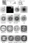
X-ray fibre diffraction provides the characteristic cross-β pattern for amyloid. Top panel shows a schematic showing the features of the cross-β pattern and structure. Lower panels show the cross-β diffraction patterns collected from amyloid fibrils formed by a diverse range of amyloidogenic proteins and peptides. Aβ11–25, AAAKKFFEAAAK, silk, hIAPP, NM Sup35, ccβ, Met30 TTR, Tau, Core PHF, β2M, Aβ42,, GNNQQNY, Fibrinogen, RVFNIM. Reproduced with permission from ref. , copyright 2000 The American Chemical Society. Reproduced with permission from ref. , copyright 2003 Elsevier. Reproduced with permission from ref. , copyright 2000 Elsevier. Reproduced with permission from ref. , copyright 2005 National Academy of Sciences. Reproduced with permission from ref. , copyright 2007 Wiley-VCH. Reproduced with permission from ref. , copyright 2004 Elsevier. Reproduced with permission from ref. , copyright 2000 American Association for the Advancement of Science. Reproduced with permission from ref. , copyright 2008 Elsevier. Reproduced with permission from ref. , copyright 1996 Ciba Foundation. Reproduced with permission from ref. , copyright 2003 National Academy of Sciences. Reproduced with permission from ref. , copyright 2017 Elsevier. Reproduced with permission from ref. , copyright 2008 American Society for Biochemistry and Molecular Biology. Reproduced with permission from ref. , copyright 2012 American Society for Biochemistry and Molecular Biology. Reproduced with permission from ref. , copyright 2010 Elsevier. Reproduced with permission from ref. , copyright 2007 Informa Healthcare. Reproduced with permission from ref. , copyright 2013 Portland Press.
Fig. 5
Fig. 5. Atomic-resolution crystal structures of two adhesive segments and two amyloid fibrils of the protein α-synuclein associated with PD.
Upper left: crystal structure of the PreNAC segment with sequence 47GVVHGVTTVA (the first T in this sequence is a hereditary early-onset disease mutation A52T). The upper view is down the axis of this steric zipper, showing atoms with van der Waals radii forming a tight, dry interface. Upper right: crystal structure of the NACore segment with sequence 68GAVVTGVTAVA. The center shows two amyloid-like fibrils formed by α-synuclein (αS). In the top of the left center, one layer of the Rod polymorph is viewed down the fibril axis, showing that it contains two identical αS chains each bent into a double hairpin shape. The two chains meet at a steric zipper formed by the PreNAC segments of the two chains. Identical layers are stacked on each other, forming a two-protofilament, slowly twisting fibril. In the top right center, one layer of the Twister polymorph is viewed down the fibril axis. The fold of the two αS molecules is similar to that of the Rod polymorph but the two chains meet at a different point than those of the Rod polymorph. They meet at an interface similar to that of the NACore crystal structure. That is, the steric zipper interfaces that pair the β-sheets in the crystal structures are similar to the interfaces between paired protofilaments in the fibrils. Notice that the slowly twisting Twister fibril is formed by stacking identical layers on each other, with a slight twist. Reproduced with permission from ref. , copyright 2018 Springer Nature.
Fig. 6
Fig. 6. Example ssNMR and cryoTEM structures for amyloid fibrils.
Upper panel: ssNMR structure of Aβ42., Two S-shaped molecules of Aβ42 (black and gray) are related by a twofold axis (marked by a circle), which runs down the center of the fibril. The N-terminal 14 residues are disordered; one possible conformation is shown here by dotted lines. Many of the known hereditary mutations are carried by residues located on the outer surface (red). The surface hydrophobic patch formed by residues V40 and A42 (orange) may explain the greater rate of secondary nucleation by the 1–42 species compared with 1–40., Bottom panel: CryoTEM structures of two amyloid fibrils of Tau. These two polymorphs of Tau amyloid fibrils were purified from the autopsied brains of AD patients. In both polymorphs, individual Tau proteins form C shapes, as shown by the cartoon ribbons with arrows that lie nearly in a plane perpendicular to the fibril axis. The protein layers are stacked up to form a protofilament. For each polymorph, there are two protofilaments, but they meet at different interfaces. Steric zippers are noted in the straight filament polymorph. The β-helical feature is enlarged in the right-hand panel where it is shown in yellow. Reproduced with permission from ref. , copyright 2016 National Academy of Sciences. Reproduced with permission from ref. , copyright 2017 Springer Nature. Reproduced with permission from ref. , copyright 2017 Annual Reviews.
Fig. 7
Fig. 7
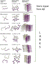
Atomic-resolution crystal structures of five LARKS contrasted with the structure of a steric zipper from the segment with sequence NKGAII from Aβ. The right-hand column shows the paired β-sheets of the steric zipper at the top and of the five LARKS below. For each structure, five layers are shown of the thousands in the crystals, with the fibril axes vertical. The view in the middle column is down the fibril axis and shows all atoms of the interfaces. The view in the left column is also down the fibril axis and shows the tracings of the protein backbones. The tight interface of the steric zipper offers a strong interaction. The kinked interfaces of the LARKS are weaker. Each interface is characterized by its shape complementarity score (Sc = 1.0 for perfect complementarity) and buried solvent-accessible surface area (Aβ) in Å2 between the mated sheets. Nitrogen atoms are blue, and oxygen atoms are red. Reproduced with permission from ref. , copyright 2018 American Association for the Advancement of Science.
Fig. 8
Fig. 8
Structure of amyloid fibrils as a function of the constitutive number of protofilaments in HEWL lysozyme as observed by AFM. The number of protofilaments is indicated in each panel. Up to 3 protofilaments, the fibrils remain straight and in a twisted ribbon configuration (H = 0). Starting from 4 protofilaments, amyloid fibrils change into a helical ribbon configuration, as revealed by the characteristic zig-zag contour shape associated with a non-zero mean curvature (H ≠ 0). Reproduced with permission from ref. , copyright 2011 American Chemical Society.
Fig. 9
Fig. 9
(Left) Schematic representation of the main mesoscopic polymorphs observed for amyloid fibrils and their approximate Mean (H) and Gaussian (K) curvatures. (Right) Sketch of the protein folding landscape in the region around the amyloid minimum: different polymorphs occupy different energy levels, with amyloid crystals populating the absolute minimum. The right panel is redrawn with permission from Adamcik et al. Reproduced with permission from ref. , copyright 2018 Wiley-VCH.
Fig. 10
Fig. 10
Primary and secondary nucleation and their verification with microfluidics. (A) Illustration of the power of chemical kinetics to elucidate microscopic mechanisms. Experimental data for the aggregation of the Aβ42 peptide fitted to an integrated rate law where the dominant source of new aggregates is, from left to right, primary nucleation, fragmentation and secondary nucleation, respectively. (B) Schematic illustration of the microfluidic strategy to detect directly single primary nucleation events and monitor the aggregation reaction in both time and space. (C) Time-lapse microscopy of a single microdroplet trapped in the array shown in panel B. (D) Schematic illustration of the primary and secondary nucleation events and subsequent aggregate multiplication which can be measured directly in microfluidic experiments. Reproduced with permission from ref. , copyright 2013 National Academy of Sciences. Panels B-D adapted from ref. , copyright 2011 National Academy of Sciences.
Fig. 11
Fig. 11
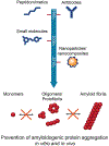
Major anti-amyloidosis strategies with peptidomimetics, antibodies, small molecules and nanoparticles/nanocomposites. The main purpose of such intervention is to stabilize the monomers, suppress the population of oligomers/protofibrils, or remodel amyloid fibrils. Such strategies have shown, to various degrees, potency and failures against amyloidosis and their associated toxicity in vitro and in vivo.
Fig. 12
Fig. 12
Schematic contrasting homologous seeding with heterologous seeding where the seed oligomer (in red) stimulates the growth of oligomers and ultimately fibrils of a different protein or peptide. Reproduced with permission from ref. , copyright 2013 Morales et al.
Fig. 13
Fig. 13. Formation of ordered fibrils and oligomers by metabolites.
Malfunction of specific enzyme due to genetic mutation results in a significant increase in the amount of specific metabolite substrate in the blood and tissues and lack of the metabolic product. In the case of PKU, the increase in phenylalanine concertation is about 30–60 fold in PKU patients as compared to individuals with normal phenylalanine metabolism. The metabolite could form fibrils or oligomers under physiological pH and the process could be inhibited by doxycycline or generic polyphenol amyloid formation inhibitors. The assembled structures, most likely the oligomers, can interact with the membranes to lead to membrane destabilization and finally apoptosis and damage to cells and tissues.
Fig. 14
Fig. 14
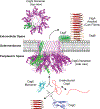
Model of curli secretion. CsgE and CsgG facilitate secretion of unfolded CsgA across the outer membrane. Outside the cell, CsgA assembles into an amyloid via a nucleation precipitation reaction assisted by CsgB and CsgF. The CsgE and CsgC proteins have been shown to prevent CsgA aggregation in vitro. The CsgG nonamer is shown in purple, and one of the CsgG subunits is colored green.
Fig. 15
Fig. 15
Schematic illustration of the binding process of gold ions with the β-sheets of amyloid fibrils. The β-sheet structure is based on the molecular dynamic simulations of the self-assembly process of the LACQCL sequence, a fragment found on the β-lactoglobulin amyloid fibrils used for water purification properties. Image rights: R. Mezzenga.

Similar articles

Cited by

References

    1. Eanes ED and Glenner GG, J. Histochem. Cytochem, 1968, 16, 673–677. - PubMed
    1. Astbury WT, Woods HJ and Bragg WL, Philos. Trans. R. Soc. A, 1934, 232, 333–394.
    1. Westermark P, Sletten K, Johansson B and Cornwell GG 3rd, Proc. Natl. Acad. Sci. U.S.A, 1990, 87, 2843–2845. - PMC - PubMed
    1. Colon W and Kelly JW, Biochemistry, 1992, 31, 8654–8660. - PubMed
    1. Horvath I and Wittung-Stafshede P, Proc. Natl. Acad. Sci. U.S.A, 2016, 113, 12473–12477. - PMC - PubMed