Oncolytic virus promotes tumor-reactive infiltrating lymphocytes for adoptive cell therapy
- PMID: 32632271
- PMCID: PMC9718357
- DOI: 10.1038/s41417-020-0189-4
Oncolytic virus promotes tumor-reactive infiltrating lymphocytes for adoptive cell therapy
Abstract
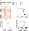
Adoptive cell therapy (ACT) using tumor-specific tumor-infiltrating lymphocytes (TILs) has demonstrated success in patients where tumor-antigen specific TILs can be harvested from the tumor, expanded, and re-infused in combination with a preparatory regimen and IL2. One major issue for non-immunogenic tumors has been that the isolated TILs lack tumor specificity and thus possess limited in vivo therapeutic function. An oncolytic virus (OV) mediates an immunogenic cell death for cancer cells, leading to elicitation and dramatic enhancement of tumor-specific TILs. We hypothesized that the tumor-specific TILs elicited and promoted by an OV would be a great source for ACT for solid cancer. In this study, we show that a local injection of oncolytic poxvirus in MC38 tumor with low immunogenicity in C57BL/6 mice, led to elicitation and accumulation of tumor-specific TILs in the tumor tissue. Our analyses indicated that IL2-armed OV-elicited TILs contain lower quantities of exhausted PD-1hiTim-3+ CD8+ T cells and regulatory T cells. The isolated TILs from IL2-expressing OV-treated tumor tissue retained high tumor specificity after expansion ex vivo. These TILs resulted in significant tumor regression and improved survival after adoptive transfer in mice with established MC38 tumor. Our study showcases the feasibility of using an OV to induce tumor-reactive TILs that can be expanded for ACT.
Conflict of interest statement
Conflicts of Interests: MF, ZL, ZSG and BLB filed a patent partly based on this work. Other authors declare that they have no conflict of interest.
Figures


Similar articles
-
In Vivo Priming of Peritoneal Tumor-Reactive Lymphocytes With a Potent Oncolytic Virus for Adoptive Cell Therapy.Front Immunol. 2021 Feb 18;12:610042. doi: 10.3389/fimmu.2021.610042. eCollection 2021. Front Immunol. 2021. PMID: 33679747 Free PMC article.
-
Expansion of Tumor-Infiltrating CD8+ T cells Expressing PD-1 Improves the Efficacy of Adoptive T-cell Therapy.Cancer Res. 2017 Jul 1;77(13):3672-3684. doi: 10.1158/0008-5472.CAN-17-0236. Epub 2017 May 18. Cancer Res. 2017. PMID: 28522749
-
Oncolytic adenovirus shapes the ovarian tumor microenvironment for potent tumor-infiltrating lymphocyte tumor reactivity.J Immunother Cancer. 2020 Jan;8(1):e000188. doi: 10.1136/jitc-2019-000188. J Immunother Cancer. 2020. PMID: 31940588 Free PMC article.
-
Multidirectional Strategies for Targeted Delivery of Oncolytic Viruses by Tumor Infiltrating Immune Cells.Pharmacol Res. 2020 Nov;161:105094. doi: 10.1016/j.phrs.2020.105094. Epub 2020 Aug 12. Pharmacol Res. 2020. PMID: 32795509 Review.
-
Adoptive CD8+ T cell therapy against cancer:Challenges and opportunities.Cancer Lett. 2019 Oct 10;462:23-32. doi: 10.1016/j.canlet.2019.07.017. Epub 2019 Jul 26. Cancer Lett. 2019. PMID: 31356845 Review.
Cited by
-
Enhanced cellular therapy: revolutionizing adoptive cellular therapy.Exp Hematol Oncol. 2024 Apr 25;13(1):47. doi: 10.1186/s40164-024-00506-6. Exp Hematol Oncol. 2024. PMID: 38664743 Free PMC article. Review.
-
SOCS3 inhibiting JAK-STAT pathway enhances oncolytic adenovirus efficacy by potentiating viral replication and T-cell activation.Cancer Gene Ther. 2024 Mar;31(3):397-409. doi: 10.1038/s41417-023-00710-2. Epub 2023 Dec 15. Cancer Gene Ther. 2024. PMID: 38102464
-
Current challenges and therapeutic advances of CAR-T cell therapy for solid tumors.Cancer Cell Int. 2024 Apr 15;24(1):133. doi: 10.1186/s12935-024-03315-3. Cancer Cell Int. 2024. PMID: 38622705 Free PMC article. Review.
-
Harnessing novel strategies and cell types to overcome immune tolerance during adoptive cell therapy in cancer.J Immunother Cancer. 2023 Apr;11(4):e006434. doi: 10.1136/jitc-2022-006434. J Immunother Cancer. 2023. PMID: 37100458 Free PMC article. Review.
-
In Vivo Priming of Peritoneal Tumor-Reactive Lymphocytes With a Potent Oncolytic Virus for Adoptive Cell Therapy.Front Immunol. 2021 Feb 18;12:610042. doi: 10.3389/fimmu.2021.610042. eCollection 2021. Front Immunol. 2021. PMID: 33679747 Free PMC article.
References
-
- Andersen R, Donia M, Ellebaek E, Borch TH, Kongsted P, Iversen TZ et al.: Long-Lasting Complete Responses in Patients with Metastatic Melanoma after Adoptive Cell Therapy with Tumor-Infiltrating Lymphocytes and an Attenuated IL2 Regimen. Clin Cancer Res 2016; 22:3734–3745. - PubMed
Publication types
MeSH terms
Grants and funding
LinkOut - more resources
Full Text Sources
Other Literature Sources
Research Materials

