. 2020 Jun 11:14:540.
doi: 10.3389/fnins.2020.00540. eCollection 2020.

TDP-43 Is Efficiently Transferred Between Neuron-Like Cells in a Manner Enhanced by Preservation of Its N-Terminus but Independent of Extracellular Vesicles

Affiliations

TDP-43 Is Efficiently Transferred Between Neuron-Like Cells in a Manner Enhanced by Preservation of Its N-Terminus but Independent of Extracellular Vesicles

Christopher Sackmann et al. Front Neurosci. .

Abstract

The misfolding of transactive response DNA-binding protein (TDP-43) is a major contributor to the pathogenesis of TDP-43 proteinopathies, including amyotrophic lateral sclerosis and frontotemporal lobar degeneration with TDP-43 inclusions, but also plays a role in other neurodegenerative diseases including Alzheimer disease. It is thought that different truncations at the N- and C-termini of TDP-43 contribute to its misfolding and aggregation in the brain, and that these aberrant TDP-43 fragments contribute to disease. Despite this, little is known about whether different truncation events influence the protein's transmissibility between cells and how this cell-to-cell transfer occurs. In this study, we use a well-established cellular model to study the efficiency by which full-length and truncated TDP-43 fragments are transferred between neuron-like cells. We demonstrate that preservation of the N-terminus of TDP-43 enhances its transmissibility between cells and that this protein transmission occurs in a manner exclusive of extracellular vesicles, instead requiring cellular proximity for efficient propagation. These data indicate that the N-terminus of TDP-43 might be a useful target in the generation of therapeutics to limit the spread of TDP-43 pathology.

Keywords: C-terminus; N-terminus; TDP-43; amyotrophic lateral sclerosis; cell-to-cell; extracellular vesicles; frontotemporal lobar degeneration; protein transfer.


Figures

FIGURE 1
FIGURE 1
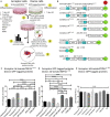
Preservation of the N-terminus of TDP-43 promotes its transmissibility between cells. A visualization of the coculture model is provided in (A). Cells are differentiated in ECM gel with growth factors according to the scheme described in (A) prior to formation of the coculture. After 24 h of coculture, cells are analyzed and separated using FACS and prepared for microscopy. Experiments involve the use of full-length TDP-43 and TDP-43 truncated fragments, all of which are fused to fluorescent proteins at the C-terminus (B). Two full-length TDP-43 proteins were used, td-tomatoTDP431– 414 and WT-TDP431– 414 (GFP), whereas all TDP-43 protein fragments were fused with EGFP. All fragments of TDP-43 transferred directly from donor cells (GFP) to td-tomatoTDP431– 414 acceptor cells with differing degrees of efficiency (C). Full-length td-tomatoTDP431– 414 donor cells transferred protein at similar levels to TDP-43 fragment expressing acceptor cells (D). Construct 61– 314 was transferred most readily among the different TDP-43 fragments, indicating that preservation of the N-terminus (and/or loss of the extreme C-terminus) of TDP-43 is influential to the transmissibility of TDP-43 between cells. Cell toxicity, as measured by LDH release, indicated that the cocultures did not exhibit cytotoxicity over 24 h (E). Images corresponding to data set C can be found in Figure 2, whereas images from data set D are presented in Supplementary Figure S3. Full ANOVA tables are available in Supplementary Tables S3, S4. Data are presented as the proportion of all detected acceptor cells that are double labeled, provided as the mean ± SEM. Significance determined by one-way ANOVA with Tukey post hoc test, n = 6–10 (C), and n = 4–7 (D). *p < 0.05, **p < 0.01, ***p < 0.001, ****p < 0.0001, n.s., not significant. BDNF, brain-derived neurotrophic factor; NRGβ1, neuregulin-β1; NGF, nerve growth factor; RA, retinoic acid; FACS, fluorescence-activated cell sorting; NLS, nuclear localization sequence; NES, nuclear export sequence; RRM, RNA recognition motif.
FIGURE 2
FIGURE 2
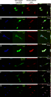
Representative images depicting cellular localization of GFP-tagged TDP-43 fragments (donor cells; green) after transfer to full-length td-tomatoTDP431– 414 acceptor cells (acceptor cells; red). Acceptor cells (td-tomatoTDP431– 414), which also displayed GFP positivity, were sorted and fixed for confocal microscopy. Association of full-length TDP-43 from acceptor cells was observed with varying degrees to each TDP fragment (A–E) as well as with actin (F) and CD63 (G). Construct 101– 105 (D) shows localization exclusively to the nucleus, whereas constructs 286– 414, 5257– 414, 61– 314, and WT-TDP-GFP1– 414 (A–C,E, respectively) could be found in the cytoplasm, as well as the nucleus. The final column contains Z-projections. Scale bar = 10 μm.
FIGURE 3
FIGURE 3
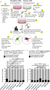
The majority of TDP-43 transfers occur from donor to acceptor cells (“anterograde”); however, there is a lesser degree of transfer from the acceptor to the donor cells (“retrograde”). A visualization of the Qdot-800 coculture model is provided in (A). Cells are differentiated in ECM gel with growth factors according to the scheme described. Prior to formation of the coculture, donor cells (shown here as GFP) were further labeled with Qdot-800 and selected by FACS to collect double-positive donor cells (+GFP and +Qdot-800) and plated directly onto fully differentiated acceptor cells. After 24 h of coculture, donor cells displaying retrograde transfer and acceptor cells displaying anterograde transfer were analyzed and separated using FACS and prepared for microscopy. Of the total transfer events, the majority of TDP-43 was transferred from donor to acceptor cells (anterograde; gray) (B,C, presented as proportions of detected transfer events). TDP-43 and TDP-43 truncated fragments also transferred from acceptor cells to donor cells (retrograde; black); however, retrograde transfer was comparatively minimal. Cell images corresponding to these data can be found in Supplementary Figure S4 (for data set B), and Supplementary Figure S5 (for data set C). Full ANOVA tables are provided in Supplementary Tables S5, S6. Data are presented as mean with significance determined by one-way ANOVA with Tukey post hoc test, n = 3–4 for each permutation. *p < 0.05, **p < 0.01, ***p < 0.001, ****p < 0.0001, ns, not significant. The data represent the detected retrograde and anterograde transfer events as a proportion of all detected transfer events. BDNF, brain-derived neurotrophic factor; NRGβ1, neuregulin-β1; NGF, nerve growth factor; RA, retinoic acid; FACS, fluorescence-activated cell sorting.
FIGURE 4
FIGURE 4
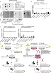
Transfer of TDP-43 and TDP-43 fragments requires physical connectivity between cells. Extracellular vesicles isolated from stably expressing TDP-43 cell lines and human tissues indicate that the TDP-43 fragments are incorporated into EVs, albeit with a low degree of efficiency (A). The TDP-43 antibody used here recognizes an epitope at the extreme C-terminus of the WT-TDP-GFP1– 414, which is absent in some constructs or obfuscated by the C-terminal GFP tags but can be observed with the anti-GFP antibody (e.g., construct 61– 314). Isolated EVs were added directly onto WT SH-SY5Y cells but failed to transfer any appreciable protein (B). Further, donor and acceptor cells were physically separated during coculture using Transwell inserts, allowing the distribution of cell products within a shared medium while creating a physical barrier between the cells (C,D), but proteins also fail to transfer by this method. Graphical representations of the experimental setup are shown in (E,F). Full ANOVA tables of (B–D) are available in Supplementary Tables S7–S9. Data in (B) are presented as the proportion of all detected cells that displayed detectable fluorescence, provided as mean ± SEM. Data in (C,D) are presented as the proportion of all detected acceptor cells that are double labeled, provided here mean ± SEM. Significance determined by one-way ANOVA with Tukey post hoc test, n = 6–9 (B), and n = 3 (C,D). *p < 0.05, **p < 0.0, ***p < 0.001, ****p < 0.0001, ns, not significant.

Similar articles

Cited by

References

    1. Afroz T., Hock E. M., Ernst P., Foglieni C., Jambeau M., Gilhespy L. A. B., et al. (2017). Functional and dynamic polymerization of the ALS-linked protein TDP-43 antagonizes its pathologic aggregation. Nat. Commun. 8 1–14. 10.1038/s41467-017-00062-0 - DOI - PMC - PubMed
    1. Agholme L., Lindström T., Kågedal K., Marcusson J., Hallbeck M. (2010). An in vitro model for neuroscience: differentiation of SH-SY5Y cells into cells with morphological and biochemical characteristics of mature neurons. J. Alzheimers. Dis. 20 1069–1082. 10.3233/JAD-2010-091363 - DOI - PubMed
    1. Asai H., Ikezu S., Tsunoda S., Medalla M., Luebke J., Haydar T., et al. (2015). Depletion of microglia and inhibition of exosome synthesis halt tau propagation. Nat. Neurosci. 18 1584–1593. 10.1038/nn.4132 - DOI - PMC - PubMed
    1. Berning B. A., Walker A. K. (2019). The pathobiology of TDP-43 C-terminal fragments in ALS and FTLD. Front. Neurosci. 13:335. 10.3389/fnins.2019.00335 - DOI - PMC - PubMed
    1. Brettschneider J., Del Tredici K., Toledo J. B., Robinson J. L., Irwin D. J., Grossman M., et al. (2013). Stages of pTDP-43 pathology in amyotrophic lateral sclerosis. Ann. Neurol. 74 20–38. 10.1002/ana.23937 - DOI - PMC - PubMed

LinkOut - more resources