. 2020 Jun 19;11(1):3116.
doi: 10.1038/s41467-020-16922-1.

Talin dissociates from RIAM and associates to vinculin sequentially in response to the actomyosin force

Affiliations

Talin dissociates from RIAM and associates to vinculin sequentially in response to the actomyosin force

Clémence Vigouroux et al. Nat Commun. .

Abstract

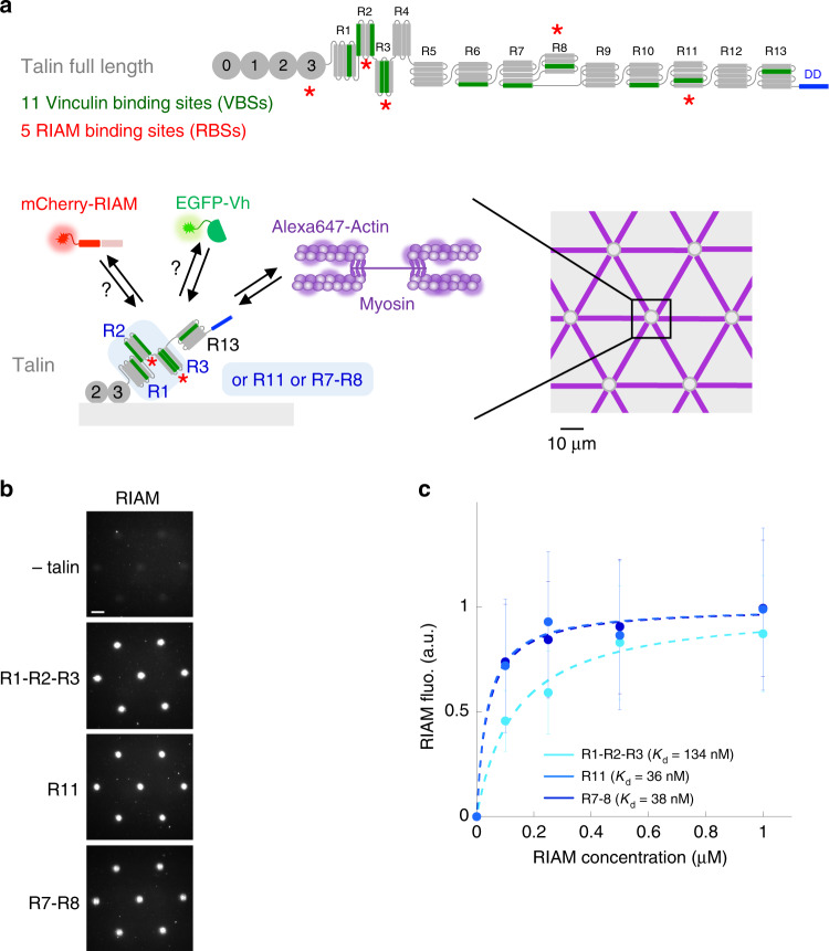
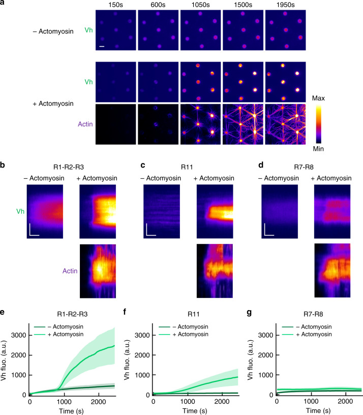
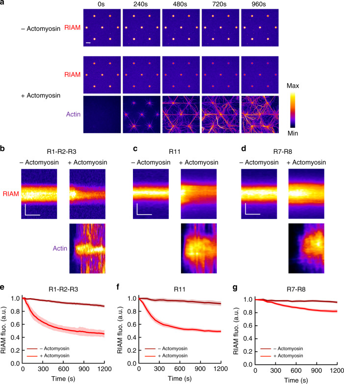
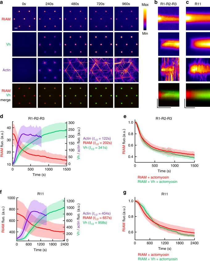
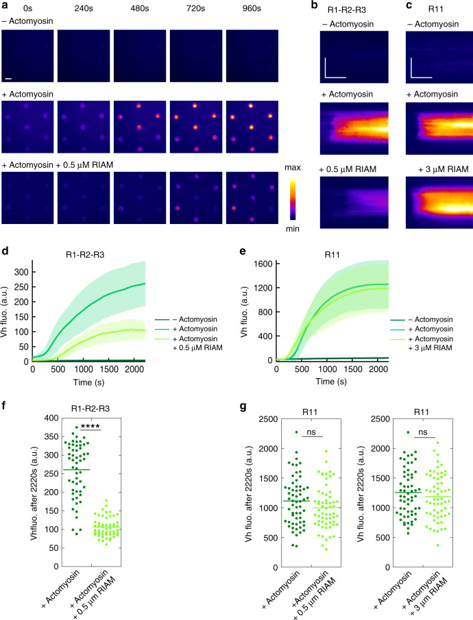
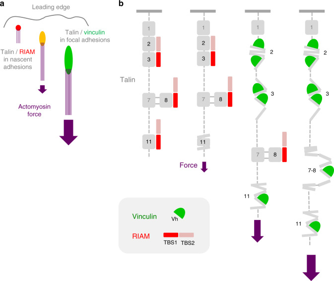
Cells reinforce adhesion strength and cytoskeleton anchoring in response to the actomyosin force. The mechanical stretching of talin, which exposes cryptic vinculin-binding sites, triggers this process. The binding of RIAM to talin could regulate this mechanism. However, the mechanosensitivity of the talin-RIAM complex has never been tested. It is also not known whether RIAM controls the mechanosensitivity of the talin-vinculin complex. To address these issues, we designed an in vitro microscopy assay with purified proteins in which the actomyosin force controls RIAM and vinculin-binding to talin. We demonstrate that actomyosin triggers RIAM dissociation from several talin domains. Actomyosin also provokes the sequential exchange of RIAM for vinculin on talin. The effect of RIAM on this force-dependent binding of vinculin to talin varies from one talin domain to another. This mechanism could allow talin to biochemically code a wide range of forces by selecting different combinations of partners.


Conflict of interest statement

The authors declare no competing interests.

Figures

Fig. 1
Fig. 1. In vitro reconstitution of talin–RIAM complexes.
a Top panel: organization of full-length talin featuring RIAM- and vinculin-binding sites. The vinculin-binding sites (VBSs) are the dark green helices. RIAM binds to the R2, R3, R8, and R11 domains of talin. R13 is the C-terminal actin-binding domain (ABD) of talin. Bottom panels: basic principle of the in vitro microscopy assay. Alexa647-labeled actin and myosin II self-assemble to apply force to talin R1–R2–R3, R11, or R7–R8 immobilized in micropatterns, which controls the binding of EGFP-vinculin head (EGFP-Vh) and mCherry-RIAM. Talin is represented as a monomer for convenience but it contains a dimerization domain (DD). b Representative images of the fluorescence of mCherry-RIAM 1-306 (1 µM) in non-coated control disks and disks coated with 1 µM talin R1–R2–R3 or R11, or R7–R8. Scale bar = 10 µm. This experiment was repeated three times independently with the same results. c Binding of RIAM to disks coated with talin R1–R2–R3, R11, and R7–R8. Conditions: 0–1 µM mCherry-RIAM 1-306, 1 µM of talin during the coating step. Data are mean ± SD. n = 150 disks all points, except (R1–R2–R3 + 0.5 µM RIAM) n = 137 disks. Source data are provided as a Source data file.
Fig. 2
Fig. 2. Talin domains bind to vinculin differently in response to the actomyosin force.
a Time lapses showing the recruitment of Vh to talin R1–R2–R3-coated disks in the absence (top) or presence of actomyosin (Vh is shown on the middle and actin on the bottom panel). This experiment was repeated five times independently with the same results. bd Kymographs of EGFP-Vh (top) and actin (bottom) along a cross-section of a disk coated with talin R1–R2–R3 (b) or R11 (c) or R7–R8 (d) in the absence (left) or presence (right) of actomyosin. Conditions: 100 nM EGFP-Vh, 2.4 µM actin (2% Alexa594-labeled), 50 nM myosin, and 1 µM talin during the coating step. The images are color coded using the fire LUT of ImageJ. Scale bar in time lapses = 10 µm. In kymographs, horizontal bar = 500 s, vertical bar = 5 µm. eg Kinetics of the mean fluorescence of EGFP-Vh corresponding to the conditions described in (bd). Data are mean ± SD. e n = 80 (−actomyosin), n = 60 disks (+actomyosin). f n = 60 (−actomyosin), n = 75 disks (+actomyosin). g n = 80 (−actomyosin), n = 76 disks (+actomyosin). Source data are provided as a Source data file. See Supplementary Movie 1, Supplementary Movie 2, and Supplementary Movie 3.
Fig. 3
Fig. 3. The actomyosin force provokes RIAM dissociation from several talin domains.
a Time lapses showing the binding of RIAM 1-306 to talin R1–R2–R3-coated disks in the absence (top) or presence of actomyosin (RIAM is shown on the middle and actin on the bottom panel). This experiment was repeated 11 times independently with the same results. bd Kymographs of mCherry-RIAM 1-306 (top) and actin (bottom) along a cross-section of a disk coated with talin R1–R2–R3 (b) R11 (c) or R7–R8 (d) in the absence (left) or presence (right) of actomyosin. For better comparison, the fluorescence of mCherry-RIAM 1-306 in kymographs was normalized as the maximal fluorescence in the R11- and R7–R8-coated disks. Conditions: 100 nM mCherry-RIAM 1-306, 2.4 µM actin (1% Alexa647-labeled for R1–R2–R3, 2% Alexa488 for R11 and R7–R8), 50 nM myosin, 1 µM talin during the coating step. The images are color coded using the fire LUT of ImageJ. Scale bar in time lapses = 10 µm. In kymographs, horizontal bar = 500 s, vertical bar =  5 µm. eg Kinetics of the mean fluorescence of mCherry-RIAM 1-306 corresponding to the conditions described in (bd). Data are mean ± SD. e n = 63 (−actomyosin), n = 62 disks (+actomyosin). f n = 50 (−actomyosin), n = 60 disks (+actomyosin). g n = 49 (−actomyosin), n = 60 disks (+actomyosin). Data were first normalized to 1 as the maximal mCherry-RIAM 1-306 fluorescence and synchronized using this maximal value as t0 before being averaged. Source data are provided as a Source data file. See Supplementary Movie 4, Supplementary Movie 5, and Supplementary Movie 6.
Fig. 4
Fig. 4. The actomyosin force provokes the sequential exchange of RIAM for vinculin on talin.
a Time lapses showing the concomitant dissociation of RIAM 1-306, association of Vh, accumulation of actomyosin, and a Vh/RIAM merge in the same disks coated with talin R1–R2–R3. This experiment was repeated 4 times independently with the same results. b, c From top to bottom: kymographs of RIAM, Vh, actin, and Vh/RIAM merge along the cross-section of a disk coated with talin R1–R2–R3 (b) or R11 (c). Conditions: 100 nM mCherry-RIAM, 100 nM EGFP-Vh, 2.4 µM actin (1% Alexa647-labeled), 50 nM myosin, and 1 µM talin during the coating step. The images are color coded using the fire LUT of ImageJ. Scale bar in time lapses = 10 µm. In kymographs, horizontal bar = 1000 s, vertical bar = 5 µm. d, f Kinetics of the mean fluorescence of mCherry-RIAM 1-306, EGFP-Vh and Alexa647-labeled actin in disks coated with talin R1–R2–R3 (d) and R11 (f) corresponding to the conditions described in (b) and (c). Actin fluorescence is multiplied by 3. Data are mean ± SD. n = 63 (d), n = 59 disks (f). e Kinetics of mCherry-RIAM 1-306 dissociation from disks coated with talin R1–R2–R3 in the presence of actomyosin with or without 100 nM EGFP-Vh as described in Fig. 3b and b respectively. n = 62 (RIAM + actomyosin), n = 63 disks (RIAM + Vh + actomyosin). g Kinetics of mCherry-RIAM 1-306 dissociation from disks coated with talin R11 in the presence of actomyosin with or without 100 nM EGFP-Vh as described in Fig. 3c and c respectively. n = 59 disks. e, g Data are mean ± SD. Data were first normalized to 1 as the maximal mCherry-RIAM 1-306 fluorescence and synchronized using this maximal value as t0 before being averaged. Source data are provided as a Source data file. See Supplementary Movie 7 and Supplementary Movie 8.
Fig. 5
Fig. 5. RIAM inhibits the actomyosin-dependent binding of vinculin to talin R1–R2–R3 but not to R11.
a Time lapse showing the recruitment of Vh in disks coated with talin R1–R2–R3 in the absence of actomyosin (top), presence of actomyosin (middle), and presence of actomyosin and RIAM (bottom). This experiment was repeated twice independently with the same results. b, c Kymographs of EGFP-Vh along a cross-section of a disk coated with talin R1–R2–R3 (b) and R11 (c). Conditions: 100 nM EGFP-Vh, 2.4 µM actin, 50 nM myosin, 500 nM (b) or 3 µM (c) mCherry-RIAM, 1 µM talin during the coating step. The images are color coded using the fire LUT of ImageJ. Scale bar in time lapses = 10 µm. In kymographs, horizontal bar = 1000 s, vertical bar = 5 µm. d, e Kinetics of the mean fluorescence of EGFP-Vh corresponding to the conditions described in (b, c). Data are mean ± SD. d n = 54 (−actomyosin) and (+actomyosin), n = 51 disks (+actomyosin + 0.5 µM RIAM). e n = 59 disks. f, g Steady-state binding of Vh (2220 s after sealing the chamber) in disks coated with talin R1–R2–R3 (f) or R11 (g) in the absence and presence of RIAM. f, g Same conditions as in (b, c). Each data point represents the mean fluorescence of Vh in one disk. The bar shows the mean. f n = 54 (+actomyosin), n = 51 disks (+actomyosin + 0.5 µM RIAM). A significant difference was found using a two-tailed t test (P = 3.95 × 10−22). g Left panel: n = 60. No significant difference was found using a two-tailed t test (P = 0.1126). Right panel: n = 59 disks. No significant difference was found using a two-tailed t test (P = 0.3575). ****P < 0.0001 using a two-tailed t test; ns nonsignificant. Source data are provided as a Source data file. See Supplementary Movie 9 and Supplementary Movie 10.
Fig. 6
Fig. 6. Model for the actomyosin-dependent binding of RIAM and vinculin to talin.
a Scheme illustrating that RIAM, which is initially enriched in nascent adhesions, is replaced by vinculin in mature FAs in response to the force exerted by the actomyosin stress fibers. b Model describing how talin dissociates from RIAM and associates to vinculin sequentially in response to the actomyosin force.

Similar articles

Cited by

References

    1. Le Clainche C, Carlier M-F. Regulation of actin assembly associated with protrusion and adhesion in cell migration. Physiol. Rev. 2008;88:489–513. - PubMed
    1. Wehrle-Haller B. Structure and function of focal adhesions. Curr. Opin. Cell Biol. 2012;24:116–124. - PubMed
    1. Ciobanasu C, Faivre B, Le Clainche C. Integrating actin dynamics, mechanotransduction and integrin activation: the multiple functions of actin binding proteins in focal adhesions. Eur. J. Cell Biol. 2013;92:339–348. - PubMed
    1. Schwarz US, Gardel ML. United we stand – integrating the actin cytoskeleton and cell–matrix adhesions in cellular mechanotransduction. J. Cell Sci. 2012;125:3051–3060. - PMC - PubMed
    1. Bachmann M, Kukkurainen S, Hytönen VP, Wehrle-Haller B. Cell adhesion by integrins. Physiol. Rev. 2019;99:1655–1699. - PubMed

Publication types

MeSH terms

LinkOut - more resources