

Regulation of Angiogenesis Discriminates Tissue Resident MSCs from Effective and Defective Osteogenic Environments
- PMID: 32481579
- PMCID: PMC7355658
- DOI: 10.3390/jcm9061628
Regulation of Angiogenesis Discriminates Tissue Resident MSCs from Effective and Defective Osteogenic Environments
Abstract
Background: The biological mechanisms that contribute to atrophic long bone non-union are poorly understood. Multipotential mesenchymal stromal cells (MSCs) are key contributors to bone formation and are recognised as important mediators of blood vessel formation. This study examines the role of MSCs in tissue formation at the site of atrophic non-union.
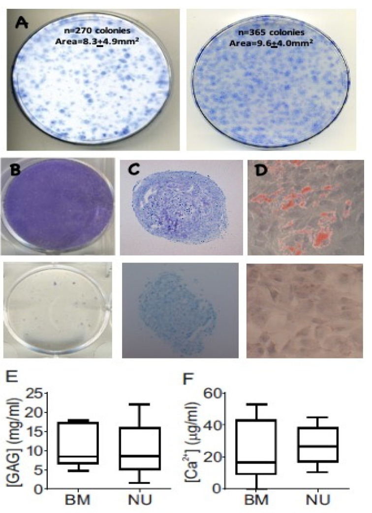
Materials and methods: Tissue and MSCs from non-union sites (n = 20) and induced periosteal (IP) membrane formed following the Masquelet bone reconstruction technique (n = 15) or bone marrow (n = 8) were compared. MSC content, differentiation, and influence on angiogenesis were measured in vitro. Cell content and vasculature measurements were performed by flow cytometry and histology, and gene expression was measured by quantitative polymerase chain reaction (qPCR).
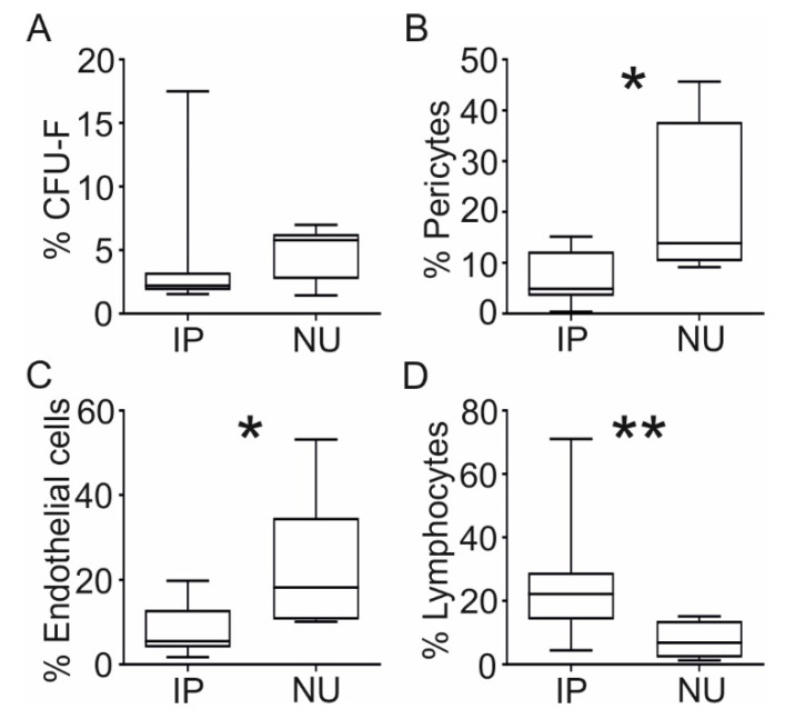
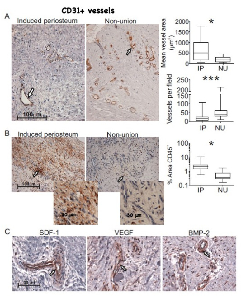
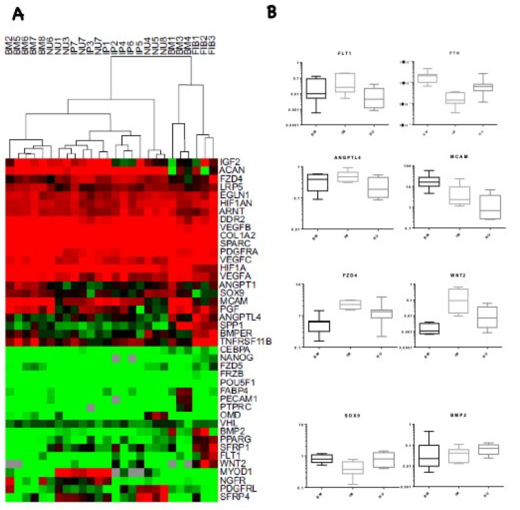
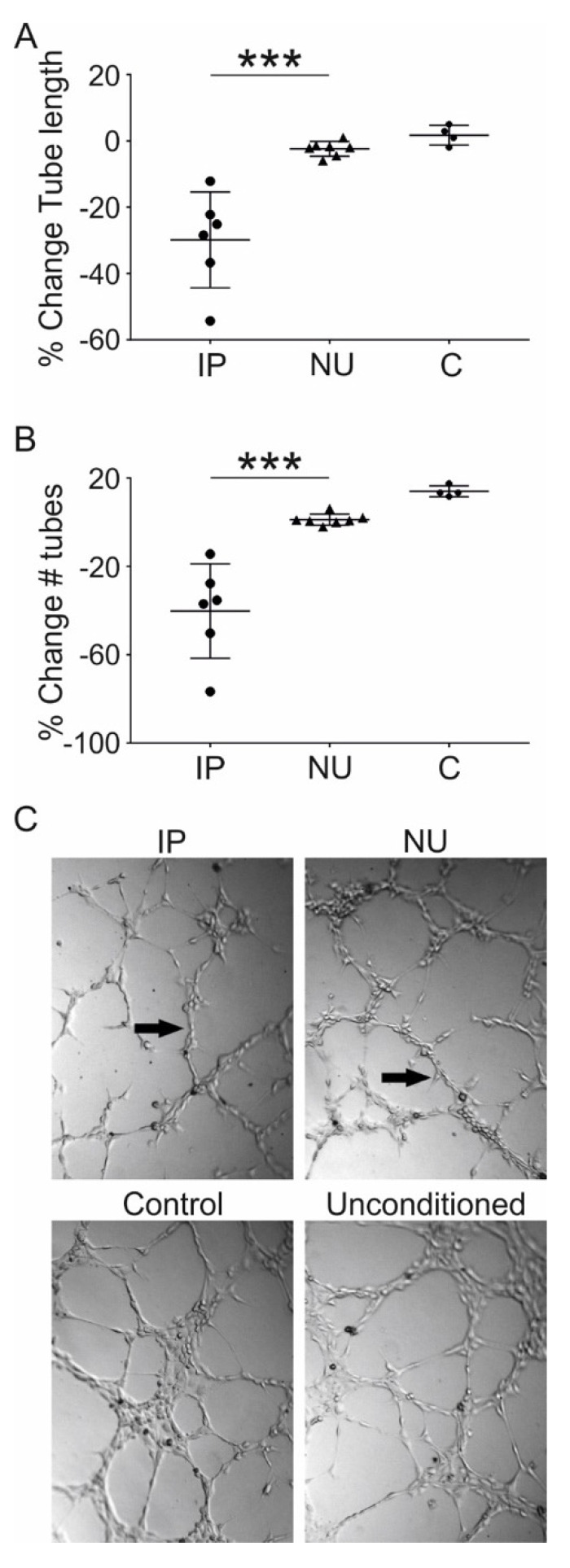
Results: MSCs from non-union sites had comparable differentiation potential to bone marrow MSCs. Compared with induced periosteum, non-union tissue contained similar proportion of colony-forming cells, but a greater proportion of pericytes (p = 0.036), and endothelial cells (p = 0.016) and blood vessels were more numerous (p = 0.001) with smaller luminal diameter (p = 0.046). MSCs showed marked differences in angiogenic transcripts depending on the source, and those from induced periosteum, but not non-union tissue, inhibited early stages of in vitro angiogenesis.
Conclusions: In vitro, non-union site derived MSCs have no impairment of differentiation capacity, but they differ from IP-derived MSCs in mediating angiogenesis. Local MSCs may thus be strongly implicated in the formation of the immature vascular network at the non-union site. Attention should be given to their angiogenic support profile when selecting MSCs for regenerative therapy.
Keywords: MSCs; fracture healing; induced periosteum; non-union; osteogenesis; regenerative medicine.
Conflict of interest statement
The authors declare no conflict of interest.
Figures
Similar articles
-
RIA fractions contain mesenchymal stroma cells with high osteogenic potency.Injury. 2015 Dec;46 Suppl 8:S23-32. doi: 10.1016/S0020-1383(15)30051-6. Injury. 2015. PMID: 26747914
-
Colony Formation, Migratory, and Differentiation Characteristics of Multipotential Stromal Cells (MSCs) from "Clinically Accessible" Human Periosteum Compared to Donor-Matched Bone Marrow MSCs.Stem Cells Int. 2019 Nov 21;2019:6074245. doi: 10.1155/2019/6074245. eCollection 2019. Stem Cells Int. 2019. PMID: 31871468 Free PMC article.
-
17β-estradiol improves the efficacy of exploited autologous bone marrow-derived mesenchymal stem cells in non-union radial defect healing: A rabbit model.Res Vet Sci. 2018 Jun;118:11-18. doi: 10.1016/j.rvsc.2017.12.024. Epub 2017 Dec 28. Res Vet Sci. 2018. PMID: 29334646
-
Biological and molecular profile of fracture non-union tissue: A systematic review and an update on current insights.J Cell Mol Med. 2022 Feb;26(3):601-623. doi: 10.1111/jcmm.17096. Epub 2022 Jan 4. J Cell Mol Med. 2022. PMID: 34984803 Free PMC article. Review.
-
Frontiers in non-union research.EFORT Open Rev. 2020 Oct 26;5(10):574-583. doi: 10.1302/2058-5241.5.190062. eCollection 2020 Oct. EFORT Open Rev. 2020. PMID: 33204499 Free PMC article. Review.
Cited by
-
Clinical Trials Based on Mesenchymal Stromal Cells are Exponentially Increasing: Where are We in Recent Years?Stem Cell Rev Rep. 2022 Jan;18(1):23-36. doi: 10.1007/s12015-021-10231-w. Epub 2021 Aug 16. Stem Cell Rev Rep. 2022. PMID: 34398443 Free PMC article. Review.
-
Current Aspects on the Pathophysiology of Bone Metabolic Defects during Progression of Scoliosis in Neurofibromatosis Type 1.J Clin Med. 2022 Jan 15;11(2):444. doi: 10.3390/jcm11020444. J Clin Med. 2022. PMID: 35054138 Free PMC article. Review.
-
Mesenchymal stem cell-based therapy and exosomes in COVID-19: current trends and prospects.Stem Cell Res Ther. 2021 Aug 21;12(1):469. doi: 10.1186/s13287-021-02542-z. Stem Cell Res Ther. 2021. PMID: 34419143 Free PMC article. Review.
-
Mesenchymal stem cells derived from the fibrotic tissue of atrophic nonunion or the bone marrow of iliac crest: A donor-matched comparison.Regen Ther. 2023 Sep 12;24:398-406. doi: 10.1016/j.reth.2023.08.005. eCollection 2023 Dec. Regen Ther. 2023. PMID: 37719889 Free PMC article.
-
Bone Healing Gone Wrong: Pathological Fracture Healing and Non-Unions-Overview of Basic and Clinical Aspects and Systematic Review of Risk Factors.Bioengineering (Basel). 2023 Jan 9;10(1):85. doi: 10.3390/bioengineering10010085. Bioengineering (Basel). 2023. PMID: 36671657 Free PMC article. Review.
References
-
- Megas P. Classification of non-union. Injury. 2005;36(Suppl. 4):S30–S37. - PubMed
LinkOut - more resources
Full Text Sources
