

Diversity and Biology of Cancer-Associated Fibroblasts
- PMID: 32466724
- PMCID: PMC7864232
- DOI: 10.1152/physrev.00048.2019
Diversity and Biology of Cancer-Associated Fibroblasts
Abstract
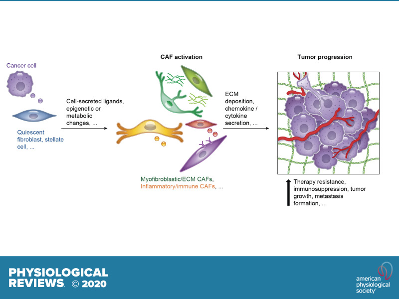
Efforts to develop anti-cancer therapies have largely focused on targeting the epithelial compartment, despite the presence of non-neoplastic stromal components that substantially contribute to the progression of the tumor. Indeed, cancer cell survival, growth, migration, and even dormancy are influenced by the surrounding tumor microenvironment (TME). Within the TME, cancer-associated fibroblasts (CAFs) have been shown to play several roles in the development of a tumor. They secrete growth factors, inflammatory ligands, and extracellular matrix proteins that promote cancer cell proliferation, therapy resistance, and immune exclusion. However, recent work indicates that CAFs may also restrain tumor progression in some circumstances. In this review, we summarize the body of work on CAFs, with a particular focus on the most recent discoveries about fibroblast heterogeneity, plasticity, and functions. We also highlight the commonalities of fibroblasts present across different cancer types, and in normal and inflammatory states. Finally, we present the latest advances regarding therapeutic strategies targeting CAFs that are undergoing preclinical and clinical evaluation.
Keywords: fibroblasts; heterogeneity; tumor microenvironment.
Conflict of interest statement
D. A. Tuveson reports receiving commercial research grants from Fibrogen and ONO, has ownership interest (including stock, patents, etc.) in Leap Therapeutics and Surface Oncology, and is a consultant/advisory board member for Leap Oncology and Surface Oncology, Cygnal, and Merck. G. Biffi has no conflicts of interest, financial or otherwise.
Figures
Similar articles
-
Architecture of Cancer-Associated Fibroblasts in Tumor Microenvironment: Mapping Their Origins, Heterogeneity, and Role in Cancer Therapy Resistance.OMICS. 2020 Jun;24(6):314-339. doi: 10.1089/omi.2020.0023. OMICS. 2020. PMID: 32496970
-
Metabolic reprogramming and crosstalk of cancer-related fibroblasts and immune cells in the tumor microenvironment.Front Endocrinol (Lausanne). 2022 Aug 15;13:988295. doi: 10.3389/fendo.2022.988295. eCollection 2022. Front Endocrinol (Lausanne). 2022. PMID: 36046791 Free PMC article. Review.
-
Heterogeneity of cancer-associated fibroblasts: Opportunities for precision medicine.Cancer Sci. 2020 Aug;111(8):2708-2717. doi: 10.1111/cas.14537. Epub 2020 Jul 11. Cancer Sci. 2020. PMID: 32573845 Free PMC article. Review.
-
Active autophagy in cancer-associated fibroblasts: Recent advances in understanding the novel mechanism of tumor progression and therapeutic response.J Cell Physiol. 2021 Nov;236(11):7887-7902. doi: 10.1002/jcp.30419. Epub 2021 May 18. J Cell Physiol. 2021. PMID: 34008184 Review.
-
Regulation of heterogeneous cancer-associated fibroblasts: the molecular pathology of activated signaling pathways.J Exp Clin Cancer Res. 2020 Jun 16;39(1):112. doi: 10.1186/s13046-020-01611-0. J Exp Clin Cancer Res. 2020. PMID: 32546182 Free PMC article. Review.
Cited by
-
HSPD1 Supports Osteosarcoma Progression through Stabilizing ATP5A1 and thus Activation of AKT/mTOR Signaling.Int J Biol Sci. 2024 Sep 23;20(13):5162-5190. doi: 10.7150/ijbs.100015. eCollection 2024. Int J Biol Sci. 2024. PMID: 39430254 Free PMC article.
-
Tumor Microenvironment and its Implications for Antitumor Immunity in Cholangiocarcinoma: Future Perspectives for Novel Therapies.Int J Biol Sci. 2022 Aug 21;18(14):5369-5390. doi: 10.7150/ijbs.73949. eCollection 2022. Int J Biol Sci. 2022. PMID: 36147461 Free PMC article. Review.
-
Immunological signatures from irradiated cancer-associated fibroblasts.Front Immunol. 2024 Sep 6;15:1433237. doi: 10.3389/fimmu.2024.1433237. eCollection 2024. Front Immunol. 2024. PMID: 39308864 Free PMC article.
-
Exosomal ITGB6 from dormant lung adenocarcinoma cells activates cancer-associated fibroblasts by KLF10 positive feedback loop and the TGF-β pathway.Transl Lung Cancer Res. 2023 Dec 26;12(12):2520-2537. doi: 10.21037/tlcr-23-707. Epub 2023 Dec 14. Transl Lung Cancer Res. 2023. PMID: 38205211 Free PMC article.
-
Tracing the Origin of Fibroblasts in Pancreatic Cancer.Cell Mol Gastroenterol Hepatol. 2020;10(3):645-646. doi: 10.1016/j.jcmgh.2020.06.008. Epub 2020 Jul 5. Cell Mol Gastroenterol Hepatol. 2020. PMID: 32640201 Free PMC article. No abstract available.
References
-
- Albrengues J, Bertero T, Grasset E, Bonan S, Maiel M, Bourget I, Philippe C, Herraiz Serrano C, Benamar S, Croce O, Sanz-Moreno V, Meneguzzi G, Feral CC, Cristofari G, Gaggioli C. Epigenetic switch drives the conversion of fibroblasts into proinvasive cancer-associated fibroblasts. Nat Commun 6: 10204, 2015. doi:10.1038/ncomms10204. - DOI - PMC - PubMed
-
- Alkasalias T, Flaberg E, Kashuba V, Alexeyenko A, Pavlova T, Savchenko A, Szekely L, Klein G, Guven H. Inhibition of tumor cell proliferation and motility by fibroblasts is both contact and soluble factor dependent. Proc Natl Acad Sci USA 111: 17188–17193, 2014. doi:10.1073/pnas.1419554111. - DOI - PMC - PubMed
Publication types
MeSH terms
Substances
Grants and funding
LinkOut - more resources
Full Text Sources
Other Literature Sources