

Evaluation of LIBRA Software for Fully Automated Mammographic Density Assessment in Breast Cancer Risk Prediction
- PMID: 32396041
- PMCID: PMC7325699
- DOI: 10.1148/radiol.2020192509
Evaluation of LIBRA Software for Fully Automated Mammographic Density Assessment in Breast Cancer Risk Prediction
Abstract
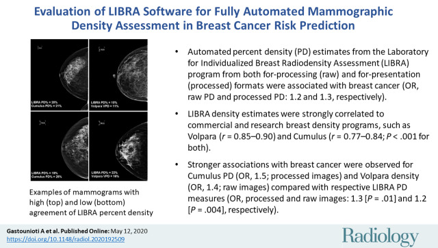
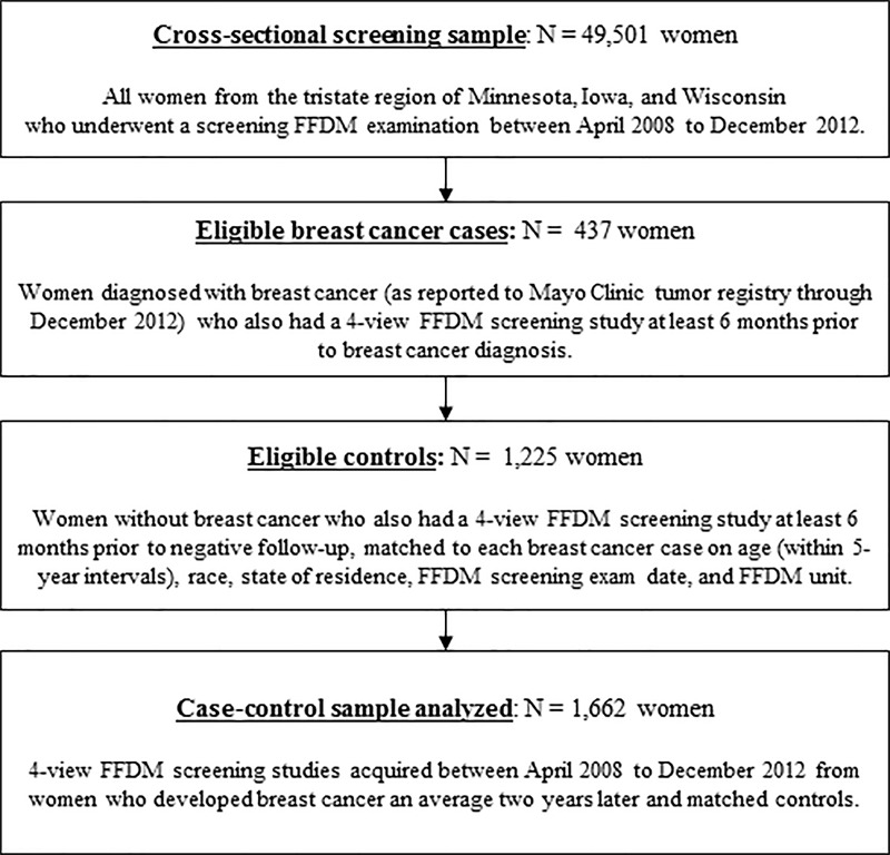
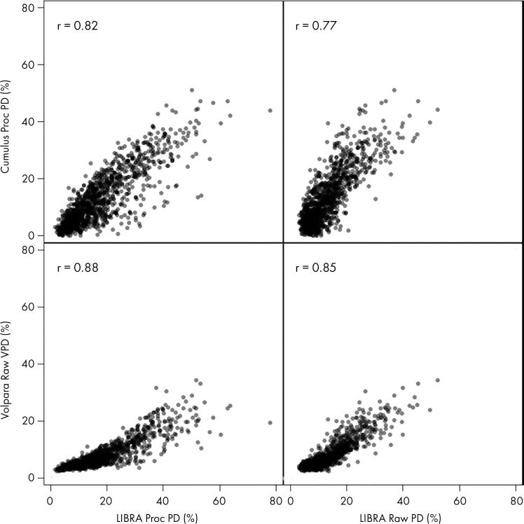
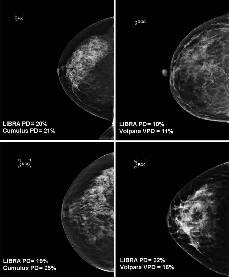
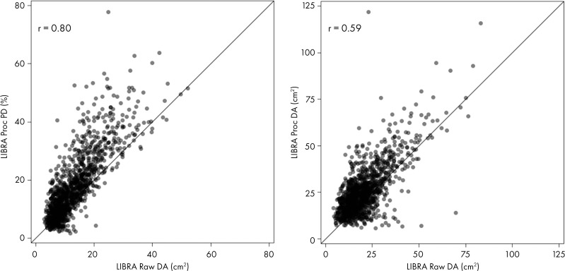
Background The associations of density measures from the publicly available Laboratory for Individualized Breast Radiodensity Assessment (LIBRA) software with breast cancer have primarily focused on estimates from the contralateral breast at the time of diagnosis. Purpose To evaluate LIBRA measures on mammograms obtained before breast cancer diagnosis and compare their performance to established density measures. Materials and Methods For this retrospective case-control study, full-field digital mammograms in for-processing (raw) and for-presentation (processed) formats were obtained (March 2008 to December 2011) in women who developed breast cancer an average of 2 years later and in age-matched control patients. LIBRA measures included absolute dense area and area percent density (PD) from both image formats. For comparison, dense area and PD were assessed by using the research software (Cumulus), and volumetric PD (VPD) and absolute dense volume were estimated with a commercially available software (Volpara). Density measures were compared by using Spearman correlation coefficients (r), and conditional logistic regression (odds ratios [ORs] and 95% confidence intervals [CIs]) was performed to examine the associations of density measures with breast cancer by adjusting for age and body mass index. Results Evaluated were 437 women diagnosed with breast cancer (median age, 62 years ± 17 [standard deviation]) and 1225 matched control patients (median age, 61 years ± 16). LIBRA PD showed strong correlations with Cumulus PD (r = 0.77-0.84) and Volpara VPD (r = 0.85-0.90) (P < .001 for both). For LIBRA, the strongest breast cancer association was observed for PD from processed images (OR, 1.3; 95% CI: 1.1, 1.5), although the PD association from raw images was not significantly different (OR, 1.2; 95% CI: 1.1, 1.4; P = .25). Slightly stronger breast cancer associations were seen for Cumulus PD (OR, 1.5; 95% CI: 1.3, 1.8; processed images; P = .01) and Volpara VPD (OR, 1.4; 95% CI: 1.2, 1.7; raw images; P = .004) compared with LIBRA measures. Conclusion Automated density measures provided by the Laboratory for Individualized Breast Radiodensity Assessment from raw and processed mammograms correlated with established area and volumetric density measures and showed comparable breast cancer associations. © RSNA, 2020 Online supplemental material is available for this article.
Figures
Similar articles
-
Fully Automated Volumetric Breast Density Estimation from Digital Breast Tomosynthesis.Radiology. 2021 Dec;301(3):561-568. doi: 10.1148/radiol.2021210190. Epub 2021 Sep 14. Radiology. 2021. PMID: 34519572 Free PMC article.
-
Preliminary evaluation of the publicly available Laboratory for Breast Radiodensity Assessment (LIBRA) software tool: comparison of fully automated area and volumetric density measures in a case-control study with digital mammography.Breast Cancer Res. 2015 Aug 25;17:117. doi: 10.1186/s13058-015-0626-8. Breast Cancer Res. 2015. PMID: 26303303 Free PMC article.
-
Impact of type of full-field digital image on mammographic density assessment and breast cancer risk estimation: a case-control study.Breast Cancer Res. 2016 Sep 26;18(1):96. doi: 10.1186/s13058-016-0756-7. Breast Cancer Res. 2016. PMID: 27670914 Free PMC article.
-
Detection of potential microcalcification clusters using multivendor for-presentation digital mammograms for short-term breast cancer risk estimation.Med Phys. 2019 Apr;46(4):1938-1946. doi: 10.1002/mp.13450. Epub 2019 Mar 7. Med Phys. 2019. PMID: 30801718 Free PMC article. Review.
-
Repeated measures of mammographic density and texture to evaluate prediction and risk of breast cancer: a systematic review of the methods used in the literature.Cancer Causes Control. 2023 Nov;34(11):939-948. doi: 10.1007/s10552-023-01739-2. Epub 2023 Jun 20. Cancer Causes Control. 2023. PMID: 37340148 Free PMC article. Review.
Cited by
-
Genome-Wide Association Study of Breast Density among Women of African Ancestry.Cancers (Basel). 2023 May 16;15(10):2776. doi: 10.3390/cancers15102776. Cancers (Basel). 2023. PMID: 37345113 Free PMC article.
-
Concordant and discordant breast density patterns by different approaches for assessing breast density and breast cancer risk.Breast Cancer Res Treat. 2025 Feb;210(1):105-114. doi: 10.1007/s10549-024-07541-1. Epub 2024 Nov 1. Breast Cancer Res Treat. 2025. PMID: 39482557
-
Screening mammography performance according to breast density: a comparison between radiologists versus standalone intelligence detection.Breast Cancer Res. 2024 Apr 22;26(1):68. doi: 10.1186/s13058-024-01821-w. Breast Cancer Res. 2024. PMID: 38649889 Free PMC article.
-
A Nomogram Using Imaging Features to Predict Ipsilateral Breast Tumor Recurrence After Breast-Conserving Surgery for Ductal Carcinoma In Situ.Korean J Radiol. 2024 Oct;25(10):876-886. doi: 10.3348/kjr.2024.0268. Korean J Radiol. 2024. PMID: 39344545 Free PMC article.
-
Examination of fully automated mammographic density measures using LIBRA and breast cancer risk in a cohort of 21,000 non-Hispanic white women.Breast Cancer Res. 2023 Aug 6;25(1):92. doi: 10.1186/s13058-023-01685-6. Breast Cancer Res. 2023. PMID: 37544983 Free PMC article.
References
-
- McCormack VA, dos Santos Silva I. Breast density and parenchymal patterns as markers of breast cancer risk: a meta-analysis. Cancer Epidemiol Biomarkers Prev 2006;15(6):1159–1169. - PubMed
-
- Mandelson MT, Oestreicher N, Porter PL, et al. Breast density as a predictor of mammographic detection: comparison of interval- and screen-detected cancers. J Natl Cancer Inst 2000;92(13):1081–1087. - PubMed
-
- Pisano ED, Gatsonis C, Hendrick E, et al. Diagnostic performance of digital versus film mammography for breast-cancer screening. N Engl J Med 2005;353(17):1773–1783 [Published correction appears in N Engl J Med 2006;355(17):1840.]. - PubMed
Publication types
MeSH terms
Grants and funding
LinkOut - more resources
Full Text Sources
Medical
