. 2020 Oct;69(10):1973-1987.
doi: 10.1007/s00262-020-02598-5. Epub 2020 May 9.

Activated HIF1α of tumor cells promotes chemoresistance development via recruiting GDF15-producing tumor-associated macrophages in gastric cancer

Affiliations

Activated HIF1α of tumor cells promotes chemoresistance development via recruiting GDF15-producing tumor-associated macrophages in gastric cancer

Shan Yu et al. Cancer Immunol Immunother. 2020 Oct.

Abstract

Chemotherapy is the preferred treatment for advanced stage gastric cancer (GC) patients, and developing chemoresistance is a tremendous challenge to efficacy of GC treatment. The treatments of anti-tumor chemo-agents recruit more tumor-associated macrophages (TAMs) which are highly implicated in the chemoresistance development, but the underlying molecular mechanism is unclear. Here, we demonstrate that hypoxia-inducible factor 1α (HIF1α) in GC cells is activated upon 5-fluorouracil (5-FU) treatment and results in much more accumulation of M2-type TAMs which protect tumor cells from chemo-agents. Mechanistically, in the GC cells under the 5-FU treatment, reactive oxygen species is accumulated and then induces the activation of HIF1α signaling to drive the expression of high-mobility group box 1, which leads to more macrophage's infiltration into GC tumor. In turn, the recruited TAMs exhibit tumor-protected M2-type phenotype and promote the chemoresistance of GC cells via producing growth differentiation factor 15 (GDF15) to exacerbate the fatty acid β-oxidation in tumor cells. Blocking GDF15 using antibody or inhibiting FAO of tumor cells by etomoxir efficiently gave rise to the tumor cell sensitivity to 5-FU. Therefore, our study demonstrates a novel insight in understanding the cross talking between tumor cells and immune microenvironment and provides new therapeutic targets for clinic treatments of gastric cancer.

Keywords: Chemoresistance; GDF15; Gastric cancer; HIF1α; HMGB1; Tumor-associated macrophages.


Conflict of interest statement

The authors declare no competing financial interests.

Figures
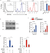
Fig. 1
Fig. 1
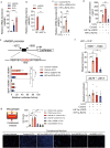
Chemo-agents activate HIF1α signaling via inducing ROS accumulation in gastric cancer cells. a Representative images of flow cytometry analysis of intracellular ROS. AGS cells were treated by 0.1 mg/ml 5-FU or 0.1 nmol/l VCR for 24 h and then stained with DCFDA for the ROS analysis by flow cytometry. ROS-positive cells were quantified. n = 5 for each group. b Representative images of immunoblotting of HIF1α and β-actin (internal control) in the cell extracts of AGS cells 24 h after treatments by VCR or 5-FU. c Luciferase activities were determined in AGS cells treated by 5-FU or VCR for indicated time. d Luciferase activities were determined in AGS cells treated with vehicle, 5-FU, 5-FU combined with NAC, VCR, or VCR combined with NAC, respectively. For c, d AGS cells were transfected with reporter constructs within which luciferase expression is driven by the activated HRE element. The expression of luciferase is driven by the HRE element. Luciferase activity is presented as fold change after normalizing values of Luc to Renilla (RLU). The values of vehicle control group were set as “1”. All data are shown as mean ± SEM. *p < 0.05; **p < 0.01; or ***p < 0.001 by one-way ANOVA
Fig. 2
Fig. 2
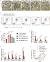
HIF1α drives HMGB1 expression in 5-FU-treated gastric cancer cells to exacerbate macrophage infiltration. a qRT-PCR analysis of HMGB1 mRNA levels of AGS cells in the presence of 5-FU (0.1 mg/ml), VCR (0.1 nmol/l) or vehicle control for 24 h. b qRT-PCR analysis of HMGB1 mRNA levels of AGSs in the presence of 5-FU or vehicle. The AGS cells were transfected with siRNA targeted at HIF1a (siHIF1a) or negative control (NC) prior to 5-FU or vehicle treatment. c, d, AGS cells were transfected with empty vector (control) or constructs expressing human HIF1a mutants HIF1a-ΔODD or HIF1a-R27G. (c) qRT-PCR analysis of HMGB1 mRNA levels of 5-FU-treated cells and d ELISA determined the HMGB1 protein levels in the cell culture of AGS cells after treated by vehicle or 5-FU. e AGS cells were transfected with reporter constructs within which luciferase expression is driven under HMGB1 full-length promoter (2000 bp upstream of TSS) or HRE-truncated promoter in combination with empty vector (control) or constructs expressing HIF1a-ΔODD or HIF1a-R27G, respectively. Luciferase activity is presented as fold change after normalizing Luc values to Renilla activity (RLU). The values of vehicle control group were set as “1”. f ChIP assays were conducted by the using of IgG or anti-HIF1α antibody in cell lysis from AGS cells which were transfected with control vector or constructs expressing HIF1a-ΔODD or HIF1a-R27G followed with treatments of 0.1 mg/ml 5-FU or vehicle. qRT-PCR analyses of immunoprecipitated DNA were performed using the primers which were designed to amplify the indicated region of the HMGB1 promoter. g Representative images of transmembraned macrophages counterstained with DAPI. Transwell assays of macrophages were conducted in the presence of conditioned media derived from the cell culture of AGS cells which were previously subjected to indicated treatments. All data are shown as mean ± SEM. *p < 0.05; **p < 0.01; or ***p < 0.001 by unpaired two-tailed Student’s t test or one-way ANOVA
Fig. 3
Fig. 3
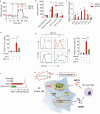
HIF1α signaling is required for recruiting and activating TAMs by 5-FU-treated GC. AGS cells were transfected with lentivirus to derive new cell lines which stably express GFP (control) or HIF1α mutants (HIF1a-ΔODD or HIF1a-R27G), respectively. 5 × 106 cells per cell line were implanted into nude mice at flank to induce tumor formation (n = 6 per group). The mice were subsequently i.p. administered with vehicle or 5-FU, or 5-FU in combination with anti-HMGB1 Ab as indicated. a Representative images of immunostaining of the xenograft tumors sections for F4/80. b Representative images of flow cytometry analysis of macrophages in xenograft tumor using anti-CD45, anti-F4/80 and anti-CD11b flow antibodies. TAMs were gated as CD45+CD11b+F4/80+ cells. c The frequencies of TAMs in CD45+ cells of each group were quantified. d qRT-PCR analysis of mRNA levels of HMGB1 and macrophage markers (ANGRE1 and CD68) in xenograft tumor. e qRT-PCR analysis of mRNA levels of indicated genes in xenograft tumor. f Tumor sizes of each group. All the data are shown as mean ± SEM. *p < 0.05; **p < 0.01; or ***p < 0.001 by one-way ANOVA
Fig. 4
Fig. 4
Accumulated TAMs elevate the chemoresistance of GC cells via secreting GDF15. a IC50 of 5-FU was determined in AGS cells which were cocultured with TAMs isolated from xenograft tumors as indicated. The tumors were derived from AGS cell lines expressing GFP (control) or HIF1α mutants, and then, the mice were treated with vehicle or 5-FU, or 5-FU in combination with anti-HMGB1 Ab prior to the sorting of macrophages. The sorted TAMs were cocultured with AGS cells in the transwell plates as indicated. b Colony-formation assays were performed using AGS cells with or without the presence of sorted TAMs followed the treatments of vehicle, 5-FU or 5-FU in combination with anti-GDF15 Ab. c IC50 of 5-FU was determined in AGS cells treated with vehicle or recombinant murine GDF15. d qRT-PCR analysis of mRNA levels of Gdf15 in TAMs isolated from the xenograft tumors as indicated. e IC50 of 5-FU was determined in AGS cells which were maintained in indicated conditioned media. Conditioned media were derived from TAMs which were isolated from indicated xenograft tumors and then incubated with IgG or anti-GDF15 Ab. All the data are shown as mean ± SEM. *p < 0.05; **p < 0.01; or ***p < 0.001 by unpaired two-tailed Student’s t test or one-way ANOVA
Fig. 5
Fig. 5
GDF15 enhances the FAO of tumor cells to reduce chemo-sensitivity as well as promote ROS production. a, b Analysis of mitochondrial FAO in vehicle- or 100 ng/ml GDF15-treated AGS cells by the using of the Seahorse Bioscience XF24e extracellular flux analyzer. c qRT-PCR analysis of FAO-associated genes expression in AGS cells treated with vehicle or 100 ng/ml GDF15. df AGS cells were treated with vehicle, 100 ng/ml GDF15 or 100 ng/ml GDF15 in combination with FAO inhibitor etomoxir (ETO, 1 μmol/l). d IC50 of 5-FU was determined in AGS cells. e ROS-positive AGS cells were analyzed using flow cytometry. f Luciferase activity using HRE reporter was determined in AGS cells. g Schematic model. Upon the chemotherapy of 5-FU, ROS is accumulated and activates HIF1α signaling to drive HMGB1 transcription in AGS cells. The increased HMGB1 exacerbates the infiltration of M2-type TAMs, which in turn secret GDF15 to enhance FAO and reduce chemo-sensitivity of 5-FU in gastric tumor cells. All data are shown as mean ± SEM. *p < 0.05; **p < 0.01; ***p < 0.001 by unpaired two-tailed Student’s t test or one-way ANOVA

Similar articles

Cited by

References

    1. Miller KD, Siegel RL, Lin CC, Mariotto AB, Kramer JL, Rowland JH, Stein KD, Alteri R, Jemal A. Cancer treatment and survivorship statistics. CA Cancer J Clin. 2016;66(4):271–289. doi: 10.3322/caac.21349. - DOI - PubMed
    1. Digklia A, Wagner AD. Advanced gastric cancer: current treatment landscape and future perspectives. World J Gastroenterol. 2016;22(8):2403–2414. doi: 10.3748/wjg.v22.i8.2403. - DOI - PMC - PubMed
    1. Davidson M, Okines AF, Starling N. Current and future therapies for advanced gastric cancer. Clin Colorectal Cancer. 2015;14(4):239–250. doi: 10.1016/j.clcc.2015.05.013. - DOI - PubMed
    1. Kitajima Y, Miyazaki K. The critical impact of HIF-1a on gastric cancer biology. Cancers (Basel) 2013;5(1):15–26. doi: 10.3390/cancers5010015. - DOI - PMC - PubMed
    1. Rohwer N, Cramer T. HIFs as central regulators of gastric cancer pathogenesis. Cancer Biol Ther. 2010;10(4):383–385. doi: 10.4161/cbt.10.4.12850. - DOI - PubMed

MeSH terms

Substances