Bone marrow mesenchymal stem cells improve bone erosion in collagen-induced arthritis by inhibiting osteoclasia-related factors and differentiating into chondrocytes
- PMID: 32381074
- PMCID: PMC7203805
- DOI: 10.1186/s13287-020-01684-w
Bone marrow mesenchymal stem cells improve bone erosion in collagen-induced arthritis by inhibiting osteoclasia-related factors and differentiating into chondrocytes
Abstract
Background: Rheumatoid arthritis (RA) is characterized by joint inflammation and damage to the cartilage and bone in collagen-induced arthritis (CIA). Mesenchymal stem cells (MSCs) can improve articular symptoms and reduce bone erosion in CIA rats; however, the underlying mechanism remains unknown. This study aimed to investigate the mechanism underlying MSC-induced improvement of bone destruction in CIA.
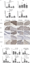
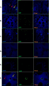
Methods: Wistar rats were divided into a normal group, CIA control group, MTX intervention group, and BMSC intervention group, each comprising 8 rats. Serum RANKL, OPG, and CXCL10 levels of all groups were determined via flow cytometry after 42 days of interventions. RANKL, OPG, TRAF6, CXCL10, and CXCR3 were detected on the synovial membrane via immunohistochemistry, and their relative mRNA levels were determined via RT-PCR analysis. BMSCs were labeled with GFP and administered to CIA rats via the tail vein. At different time points, the distribution of implanted GFP-MSCs in synovial tissues was observed using a fluorescence microscope, and the potential of GFP-MSCs to differentiate into chondrocytes was assessed via immunofluorescence analysis.
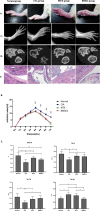
Results: BMSC transplantation improved joint inflammation and inhibited bone destruction in CIA rats. BMSCs inhibited the expression of serum CXCL10 and CXCL10 and CXCR3 expression at the synovial membrane. Moreover, protein and mRNA expression analyses revealed that BMSCs potentially regulated RANKL/OPG expression levels in the serum and synovial tissue. Upon implantation into CIA rats, GFP-MSCs were traced in the joints. GFP-positive cells were observed in the cartilage tissue from day 11 and until 42 days after transplantation. Anti-type II collagen/GFP double-positive cells were observed in the articular cartilage (especially damaged cartilage) upon immunofluorescence staining of anti-type II collagen.
Conclusions: BMSCs improve bone destruction in CIA by inhibiting the CXCL10/CXCR3 chemotactic axis, regulating the RANKL/OPG ratio, and directly differentiating into chondrocytes.
Keywords: Bone destruction; Bone marrow-derived mesenchymal stem cells; Chemokines; Green fluorescent protein; RANKL/OPG; Rheumatoid arthritis; Tissue repair.
Conflict of interest statement
The authors declare that they have no competing interests.
Figures


Similar articles
-
Bone Marrow Mesenchymal Stem Cells Decrease the Expression of RANKL in Collagen-Induced Arthritis Rats via Reducing the Levels of IL-22.J Immunol Res. 2019 Nov 7;2019:8459281. doi: 10.1155/2019/8459281. eCollection 2019. J Immunol Res. 2019. PMID: 31828174 Free PMC article.
-
[Effects of transcutaneous auricular vagus nerve stimulation on bone and cartilage destruction in rats with rheumatoid arthritis].Zhen Ci Yan Jiu. 2022 Mar 25;47(3):237-43. doi: 10.13702/j.1000-0607.20211130. Zhen Ci Yan Jiu. 2022. PMID: 35319841 Chinese.
-
[Effect of icariin on bone destruction and serum RANKL/OPG levels in type II collagen-induced arthritis rats].Zhongguo Zhong Xi Yi Jie He Za Zhi. 2013 Sep;33(9):1221-5. Zhongguo Zhong Xi Yi Jie He Za Zhi. 2013. PMID: 24273978 Chinese.
-
Bone destruction in arthritis.Ann Rheum Dis. 2002 Nov;61 Suppl 2(Suppl 2):ii84-6. doi: 10.1136/ard.61.suppl_2.ii84. Ann Rheum Dis. 2002. PMID: 12379632 Free PMC article. Review.
-
Synovial Fluid Mesenchymal Stem Cells for Knee Arthritis and Cartilage Defects: A Review of the Literature.J Knee Surg. 2021 Nov;34(13):1476-1485. doi: 10.1055/s-0040-1710366. Epub 2020 May 13. J Knee Surg. 2021. PMID: 32403148 Review.
Cited by
-
CXCL10 as a shared specific marker in rheumatoid arthritis and inflammatory bowel disease and a clue involved in the mechanism of intestinal flora in rheumatoid arthritis.Sci Rep. 2023 Jun 16;13(1):9754. doi: 10.1038/s41598-023-36833-7. Sci Rep. 2023. PMID: 37328529 Free PMC article.
-
Management of Rheumatoid Arthritis: Possibilities and Challenges of Mesenchymal Stromal/Stem Cell-Based Therapies.Cells. 2023 Jul 21;12(14):1905. doi: 10.3390/cells12141905. Cells. 2023. PMID: 37508569 Free PMC article. Review.
-
Application of methylation in the diagnosis of ankylosing spondylitis.Clin Rheumatol. 2024 Oct;43(10):3073-3082. doi: 10.1007/s10067-024-07113-0. Epub 2024 Aug 21. Clin Rheumatol. 2024. PMID: 39167325 Review.
-
Beraprost ameliorates postmenopausal osteoporosis by regulating Nedd4-induced Runx2 ubiquitination.Cell Death Dis. 2021 May 15;12(5):497. doi: 10.1038/s41419-021-03784-8. Cell Death Dis. 2021. PMID: 33993186 Free PMC article.
-
Mesenchymal stem cells and their microenvironment.Stem Cell Res Ther. 2022 Aug 20;13(1):429. doi: 10.1186/s13287-022-02985-y. Stem Cell Res Ther. 2022. PMID: 35987711 Free PMC article. Review.
References
Publication types
MeSH terms
Substances
LinkOut - more resources
Full Text Sources
Other Literature Sources

