. 2020 Jun 1;143(6):1697-1713.
doi: 10.1093/brain/awaa116.

Nogo receptor decoy promotes recovery and corticospinal growth in non-human primate spinal cord injury

Affiliations

Nogo receptor decoy promotes recovery and corticospinal growth in non-human primate spinal cord injury

Xingxing Wang et al. Brain. .

Abstract

After CNS trauma such as spinal cord injury, the ability of surviving neural elements to sprout axons, reorganize neural networks and support recovery of function is severely restricted, contributing to chronic neurological deficits. Among limitations on neural recovery are myelin-associated inhibitors functioning as ligands for neuronal Nogo receptor 1 (NgR1). A soluble decoy (NgR1-Fc, AXER-204) blocks these ligands and provides a means to promote recovery of function in multiple preclinical rodent models of spinal cord injury. However, the safety and efficacy of this reagent in non-human primate spinal cord injury and its toxicological profile have not been described. Here, we provide evidence that chronic intrathecal and intravenous administration of NgR1-Fc to cynomolgus monkey and to rat are without evident toxicity at doses of 20 mg and greater every other day (≥2.0 mg/kg/day), and far greater than the projected human dose. Adult female African green monkeys underwent right C5/6 lateral hemisection with evidence of persistent disuse of the right forelimb during feeding and right hindlimb during locomotion. At 1 month post-injury, the animals were randomized to treatment with vehicle (n = 6) or 0.10-0.17 mg/kg/day of NgR1-Fc (n = 8) delivered via intrathecal lumbar catheter and osmotic minipump for 4 months. One animal was removed from the study because of surgical complications of the catheter, but no treatment-related adverse events were noted in either group. Animal behaviour was evaluated at 6-7 months post-injury, i.e. 1-2 months after treatment cessation. The use of the impaired forelimb during spontaneous feeding and the impaired hindlimb during locomotion were both significantly greater in the treatment group. Tissue collected at 7-12 months post-injury showed no significant differences in lesion size, fibrotic scar, gliosis or neuroinflammation between groups. Serotoninergic raphespinal fibres below the lesion showed no deficit, with equal density on the lesioned and intact side below the level of the injury in both groups. Corticospinal axons traced from biotin-dextran-amine injections in the left motor cortex were equally labelled across groups and reduced caudal to the injury. The NgR1-Fc group tissue exhibited a significant 2-3-fold increased corticospinal axon density in the cervical cord below the level of the injury relative to the vehicle group. The data show that NgR1-Fc does not have preclinical toxicological issues in healthy animals or safety concerns in spinal cord injury animals. Thus, it presents as a potential therapeutic for spinal cord injury with evidence for behavioural improvement and growth of injured pathways in non-human primate spinal cord injury.

Keywords: Nogo receptor; axon; oligodendrocyte; regeneration; spinal cord injury.


Figures

Figure 1
Figure 1
Experimental design. (A) Sixteen monkeys received right C5/C6 hemisection spinal cord injury. One week after SCI and prior to randomization, two monkeys were euthanized for humane reasons, because of poor general health. The remaining 14 SCI monkeys were randomly assigned to either the vehicle (PBS)-treated or NgR-Fc-treated groups, and all handlers remained blind to the experimental condition. One monkey from the NgR-Fc-treated group was removed from the experiment 2 months after SCI because of a catheterization issue. Thirteen monkeys completed the experiment and are included in the analysis with six in the vehicle group and seven in the NgR-Fc group. Each NgR-Fc-treated monkey received NgR-Fc protein at 0.10–0.17 mg/kg/day intrathecally for 4 months. (B) Schematics of the experiment: two cohorts of animals were studied, seven monkeys in cohort 1. Animals started intrathecal treatment 1 month after SCI and received continuous intrathecal administration with either vehicle (n = 4) or NgR-Fc (n = 3) for 4 months and were sacrificed 14–16 months after SCI. In cohort 2, six monkeys received the same strategy of intrathecal treatment as cohort 1 with either vehicle (n = 2) or NgR-Fc (n = 4). Animals were sacrificed 7–9 months after SCI. The timing of SCI, randomization, behavioural studies (Beh), CST axon tracing (BDA) and the four minipump administrations are illustrated.
Figure 2
Figure 2
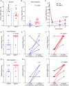
Behavioural recovery of spinal cord-injured monkeys increase by NgR-Fc treatment. (AC) Quantifications of open field voluntary feeding behaviour before and 6 months after SCI. (A) Hand usage preference 1 week before SCI. In a 10-min video analysis, the number of times that the animal uses its left hand, right hand (lesion side), or both hands to bring food to its mouth was counted. The hand usage preference was normalized to the control side (left hand, serving as internal control). (B) Hand usage preference 6 months after SCI. The attempts to use the right hand or both hands increased significantly for animals in the NgR-Fc-treated group (P = 0.047 by unpaired two-tailed t-test with Welch’s correction). Mean ± SEM with individual animals indicated. (C) Time course of hand usage preference as a function of time relative to SCI. The pre-injury and 6-month data from A and B are included in this replot. Mean ± SEM, n = 6 for control and n = 7 for NgR-Fc. By mixed-effects model, there was a significant interaction of time and treatment, P = 0.034, and by unpaired two-tailed t-test with Welch’s correction at 6 months, P = 0.047 for treatment as in B. (DI) Hindlimb functional recovery after NgR-Fc treatment. (D) In the open field video analysis of movement assay, hip, knee and ankle movements were scored 6 months after SCI. The NgR-Fc treatment group shows significant recovery of their joint’s movements in the hindlimb walking assessment (P = 0.023 by unpaired two-tailed t-test with Welch’s correction). (E and F) Comparison of hindlimb walking scores at 1 week pre-injury and 6 months post-injury for each individual monkey. There was a statistically significant difference for animals from the NgR-Fc treatment group (P = 0.0017 by unpaired two-tailed t-test with Bonferroni-Dunn correction for multiple tests) and no significant difference for animals from the vehicle treatment group (P = 0.07). Mean ± SEM with individual animals indicated. (G) Quantification of the hindlimb digital function. There was a trend of increasing hindlimb digital scores in the NgR-Fc-treated group compared to the vehicle-treated group, but no significant statistical difference. (H and I) Comparison of hindlimb digital function scores at 1 week pre-injury and 6 months post-injury for each individual monkey. There was a statistically significant difference for animals from the NgR-Fc treatment group (P = 0.0065 by unpaired two-tailed t-test with Bonferroni-Dunn correction for multiple tests) and no significant difference for animals from the vehicle treatment group (P = 0.07). Mean ± SEM with individual animals indicated.
Figure 3
Figure 3
Lesion size is similar in the vehicle and NgR-Fc groups. (A and B) Representative images of horizontal section of the lesion segment of spinal cord from both the PBS-treated (A) and NgR-Fc-treated (B) animals, stained with anti-GFAP antibody to visualize the injury site. Rostral is to the left and right side is up. Scale bar = 1000 µm. (CF) Boxed areas in A and B are magnified in CF. Scale bar = 50 μm. (G) The depth of lesion at the injury site was measured for each consecutive GFAP stained section. Mean ± SEM with individual animals indicated. There was no statistically significant difference between the two treatment groups. (H and I) Representative images of the horizontal section of the lesion segment of spinal cord from PBS- (H) and NgR-Fc-treated (I) animals, stained with anti-Laminin antibody to show the fibrotic scar. Red outlines indicate the fibrotic area. Scale bar = 1000 µm. (J) Quantification of the laminin-positive fibrotic scar area. There was no statistically significant difference between groups. Data are mean ± SEM, unpaired two-tailed t-test.
Figure 4
Figure 4
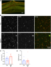
NgR-Fc treatment does not alter chronic gliosis. (A) Representative image of immunofluorescent staining for Iba1 (green) and CD68 (red) in horizontal spinal cord sections containing the SCI site. Scale bar = 500 µm. (B) High-magnification view near the lesion area stained as in A from a vehicle-treated animal (Con) and a NgR-Fc-treated animal (NgR-Fc). Scale bar = 50 µm. (C and D) Quantification of Iba1 and CD68 immunoreactive area from sections, as in A and B. There was no statistically significant difference between groups. Data are mean ± SEM, unpaired two-tailed t-test.
Figure 5
Figure 5
Intact raphespinal serotonergic innervation of the caudal spinal cord after injury. (A) Representative low-magnification transverse section of the spinal cord caudal to the lesion site at the T3-T4 level stained with anti-serotonin antibody with the ventral horn measurement area indicated by white square. Scale bar = 1000 µm. (B) 5HT fibres in the ventral horn from sections stained, as in A, are from a vehicle or NgR-Fc sample of the ventral horn on the right side or left side, as indicated. Scale bar = 50 µm. (C) The ratio of 5HT axon length per area in the ventral horn of the lesion side to the intact side was measured for sections as in B. There was no statistically significant difference of raphespinal serotonergic innervation between left and right, or between two treatment groups. Data are mean ± SEM, unpaired two-tailed t-test.
Figure 6
Figure 6
Efficiency of BDA tracing in CST. (A) A transverse section of spinal cord at the C1 level was stained for BDA-labelled CST fibres. Three high-magnification images from the regions shown by white boxes were used for quantification of total density in right dorso-lateral CST outlined in white. Scale bar = 500 µm. (B and C) Representative of high-magnification images of BDA-labelled CST axons in the right lateral column at C1 level from a vehicle-treated animal (B) and a NgR-Fc-treated animal (C). Scale bar = 50 µm. (D) Quantification of BDA-labelled CST axons at C1 spinal cord. No statistically significant difference between groups. Data are mean ± SEM, unpaired two-tailed t-test.
Figure 7
Figure 7
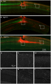
NgR-Fc treatment increases growth of CST fibres caudal to the injury. Representative images of horizontal section of spinal cord stained for BDA (green) and anti-GFAP (red) from one vehicle-treated (A) and two NgR-Fc-treated animals (B and C). Rostral to the left and right side is up. The area in the white boxes from AC (marked di) are magnified in DI. Note numerous BDA-labelled CST fibres significantly increased in the NgR-Fc treated animals. Scale bar = 2000 µm in AC, 300 µm in DI.
Figure 8
Figure 8
Quantification of BDA-labelled CST axons. (A) CST fibre counts from horizontal sections, as in Fig. 7, are reported by an axon index relative to the most rostral count, and are plotted as a function of rostral-caudal distance relative to the SCI site. Repeated measures ANOVA caudal to the lesion show a significant greater fibre count (P = 0.006) in the NgR-Fc group, with post hoc pairwise comparisons at the indicated distances *P <0.05; **P <0.01. Data are mean ± SEM. (B) Enlarged view of graph from A from the lesion centre to 12 mm caudal to the injury. (C) The average CST axon index from 7–12 mm caudal to the injury on the lesion side, on the intact side and on both sides of the spinal cord. There was statistically greater CST fibre density in the NgR-C group overall, and on the intact side. Unpaired two-tailed t-test with Holm-Sidak correction for multiple comparisons. Mean ± SEM, with individual animals indicated. (D) Transverse sections of the spinal cord caudal to the lesion site at the T3-T4 level were stained for BDA-labelled CST fibres. The CST axon length per area in the grey matter of the lesion side and the intact side was measured for control (Con) and NgR-Fc groups. Unpaired two-tailed t-test with Holm-Sidak correction for multiple comparisons. Mean ± SEM, with individual animals indicated.

Comment in

Similar articles

Cited by

References

    1. Akbik F, Cafferty WBJ, Strittmatter SM.. Myelin associated inhibitors: a link between injury-induced and experience-dependent plasticity. Exp Neurol 2012; 235: 43–52. - PMC - PubMed
    1. Akbik FV, Bhagat SM, Patel PR, Cafferty WB, Strittmatter SM.. Anatomical plasticity of adult brain is titrated by Nogo Receptor 1. Neuron 2013; 77: 859–66. - PMC - PubMed
    1. Angeli CA, Boakye M, Morton RA, Vogt J, Benton K, Chen Y, et al.Recovery of over-ground walking after chronic motor complete spinal cord injury. N Engl J Med 2018; 379: 1244–50. - PubMed
    1. Atwal JK, Pinkston-Gosse J, Syken J, Stawicki S, Wu Y, Shatz C, et al.PirB is a functional receptor for myelin inhibitors of axonal regeneration. Science 2008; 322: 967–70. - PubMed
    1. Benson MD, Romero MI, Lush ME, Lu QR, Henkemeyer M, Parada LF.. Ephrin-B3 is a myelin-based inhibitor of neurite outgrowth. Proc Natl Acad Sci USA 2005; 102: 10694–9. - PMC - PubMed

Publication types

MeSH terms