

The relationship between pancreas steatosis and the risk of metabolic syndrome and insulin resistance in Chinese adolescents with concurrent obesity and non-alcoholic fatty liver disease
- PMID: 32351030
- PMCID: PMC7507143
- DOI: 10.1111/ijpo.12653
The relationship between pancreas steatosis and the risk of metabolic syndrome and insulin resistance in Chinese adolescents with concurrent obesity and non-alcoholic fatty liver disease
Abstract
Background: The incidence of childhood obesity and associated comorbidities are on an increasing trend worldwide. More than 340 million children and adolescents aged between 5 and 19 years old were overweight or had obesity in 2016, from which over 124 million children and adolescents (6% of girls and 8% of boys) had obesity.
Objective: To describe the relationship between pancreas steatosis, body fat and the risk of metabolic syndrome, insulin resistance in Hong Kong Chinese adolescents with both obesity and non-alcoholic fatty liver disease (NAFLD).
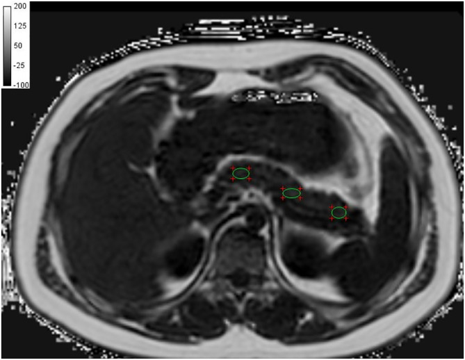
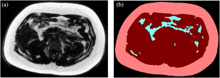
Methods: Fifty two adolescents with obesity and NAFLD were analysed (14-18 years), stratified into fatty and non-fatty pancreas groups using chemical shift encoded MRI-pancreas proton density fat fraction ≥5%. Pancreatic, abdominal subcutaneous adipose tissue (SAT)/visceral adipose tissue (VAT) volumes, biochemical and anthropometric parameters were measured. Mann-Whitney U test, multiple linear/binary logistic regression analyses and odds ratios were used.
Results: Fifty percent had fatty pancreas, 38% had metabolic syndrome and 81% had insulin resistance. Liver proton density fat fraction (PDFF) and VAT were independent predictors of insulin resistance (P = .006, .016). Pancreas and liver PDFF were both independent predictors of beta cells dysfunction (P = .015, .050) and metabolic syndrome (P = .021, .041). Presence of fatty pancreas in obesity was associated with insulin resistance (OR = 1.58, 95% CI = 0.39-6.4) and metabolic syndrome (OR = 1.70, 95% CI = 0.53-5.5).
Conclusion: A significant causal relationship exists between fatty pancreas, fatty liver, body fat and the risk of developing metabolic syndrome and insulin resistance.
Key points: Fatty pancreas is a common finding in adolescents with obesity, with a prevalence rate of 50% in this study cohort. Liver PDFF and VAT are independent predictors of insulin resistance while pancreas PDFF and liver PDFF are independent predictors of both beta cells dysfunction and metabolic syndrome. Presence of fatty pancreas at imaging should not be considered as a benign finding but rather as an imaging biomarker of emerging pancreatic metabolic and endocrine dysfunction.
Keywords: fatty liver; insulin resistance; magnetic resonance imaging; metabolic syndrome; pancreas.
© 2020 The Authors. Pediatric Obesity published by John Wiley & Sons Ltd on behalf of World Obesity Federation.
Conflict of interest statement
The authors declare that they have no conflict of interest.
Figures


Similar articles
-
Implications of Abdominal Adipose Tissue Distribution on Nonalcoholic Fatty Liver Disease and Metabolic Syndrome: A Chinese General Population Study.Clin Transl Gastroenterol. 2021 Feb 17;12(2):e00300. doi: 10.14309/ctg.0000000000000300. Clin Transl Gastroenterol. 2021. PMID: 33600104 Free PMC article.
-
Pancreatic fat and β-cell function in overweight/obese children with nonalcoholic fatty liver disease.World J Gastroenterol. 2015 Apr 21;21(15):4688-95. doi: 10.3748/wjg.v21.i15.4688. World J Gastroenterol. 2015. PMID: 25914480 Free PMC article.
-
[The correlations of abdominal adipose tissue with anthropometric and metabolic parameters in obese children by magnetic resonance imaging].Zhonghua Er Ke Za Zhi. 2022 Aug 2;60(8):798-803. doi: 10.3760/cma.j.cn112140-20220129-00099. Zhonghua Er Ke Za Zhi. 2022. PMID: 35922191 Chinese.
-
Non-Alcoholic Fatty Liver Disease in Obese Youth With Insulin Resistance and Type 2 Diabetes.Front Endocrinol (Lausanne). 2021 Apr 6;12:639548. doi: 10.3389/fendo.2021.639548. eCollection 2021. Front Endocrinol (Lausanne). 2021. PMID: 33889132 Free PMC article. Review.
-
Non-Alcoholic Fatty Pancreas Disease (NAFPD): A Silent Spectator or the Fifth Component of Metabolic Syndrome? A Literature Review.Endocr Metab Immune Disord Drug Targets. 2018;18(6):547-554. doi: 10.2174/1871530318666180328111302. Endocr Metab Immune Disord Drug Targets. 2018. PMID: 29595117 Review.
Cited by
-
Association of Incidence between Pancreatic Adipose Infiltration and Metabolic Syndrome: A Literature Review and Meta-analysis.Comput Math Methods Med. 2021 Nov 5;2021:5747558. doi: 10.1155/2021/5747558. eCollection 2021. Comput Math Methods Med. 2021. PMID: 34777565 Free PMC article.
-
Obesity, heart failure with preserved ejection fraction, and the role of glucagon-like peptide-1 receptor agonists.ESC Heart Fail. 2024 Apr;11(2):649-661. doi: 10.1002/ehf2.14560. Epub 2023 Dec 13. ESC Heart Fail. 2024. PMID: 38093506 Free PMC article. Review.
-
Test-Retest Reliability of a Physical Activity Behavior, Health and Wellbeing Questionnaire in Adolescents.Open Res Eur. 2024 Sep 5;3:154. doi: 10.12688/openreseurope.16535.3. eCollection 2023. Open Res Eur. 2024. PMID: 39246696 Free PMC article.
-
Association of pancreatic fat on imaging with pediatric metabolic co-morbidities.Pediatr Radiol. 2023 Sep;53(10):2030-2039. doi: 10.1007/s00247-023-05669-8. Epub 2023 Apr 28. Pediatr Radiol. 2023. PMID: 37106090 Free PMC article.
-
Relation Between Non-Alcoholic Fatty Pancreas and Clinical and Biochemical Parameters in Women with Polycystic Ovary Syndrome: A Multi-Centric Study.Int J Gen Med. 2022 Nov 19;15:8225-8233. doi: 10.2147/IJGM.S384073. eCollection 2022. Int J Gen Med. 2022. PMID: 36438020 Free PMC article.
References
-
- Maggio AB, Mueller P, Wacker J, et al. Increased pancreatic fat fraction is present in obese adolescents with metabolic syndrome. J Pediatr Gastroenterol Nutr. 2012;54(6):720‐726. - PubMed
-
- Hocking S, Samocha‐Bonet D, Milner K, Greenfield JR, Chisholm DJ. Adiposity and insulin resistance in humans: the role of the different tissue and cellular lipid depots. Endocr Rev. 2013;34(4):463‐500. - PubMed
-
- Gutin B, Johnson MH, Humphries MC, et al. Relationship of visceral adiposity to cardiovascular disease risk factors in black and white teens. Obesity. 2007;15(4):1029‐1035. - PubMed
-
- Lee H, Park H, Hwang J. HbA1c and glucose intolerance in obese children and adolescents. Diabet Med. 2012;29(7):e102‐e105. - PubMed
Publication types
MeSH terms
Substances
LinkOut - more resources
Full Text Sources
Medical
Research Materials
