Review
. 2021 May;10(5):234-256.
doi: 10.1089/wound.2019.1094. Epub 2020 Jun 2.

Nanotechnology Approaches in Chronic Wound Healing

Affiliations
Review

Nanotechnology Approaches in Chronic Wound Healing

Barbara Blanco-Fernandez et al. Adv Wound Care (New Rochelle). 2021 May.

Abstract

Significance: The incidence of chronic wounds is increasing due to our aging population and the augment of people afflicted with diabetes. With the extended knowledge on the biological mechanisms underlying these diseases, there is a novel influx of medical technologies into the conventional wound care market. Recent Advances: Several nanotechnologies have been developed demonstrating unique characteristics that address specific problems related to wound repair mechanisms. In this review, we focus on the most recently developed nanotechnology-based therapeutic agents and evaluate the efficacy of each treatment in in vivo diabetic models of chronic wound healing. Critical Issues: Despite the development of potential biomaterials and nanotechnology-based applications for wound healing, this scientific knowledge is not translated into an increase of commercially available wound healing products containing nanomaterials. Future Directions: Further studies are critical to provide insights into how scientific evidences from nanotechnology-based therapies can be applied in the clinical setting.

Keywords: chronic; diabetes; liposomes; nanofibers; nanoparticles; wound healing.


Conflict of interest statement

No competing financial interests exist. The content of this article was expressly written by the authors listed. No ghostwriters were used to write this article.

Figures

None
Soledad Pérez-Amodio, PhD
Figure 1.
Figure 1.
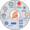
Schematic representation of nanocarriers used for chronic wound healing: self-assembled nanocarriers (liposomes, micelles, nanogels), NPs (polymeric, inorganic, lipid), and nanofibers (plain and encapsulating nanocarriers or therapeutic agents). NP, nanoparticle. Color images are available online.
Figure 2.
Figure 2.
Antibacterial mechanism of action of metallic NPs. Metal NPs provoke protein denaturalization, enzyme inactivation, DNA damage, and the disassembly of ribosomes, as well as ROS generation. Altogether, promote the programmed bacterial cell death. ROS, reactive oxygen species. Color images are available online.
Figure 3.
Figure 3.
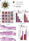
The topical administration of AuNPs conjugated to spherical nucleic acid for GM3S shows a reduction in local GM3S expression and heals the wound in 12 days in diabetic mice wounds. An increase in granulation tissue, new blood vessel formation, and IGF-1 and EGF receptor phosphorylation is observed. (A) SNA are 13-nm gold cores functionalized with thiolated siRNA duplexes (targeted to) and oligoethylene glycol for colloidal stability. (B–E) Topical application of GM3S SNA prevents the delayed wound healing in the DIO mouse. (B) Representative clinical images of wounds. (C) Computerized measurements of the open wound area. (D) Epidermal gap (the maximum distance between KCs at the leading wound edges) was measured by computerized morphometry. (E) Representative histologic images of NT and NS SNA- and GM3S SNA-treated wounds at day 12. D, dermis; E, epidermis; EG, epidermal gap; GT, granulation tissue (Scale bar: 500 μm.). (F) Granulation tissue area and vascularity (CD31+ staining) of the diabetic wounds. Adapted from 55 with permission. *p < 0.05, **p < 0.01, ***p < 0.001. AuNPs, gold nanoparticles; EGF, epidermal growth factor; GM3S, ganglioside-monosialic acid 3 synthase; IGF-1, insulin-like growth factor-1. Color images are available online.
Figure 4.
Figure 4.
NPs of fusion protein of ELP and KGF enhance wound healing in diabetic mice by promoting reepithelialization and granulation tissue formation. (A, B) TEM images of KGF-ELP (A) and ELP (B) NPs; scale bar = 100 nm. (C) Quantification of granulation tissue formation in diabetic mice wounds, after the treatment with a fibrin gel, KGF-fibrin gel, ELP-NPs in fibrin gel, ELP-KGF-NPs in fibrin gel, and KGF and ELP-NPs in fibrin gel for 14 days. (D–I) Reepithelialization enhancement of wounds of diabetic mice after treatment. Hematoxylin–eosin staining of wounds after 14 days of treatment with fibrin gel (D), KGF-fibrin gel (E), ELP-NPs in fibrin gel (F), ELP-KGF-NPs in fibrin gel (G), and KGF and ELP-NPs in fibrin gel (H). Dotted line represents the reepithelialization tissue (scale bar of 400 μm). (F) Reepithelialization quantification, normalized to the initial wound gap. Each value represents the mean thickness from 7 mice (n = 7). ** denotes p < 0.05 when compared to control or KGF. # denotes p = 0.043 when compared to ELP particles. * denotes p < 0.01 when KGF-ELP particles are compared with either ELP particles treatment or free KGF + ELP particles treatment. The up arrow indicates the edge of the created wound and the down arrow indicates the tip of the migrating tongue of the wound. Dotted line represents the extent of reepithlialization. Adapted from 84 with permission. ELP, elastin-like protein; KGF, keratinocyte growth factor.
Figure 5.
Figure 5.
SF nanofiber meshes coated with spider silk fusion proteins FN-4RC (contains motifs of fibronectin for cell adhesion) and/or Lac-4RC (contains lactoferrin, an antimicrobial peptide). When both fusion proteins are used, a faster wound healing in a diabetic model is observed, provoked by a better granulation tissue formation and reepithelialization compared to a positive control. (A) Scheme representing the fabrication and morphology of the dressing. (B, C) Wound evolution after no treatment (UNT) and treatment with Duoderm wound dressing (positive control, DD), uncoated mat (AaSF), mat coated with FN-4RC (AaSF-FN), mat coated with Lac-4RC (AaSF-Lac), and mat coated with both (AaSF-FN-Lac). **p ≤ 0.01. Scale bar 100 mm. Adapted from 102 with permission. SF, silk fibroin. Color images are available online.
Figure 6.
Figure 6.
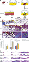
A skin-inspired 3D bilayer scaffold made of gelatin, AgNPs, and PDGF-BB had a faster wound healing, more collagen deposition, blood vessel formation, and reepithelialization in diabetic wounds. (A) Representation of the preparation process of silver and PDGF-BB coloaded bilayer 3D scaffolds. (B–D) Histological observations of skin wound healing treated with scaffold, silver-loaded scaffold, PDGF-BB-loaded scaffold, and silver and PDGF-BB coloaded scaffold. (B) Masson trichrome staining shows collagen deposition and new blood vessel formation of treated wounds. Arrows indicate newly formed blood vessels. (C) Quantification of new blood vessels. (D) Hematoxylin–eosin staining showing the reepithelialization of the treated wounds. Arrows indicate re-epithelization. Adapted from 109 with permission. *p < 0.05, **p < 0.01. AgNP, silver nanoparticle; PDGF, platelet-derived growth factor. 3D, three-dimensional. Color images are available online.
Figure 7.
Figure 7.
Nanofiber meshes made of Collagen I and PCL with different fiber organizations have different outputs in wound healing. Crossed nanofibers, which mimic the collagen pattern in skin, have a better wound healing rate and promote angiogenesis in diabetic rats. (A) Scanning Electron Microscopy images of the nanofibers with different organizations (random, aligned, and crossed). (B, C) Wound evolution in diabetic rats after treatment with the scaffolds. (D) Quantification of von Willebrand factor-positive vessels. Adapted from 111 with permission. * denotes statistical significance, p < 0.05 vs. control; # denotes statistical significance, p < 0.05 vs. random; & denotes statistical significance, p < 0.05 vs. aligned. PCL, polycaprolactone. Color images are available online.
Figure 8.
Figure 8.
Cationic liposomes made of an electrostatic complex between the plasmid of human recombinant PDGF-B, integrin receptor selective RGDK-lipopeptide, and cholesterol are able to improve wound healing in diabetic rats. The administration of RGDK-lipopeptide 1:rhPDGF-B on wound of diabetic animals shows a faster wound healing rate, reepithelialization and fibrocollagen, and keratin and vessel formation. (A) Relative wound healing and (B) wound images before the administration of the treatment (B [A–C]) and after 10 days of treatment (B [D–F]) with RGDK-lipopeptide1:rhPDGF-B complex (B [D]), RGDK-liposome (B [E]), and RGEK-lipopeptide2:rhPDGF-B complex (B [F]) in diabetic rats after the subcutaneous administration of a single dose. (C) Hematoxylin–eosin (C [A–D]) and Masson's trichrome (C [E–H]) tissue staining of wounds before the treatment (C [A, E]) and 10 days after the treatment with RGDK-lipopeptide 1:rhPDGF-B (C [B, F]); liposomes of RGDK-lipopeptide 1 (C [C, G]) and RGEK-lipopeptide 2:rhPDGF-B (C [D, H]). Adapted from 133 with permission. *p < 0.05, **p < 0.01. RGDK, Arg-Gly-Asp-Lys; rhPDGF, recombinant human platelet-derived growth factor. Color images are available online.

Similar articles

Cited by

References

    1. Han G, Ceilley R. Chronic wound healing: a review of current management and treatments. Adv Ther 2017;34:599–610 - PMC - PubMed
    1. Sen CK, Gordillo GM, Roy S, et al. . Human skin wounds: a major and snowballing threat to public health and the economy. Wound Repair Regen 2009;17:763–771 - PMC - PubMed
    1. Hurd T. Understanding the financial benefits of optimising wellbeing in patients living with a wound. Wounds Int 2013;4:13–17
    1. Martinengo LOM, Bajpai R, Soljak M, et al. . Prevalence of chronic wounds in the general population: systematic review and meta-analysis of observational studies. Ann Epidemiol 2019;29:8–15 - PubMed
    1. Malone M, Bjarnsholt T, McBain AJ, et al. . The prevalence of biofilms in chronic wounds: a systematic review and meta-analysis of published data. J Wound Care 2017;26:20–25 - PubMed

Publication types

Substances