. 2020 Apr 13;9(4):955.
doi: 10.3390/cells9040955.

The Catalytic-Dependent and -Independent Roles of Lsd1 and Lsd2 Lysine Demethylases in Heterochromatin Formation in Schizosaccharomyces pombe

Affiliations

The Catalytic-Dependent and -Independent Roles of Lsd1 and Lsd2 Lysine Demethylases in Heterochromatin Formation in Schizosaccharomyces pombe

Bahjat F Marayati et al. Cells. .

Abstract

In eukaryotes, heterochromatin plays a critical role in organismal development and cell fate acquisition, through regulating gene expression. The evolutionarily conserved lysine-specific demethylases, Lsd1 and Lsd2, remove mono- and dimethylation on histone H3, serving complex roles in gene expression. In the fission yeast Schizosaccharomyces pombe, null mutations of Lsd1 and Lsd2 result in either severe growth defects or inviability, while catalytic inactivation causes minimal defects, indicating that Lsd1 and Lsd2 have essential functions beyond their known demethylase activity. Here, we show that catalytic mutants of Lsd1 or Lsd2 partially assemble functional heterochromatin at centromeres in RNAi-deficient cells, while the C-terminal truncated alleles of Lsd1 or Lsd2 exacerbate heterochromatin formation at all major heterochromatic regions, suggesting that Lsd1 and Lsd2 repress heterochromatic transcripts through mechanisms both dependent on and independent of their catalytic activities. Lsd1 and Lsd2 are also involved in the establishment and maintenance of heterochromatin. At constitutive heterochromatic regions, Lsd1 and Lsd2 regulate one another and cooperate with other histone modifiers, including the class II HDAC Clr3 and the Sirtuin family protein Sir2 for gene silencing, but not with the class I HDAC Clr6. Our findings explore the roles of lysine-specific demethylases in epigenetic gene silencing at heterochromatic regions.

Keywords: H3K9me2; Lsd1; Lsd2; gene silencing; heterochromatin; lysine demethylase.


Conflict of interest statement

The authors declare no conflict of interest.

Figures
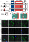
Figure 1
Figure 1
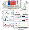
Nuclear localization of Lsd2 is required for viability. (A) Schematic representation of N-terminal mutants of Lsd2. Numbers indicate the position in the amino acid sequence of Lsd2 and Lsd1. (B) Lsd2 N-terminal mutants lsd2-N3 and lsd2-N5 are inviable. Heterozygous diploids containing one copy of the mutant allele (linked with resistance to the antibiotic G418) and one copy of the wild-type (WT) allele were sporulated and tetrads of spores were arranged in columns. Red box: sample of a tetrad that shows selective growth on rich media with or without G418. (C) Visualization of GFP-tagged full-length or N-terminal truncated Lsd2 proteins in viable heterozygous diploid strains by confocal fluorescence microscopy. Indicated strains were also stained with Hoechst to show nuclear localization.
Figure 2
Figure 2
The C-terminal domains of Lsd1 and Lsd2 are vital for proper growth and genome-wide gene expression. (A) Schematic representations of WT Lsd1 and Lsd2 compared to catalytically inactive and C-terminal truncated mutants. (B) Growth curves generated by a plate reader show the growth rates of indicated WT, lsd1, and lsd2 mutants over a 24 h period. (C) Comparison of DEGs between lsd1-ΔHMG and lsd2-ΔC using linear regression analysis. (D) Loss of silencing at peri-centromeric region, mating type locus, and sub-telomeric region in lsd1-ΔHMG and lsd2-ΔC compared to WT. Cen 2: Centromere II. Mat: Mating-type locus. Tel2R: right telomere II. Normalized RNA-seq reads are aligned with H3K9me2 enrichment as measured by ChIP, which indicate heterochromatic regions. Data are plotted along with chromosome position. (E) qRT-PCR analysis of the silenced dg/dh repeats in the peri-centromeric region and cenH (mat locus). * p ≤ 0.05 and ** p ≤ 0.01 as determined by student’s t test comparing the indicated samples with WT. Error bars represent s.e.m.
Figure 3
Figure 3
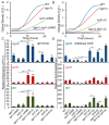
Lsd1 and Lsd2 serve overlapping, but divergent, functions. (A) Serial dilutions demonstrate the synthetic growth defects and heat sensitivities of lsd1 lsd2 double mutants. (B) qRT-PCR analysis of peri-centromeric (dg/dh) and mating-type (cenH) regions show the genetic interactions between lsd1-ao and lsd1-∆C and the double mutant allele lsd1-ao-∆C. (C) Summary of the genetic interactions between lsd1 and lsd2 mutants. (D) qRT-PCR analysis of the silenced dg/dh repeats in the peri-centromeric region and cenH (mating-type locus) show the genetic interactions between lsd2-ΔC and lsd1-ao. * p ≤ 0.05 and ** p ≤ 0.01 as determined by student’s t test comparing the indicated samples with WT values. Significance between single mutants and double mutants is indicated by horizontal lines linking the two samples. Error bars represent s.e.m.
Figure 4
Figure 4
The silencing defects of RNAi mutant ago1Δ are suppressed by Lsd1/2 amine oxidase mutants, but are exacerbated by Lsd1/2 C-terminal mutants. (A,B) Growth curves generated by a plate reader over a 24 h period show the genetic interactions between lsd1 and lsd2 C-terminal truncated strains combined with ago1Δ. (C-D) qRT-PCR (C) and qChIP (D) analyses of silent dg/dh repeats (centromeric), cenH (mating-type locus), and tlh1 (telomeric) regions demonstrate the genetic interactions between ago1Δ and lsd mutants. * p ≤ 0.05 and ** p ≤ 0.01 as determined by student’s t test comparing the indicated samples with WT for qPCR or qChIP and by horizontal lines linking the two samples. Error bars represent s.e.m.
Figure 5
Figure 5
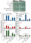
Lsd1 and Lsd2 mutants affect epigenetic maintenance and re-establishment of heterochromatin. (A) An ade6+ reporter gene replaced the K region (K∆::ade6+). Epigenetic mechanism(s) can repress ade6+ expression (K∆::ade6+ off, red colonies); loss of the epigenetic silencing causes expression of ade6+ (K∆::ade6+ on, white colonies). (B) The K∆::ade6+ off allele was introduced into cells with indicated genotypes via genetic crosses. Samples of color and shape of colonies formed by individual cells on low adenine medium are shown. Colonies were replica-plated from rich medium (YEA) to low adenine medium (YE). (C) Cells that formed red or white colonies from (B) with indicated genotypes were dissected and grown on rich medium without selection, then were transferred to low adenine medium. Samples of red (R), white (W), and color-mixed (M) colonies on YE medium are shown on the right. Bar graph to the left shows the percentage of colonies that are red, white, or color-mixed. The percentages are listed in the table for each of the indicated genotypes. (D,E) ade6+ expression in red or white cells collected in (C) were investigated by qRT-PCR (D), and relative H3K9me2 enrichment at repeat regions (versus input) (E) were compared in red or white colonies with indicated genotypes. * p ≤ 0.05 and ** p ≤ 0.01 as determined by student’s t test comparing the indicated samples with WT R for qPCR or qChIP. Error bars represent s.e.m.
Figure 6
Figure 6
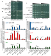
Epe1 antagonizes Lsd1 and Lsd2 functions. (A) Serial dilutions show weak synthetic growth defects when epe1Δ is combined with lsd1-ΔHMG or lsd2-ΔC at 37 °C. (B) qRT-PCR analysis demonstrates the suppressive effects of epe1Δ on lsd mutant silencing defects at all heterochromatic regions. (C) The alleviation of the silencing defects by epe1Δ on lsd mutants are correlated with the alterations of H3K9me2 detected by ChIP. * p ≤ 0.05 and ** p ≤ 0.01 as determined by student’s t test comparing the indicated samples with WT for qPCR or qChIP and by horizontal lines linking the two samples. Error bars represent s.e.m.
Figure 7
Figure 7
Lsd1 and Lsd2 mediate overlapping functions with Clr3 and Sir2, but not with Clr6, at heterochromatic regions. (AC) Serial dilutions show genetic interactions with respect to growth and heat sensitivity between lsd C-terminal mutants and mutants of histone deacetylases clr6-1 (A), clr3Δ (B), and sir2Δ (C). (D) qRT-PCR analysis shows cumulative silencing defects of Lsd mutants combined with HDAC mutants at the centromeric (dg/dh), mating-type locus (cenH), and telomeric (tlh1) heterochromatin regions. (E) H3K9me2 ChIP analysis demonstrates the alterations of heterochromatin in WT, single, and double mutant cells with the indicated genotypes. * p ≤ 0.05 and ** p ≤ 0.01 as determined by student’s t test comparing the indicated samples with WT for qPCR or qChIP and by horizontal lines linking the two samples. Error bars represent s.e.m.

Similar articles

Cited by

References

    1. Luger K., Mader A.W., Richmond R.K., Sargent D.F., Richmond T.J. Crystal structure of the nucleosome core particle at 2.8 A resolution. Nature. 1997;389:251–260. doi: 10.1038/38444. - DOI - PubMed
    1. Kouzarides T. SnapShot: Histone-modifying enzymes. Cell. 2007;128:802-e1. doi: 10.1016/j.cell.2007.02.018. - DOI - PubMed
    1. Campos E.I., Reinberg D. Histones: Annotating chromatin. Annu. Rev. Genet. 2009;43:559–599. doi: 10.1146/annurev.genet.032608.103928. - DOI - PubMed
    1. Peterson C.L., Laniel M.A. Histones and histone modifications. Curr. Biol. 2004;14:R546–R551. doi: 10.1016/j.cub.2004.07.007. - DOI - PubMed
    1. Shogren-Knaak M., Peterson C.L. Switching on chromatin: Mechanistic role of histone H4-K16 acetylation. Cell Cycle. 2006;5:1361–1365. doi: 10.4161/cc.5.13.2891. - DOI - PubMed

Publication types

MeSH terms