. 2020 Aug;27(8):2433-2450.
doi: 10.1038/s41418-020-0514-3. Epub 2020 Mar 6.

The lipogenic LXR-SREBF1 signaling pathway controls cancer cell DNA repair and apoptosis and is a vulnerable point of malignant tumors for cancer therapy

Affiliations

The lipogenic LXR-SREBF1 signaling pathway controls cancer cell DNA repair and apoptosis and is a vulnerable point of malignant tumors for cancer therapy

Bo Yang et al. Cell Death Differ. 2020 Aug.

Erratum in

Abstract

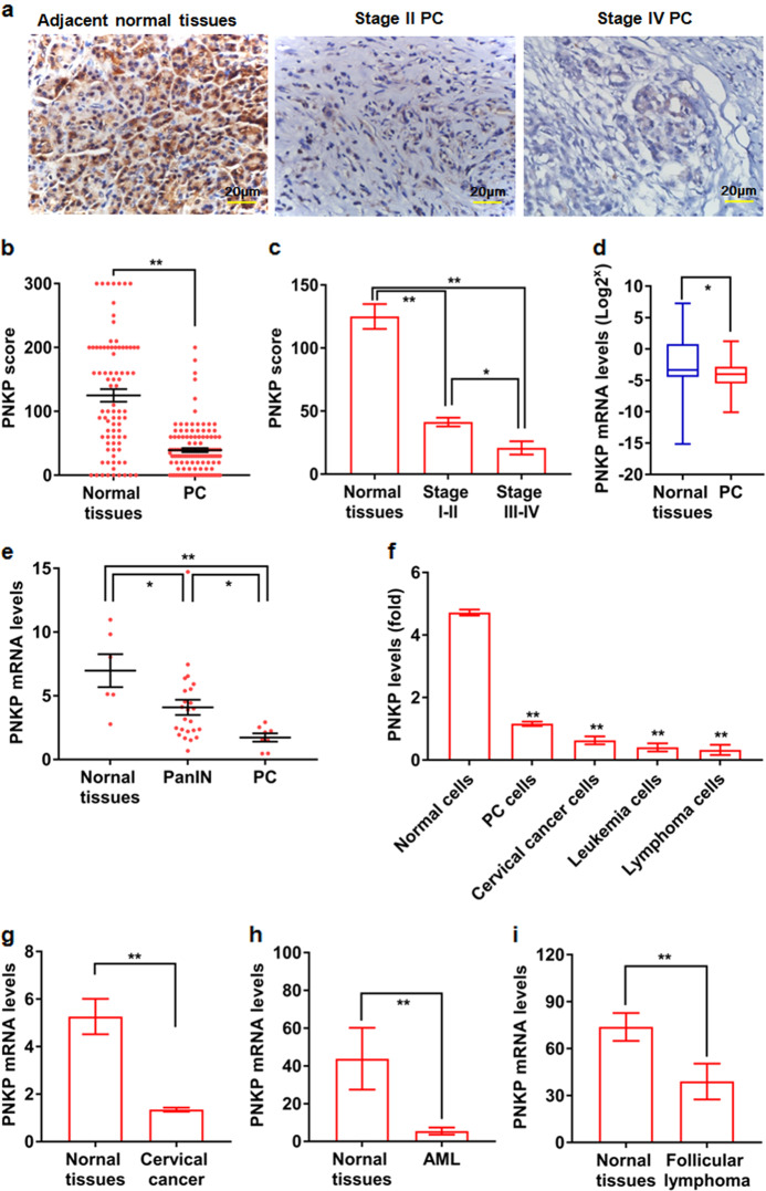
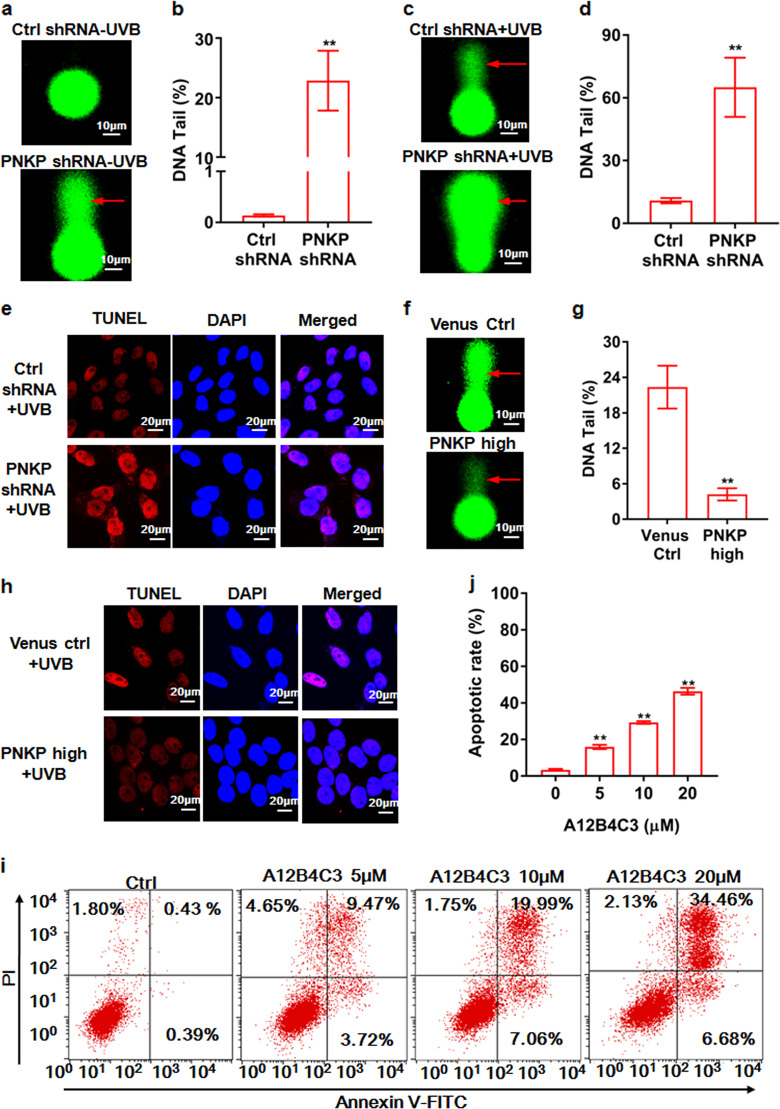
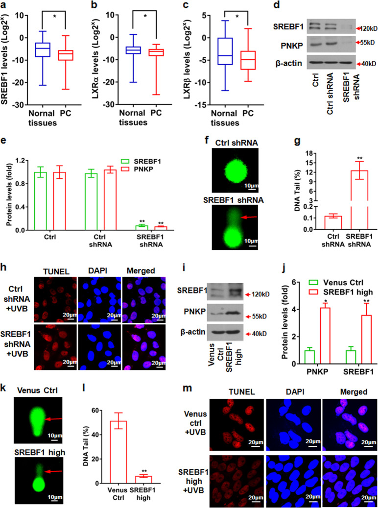
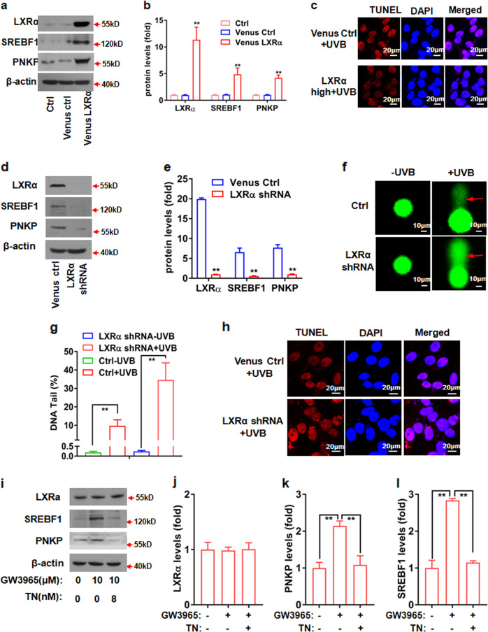
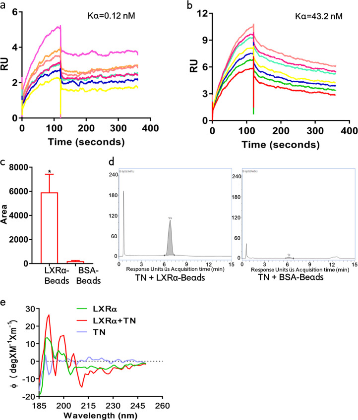
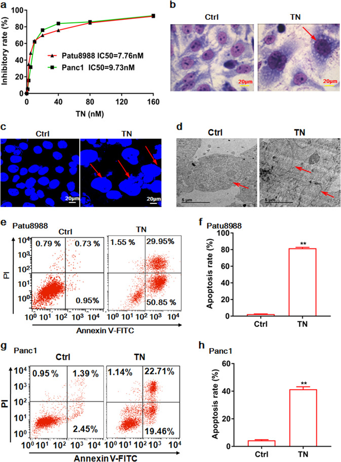
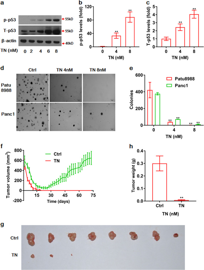
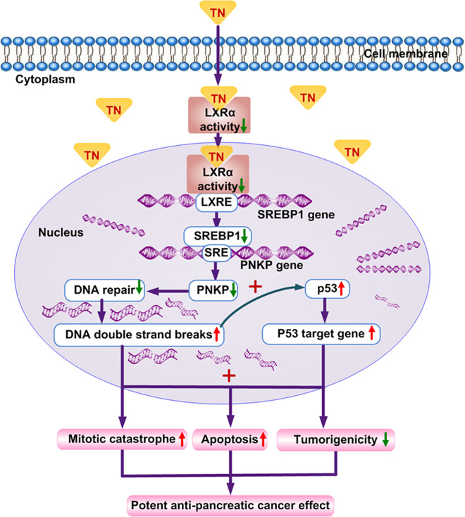
Cancer cells are defective in DNA repair, so they experience increased DNA strand breaks, genome instability, gene mutagenesis, and tumorigenicity; however, multiple classic DNA repair genes and pathways are strongly activated in malignant tumor cells to compensate for the DNA repair deficiency and gain an apoptosis resistance. The mechanisms underlying this phenomenon in cancer are unclear. We speculate that a key DNA repair gene or signaling pathway in cancer has not yet been recognized. Here, we show that the lipogenic liver X receptor (LXR)-sterol response element binding factor-1 (SREBF1) axis controls the transcription of a key DNA repair gene polynucleotide kinase/phosphatase (PNKP), thereby governing cancer cell DNA repair and apoptosis. Notably, the PNKP levels were significantly reduced in 95% of human pancreatic cancer (PC) patients, particularly deep reduction for sixfold in all of the advanced-stage PC cases. PNKP is also deficient in three other types of cancer that we examined. In addition, the expression of LXRs and SREBF1 was significantly reduced in the tumor tissues from human PC patients compared with the adjacent normal tissues. The newly identified LXR-SREBF1-PNKP signaling pathway is deficient in PC, and the defect in the pathway contributes to the DNA repair deficiency in the cancer. Strikingly, further diminution of the vulnerable LXR-SREBF1-PNKP signaling pathway using a small molecule triptonide, a new LXR antagonist identified in this investigation, at a concentration of 8 nM robustly activated tumor-suppressor p53 and readily elevated cancer cell DNA strand breaks over an apoptotic threshold, and selectively induced PC cell apoptosis, resulting in almost complete elimination of tumors in xenograft mice without obvious complications. Our findings provide new insight into DNA repair and apoptosis in cancer, and offer a new platform for developing novel anticancer therapeutics.


Conflict of interest statement

The authors declare that they have no conflict of interest.

Figures

Fig. 1
Fig. 1. PNKP is deficient in tumors from the patients suffered from pancreatic cancer and other three types of malignant tumors.
Polynucleotide kinase/phosphatase (PNKP) protein in tumor tissues and matched adjacent normal tissues from 124 human pancreatic ductal adenocarcinoma (PDA) patients at the early (I–II) and advanced (III-IV) stages was detected with immunohistochemical (IHC) staining a and analyzed b, c. Results indicated that PNKP is deficient in pancreatic cancer (PC) tumor tissues in 95% of PC cases b, with a particularly severe deficiency in all of the advanced-stage PC patients (III and IV) c. In addition, PNKP mRNA levels in the tumor tissues and adjacent normal tissues from 22 cases of pancreatic cancer patients d and in the samples from four immortalized normal cell lines, six PC cell lines, three cervical cancer cell lines, three leukemia cell lines, and three lymphoma cell lines f were measured by qPCR. Data are shown in 1f as means ± S.E of three independent replicates. In addition, the data on PNKP mRNA levels in the patients suffering from pancreatic intraepithelial neoplasia (PanIN) and pancreatic cancer (PC) e, cervical cancer g, acute myeloid leukemia (AML, h), and follicular lymphoma i, were detected by DNA microarray, were retrieved from the publicly available cancer database Oncomine (https://www.oncomine.org), and statistically analyzed. The data are shown as mean ± S.E., the p value was determined by Student’s t test. Scale bar, 200 μm.
Fig. 2
Fig. 2. PNKP controls the levels of intracellular DNA double-strand breaks and apoptosis as well as tumorigenicity of pancreatic cancer cells.
Silencing of PNKP by shRNA along (see Fig. S1c, d) markedly increased DNA single- and double-strand breaks (DSBs) in pancreatic cancer (PC) cell line Patu8988 measured by a comet assay a, b and further significantly increased DSBs upon radiation of the PC cells with a very low dose (1 mJ/cm2) ultraviolet beam (UVB) c, d as indicated by the red arrow a and c, the apoptotic cells (red) were detected by TUNEL assay e. In contrast, overexpression of PNKP (PNKP high, see Fig. S1e, f) diminished DSBs upon radiation with 2 mJ/cm2 UVB f, g and reduced apoptotic cells (red) as measured by TUNEL assay h. Inhibition of PNKP activity using its specific inhibitor A12B4C3-induced PC cell apoptosis as measured by PI-Annexin V double staining i, j. Data are shown as means ± S.D. of three independent replicates. Scale bar, 20 μm.
Fig. 3
Fig. 3. SREBF1 regulates PNKP gene expression and governs pancreatic cancer cell DNA repair and apoptosis.
The mRNA levels of SREBF1 a, LXRα b, and LXRβ c in the tumor tissues and adjacent normal tissues from 22 cases of pancreatic cancer patients were measured by qPCR. Silencing of the transcription factor SREBF1 expression by shRNA significantly diminished PNKP expression levels d, e, increased DNA single- and double-strand breaks (DSBs) in pancreatic cancer (PC) cells (red arrows in f, g) and apoptotic cells (red) as detected by TUNEL assay h. By contrast, overexpression of SREBF1 significantly increased expression of the PNKP gene i, j, decreased intracellular DSBs k, l and apoptotic cells m. Data are shown as mean ± S.D. based on three independent replicates. Scale bar, 20 μm.
Fig. 4
Fig. 4. The LXRα controls pancreatic cancer cell DNA repair and apoptosis by the regulation of downstream SREBF1 and PNKP expression.
Overexpression of LXRα (LXRα high) significantly increased expression of the SREBF1 and PNKP in pancreatic cancer cells a, b, but reduced apoptotic cells (red) as detected by TUNEL assay c; In contrast, silencing LXRα (LXRα shRNA) resulted in reduction of SREBF1 and PNKP expression levels d, e, but significantly increased the levels of DNA single- and double-strand breaks (DSBs, red arrows, f, g) and apoptotic cells h. Activation of LXRα and/or LXRβ by its specific agonist GW3965 promoted SREBF1 and PNKP expression in pancreatic cancer cells (i middle lane, k, l). Strikingly, GW3965-induced expression of SREBF1 and PNKP was almost completely abolished by triptonide (TN) in pancreatic cancer Patu8988 cells (i right lane, k, l), whereas the levels of LXRα were not significantly affected (i right lane, j), similar results were observed at mRNA levels in pancreatic cancer Panc1 cells (Figs. S3, S4). Scale bar, 20 μm. The complete western blotting gels in Fig. 4 are presented in Fig. S10.
Fig. 5
Fig. 5. Triptonide directly binds to its receptor LXRα with extreme high affinity.
The direct binding between triptonide (TN) and LXRα was ascertained by the surface plasmon resonance (SPR) assay and displayed extremely high affinity constants (Kd) of 0.12 nM a and 43.2 nM b, for the fast and slow binding phases, respectively. The binding of triptonide to LXRα was further confirmed by pull-down assay, followed by high-performance liquid chromatography (HPLC) and mass spectrometry c, d, and circular dichroism (CD) analysis e. Apparently, much more TN bound to the LXRα-beads than the control BSA-beads c, and HPLC showed a high pick in the group of TN + LXRα-beads d, and CD analysis indicated three distinct high peaks and low peak after incubation of LXRα with TN e.
Fig. 6
Fig. 6. Triptonide promotes tumor cell mitotic catastrophe and apoptosis.
Triptonide (TN) strongly inhibited pancreatic cancer cell growth with an extremely low IC50 of 7.76 nM for pancreatic cancer cell line Patu8988, and 9.73 nM for Panc1 a, respectively, and caused enlargement of the cell nuclei (red arrows) as shown by Giemsa–Wright staining (b, scale bar, 50 µm), DAPI staining (c, scale bar, 20 µm), and electronic microscope imaging (d, scale bar, 5 µm). After treatment of pancreatic cancer Patu8988 (e, f) and Panc1 (g, h) cell lines with 8 nM TN for 9 days, the cells succumbed to apoptosis (red) as detected by and Annexin V double staining and flow cytometry. Data are shown as mean ± S.E. of three independent replicates.
Fig. 7
Fig. 7. Triptonide strongly activates p53 and markedly suppresses pancreatic cancer cell tumorigenicity and tumor growth in vivo and significantly prolongs the survival of tumor-bearing xenograft mice.
Triptonide (TN) at 8 nM markedly increased the protein levels of phosphorylated tumor-suppressor p53 (p-p53) a, b and also significantly promoted expression of total p53 protein (T-p53) in pancreatic cancer (PC) cells a, c. PC cell colony-forming capability was inhibited by treatment with 8 nM TN (d, quantitation in e). The anti-PC efficacy of TN was evaluated in a mouse model. Of note, human pancreatic cancer Patu8988 cells and Matrigel were first incubated at 4 °C for 10 min and mixed, and then subcutaneously injected 300 μl of the mixture to the back each mouse. The cold Matrigel is a liquid, but it becomes solid at 37 °C in mice. The tumor volume was monitored using digital calipers every other day f, the initial tumor volume reflects the mixture of injected Matrigel and tumor cells. The Matrigel was gradually degraded in the xenograft mice within 2 weeks after injection, thereby gradually decreasing the tumor volume during the initial 2 weeks f. The tumors in five of eight xenograft mice were completely eliminated after TN treatment for 73 days at 5 mg/kg/d, and tumors in the remaining three mice were markedly reduced (g, quantitation in h). Data in 7a–e are shown as means ± S.E. of three independent replicates.
Fig. 8
Fig. 8. Schematic diagram showing that the LXRs-SREBF1-PNKP axis is essential for DNA repair in cancer cells and is a vulnerable point of pancreatic cancer for effective cancer therapy.
Hydrophobic triptonide (TN) passes through the cell membrane and directly binds to its receptor liver X receptors (LXRα and/or LXRβ), inhibits the transcriptional activity of LXRs and reduces the expression of LXR downstream transcription factor SREBF1 gene, in turn diminishing SREBF1-downstream PNKP gene expression. The LXRs-SREBF1-PNKP signaling axis controls the levels of intracellular DNA single- and double-strand breaks (DSBs) and DNA repair in pancreatic cancer (PC) cells. Reduction of the LXRs-SREBF1-PNKP axis by triptonide causes a severe defect in DNA repair, marked elevation of intracellular DSBs, activation of p53, and induction of PC cell mitotic catastrophe and apoptosis, resulting in marked reduction of PC cell tumorigenicity and potent anti-PC effects without obvious complications in murine studies.

Similar articles

Cited by

References

    1. Khanna A. DNA damage in cancer therapeutics: a boon or a curse. Cancer Res. 2015;75:2133–38. - PubMed
    1. Basu AK. DNA damage, mutagenesis and cancer. Int J Mol Sci. 2018;19:pii: E970.. - PMC - PubMed
    1. Desai A, Yan Y, Gerson SL. Advances in therapeutic targeting of the DNA damage response in cancer. DNA Repair (Amst) 2018;66-67:24–9. - PMC - PubMed
    1. Toma M, Skorski T, Sliwinski T. DNA double strand break repair - related synthetic lethality. Curr Med Chem. 2019;26:1446–82. - PubMed
    1. Wright SM, Woo YH, Alley TL, Shirley BJ, Akeson EC, Snow KJ, et al. Complex oncogenic translocations with gene amplification are initiated by specific DNA breaks in lymphocytes. Cancer Res. 2009;69:4454–60. - PMC - PubMed

Publication types

MeSH terms