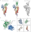
Cryo-EM structure of the 2019-nCoV spike in the prefusion conformation
- PMID: 32075877
- PMCID: PMC7164637
- DOI: 10.1126/science.abb2507
Cryo-EM structure of the 2019-nCoV spike in the prefusion conformation
Abstract
The outbreak of a novel coronavirus (2019-nCoV) represents a pandemic threat that has been declared a public health emergency of international concern. The CoV spike (S) glycoprotein is a key target for vaccines, therapeutic antibodies, and diagnostics. To facilitate medical countermeasure development, we determined a 3.5-angstrom-resolution cryo-electron microscopy structure of the 2019-nCoV S trimer in the prefusion conformation. The predominant state of the trimer has one of the three receptor-binding domains (RBDs) rotated up in a receptor-accessible conformation. We also provide biophysical and structural evidence that the 2019-nCoV S protein binds angiotensin-converting enzyme 2 (ACE2) with higher affinity than does severe acute respiratory syndrome (SARS)-CoV S. Additionally, we tested several published SARS-CoV RBD-specific monoclonal antibodies and found that they do not have appreciable binding to 2019-nCoV S, suggesting that antibody cross-reactivity may be limited between the two RBDs. The structure of 2019-nCoV S should enable the rapid development and evaluation of medical countermeasures to address the ongoing public health crisis.
Copyright © 2020 The Authors, some rights reserved; exclusive licensee American Association for the Advancement of Science. No claim to original U.S. Government Works.
Figures


Update of
-
Cryo-EM Structure of the 2019-nCoV Spike in the Prefusion Conformation.bioRxiv [Preprint]. 2020 Feb 15:2020.02.11.944462. doi: 10.1101/2020.02.11.944462. bioRxiv. 2020. Update in: Science. 2020 Mar 13;367(6483):1260-1263. doi: 10.1126/science.abb2507. PMID: 32511295 Free PMC article. Updated. Preprint.
Similar articles
-
Cryo-EM Structure of the 2019-nCoV Spike in the Prefusion Conformation.bioRxiv [Preprint]. 2020 Feb 15:2020.02.11.944462. doi: 10.1101/2020.02.11.944462. bioRxiv. 2020. Update in: Science. 2020 Mar 13;367(6483):1260-1263. doi: 10.1126/science.abb2507. PMID: 32511295 Free PMC article. Updated. Preprint.
-
On the interactions of the receptor-binding domain of SARS-CoV-1 and SARS-CoV-2 spike proteins with monoclonal antibodies and the receptor ACE2.Virus Res. 2020 Aug;285:198021. doi: 10.1016/j.virusres.2020.198021. Epub 2020 May 15. Virus Res. 2020. PMID: 32416259 Free PMC article.
-
A highly conserved cryptic epitope in the receptor binding domains of SARS-CoV-2 and SARS-CoV.Science. 2020 May 8;368(6491):630-633. doi: 10.1126/science.abb7269. Epub 2020 Apr 3. Science. 2020. PMID: 32245784 Free PMC article.
-
Progress in Studies on Structural and Remedial Aspects of Newly Born Coronavirus, SARS-CoV-2.Curr Top Med Chem. 2020;20(26):2362-2378. doi: 10.2174/1568026620666200922112300. Curr Top Med Chem. 2020. PMID: 32962613 Review.
-
ACE2, the Receptor that Enables Infection by SARS-CoV-2: Biochemistry, Structure, Allostery and Evaluation of the Potential Development of ACE2 Modulators.ChemMedChem. 2020 Sep 16;15(18):1682-1690. doi: 10.1002/cmdc.202000368. Epub 2020 Aug 11. ChemMedChem. 2020. PMID: 32663362 Free PMC article. Review.
Cited by
-
Engineered protein subunit COVID19 vaccine is as immunogenic as nanoparticles in mouse and hamster models.Sci Rep. 2024 Oct 26;14(1):25528. doi: 10.1038/s41598-024-76377-y. Sci Rep. 2024. PMID: 39462119 Free PMC article.
-
Evaluation of severe acute respiratory syndrome coronavirus 2 (SARS-CoV-2) using a high figure-of-merit plasmonic multimode refractive index optical sensor.Sci Rep. 2024 Oct 26;14(1):25499. doi: 10.1038/s41598-024-77336-3. Sci Rep. 2024. PMID: 39462024 Free PMC article.
-
Large-Scale Transient Transfection of Suspension-Adapted Chinese Hamster Ovary Cells for the Production of the Trimeric SARS-CoV-2 Spike Protein.Methods Mol Biol. 2025;2853:7-16. doi: 10.1007/978-1-0716-4104-0_2. Methods Mol Biol. 2025. PMID: 39460911
-
Navigating the Landscape of B Cell Mediated Immunity and Antibody Monitoring in SARS-CoV-2 Vaccine Efficacy: Tools, Strategies and Clinical Trial Insights.Vaccines (Basel). 2024 Sep 24;12(10):1089. doi: 10.3390/vaccines12101089. Vaccines (Basel). 2024. PMID: 39460256 Free PMC article. Review.
-
The Role of Macrophages in Airway Disease Focusing on Porcine Reproductive and Respiratory Syndrome Virus and the Treatment with Antioxidant Nanoparticles.Viruses. 2024 Oct 1;16(10):1563. doi: 10.3390/v16101563. Viruses. 2024. PMID: 39459897 Free PMC article. Review.
References
-
- Chan J. F., Yuan S., Kok K.-H., To K. K.-W., Chu H., Yang J., Xing F., Liu J., Yip C. C.-Y., Poon R. W.-S., Tsoi H.-W., Lo S. K.-F., Chan K.-H., Poon V. K.-M., Chan W.-M., Ip J. D., Cai J.-P., Cheng V. C.-C., Chen H., Hui C. K.-M., Yuen K.-Y., A familial cluster of pneumonia associated with the 2019 novel coronavirus indicating person-to-person transmission: A study of a family cluster. Lancet 395, 514–523 (2020). 10.1016/S0140-6736(20)30154-9 - DOI - PMC - PubMed
-
- Huang C., Wang Y., Li X., Ren L., Zhao J., Hu Y., Zhang L., Fan G., Xu J., Gu X., Cheng Z., Yu T., Xia J., Wei Y., Wu W., Xie X., Yin W., Li H., Liu M., Xiao Y., Gao H., Guo L., Xie J., Wang G., Jiang R., Gao Z., Jin Q., Wang J., Cao B., Clinical features of patients infected with 2019 novel coronavirus in Wuhan, China. Lancet 395, 497–506 (2020). 10.1016/S0140-6736(20)30183-5 - DOI - PMC - PubMed
-
- Lu R., Zhao X., Li J., Niu P., Yang B., Wu H., Wang W., Song H., Huang B., Zhu N., Bi Y., Ma X., Zhan F., Wang L., Hu T., Zhou H., Hu Z., Zhou W., Zhao L., Chen J., Meng Y., Wang J., Lin Y., Yuan J., Xie Z., Ma J., Liu W. J., Wang D., Xu W., Holmes E. C., Gao G. F., Wu G., Chen W., Shi W., Tan W., Genomic characterisation and epidemiology of 2019 novel coronavirus: Implications for virus origins and receptor binding. Lancet S0140-6736(20)30251-8 (2020). 10.1016/S0140-6736(20)30251-8 - DOI - PMC - PubMed
-
- Wu F., Zhao S., Yu B., Chen Y.-M., Wang W., Song Z.-G., Hu Y., Tao Z.-W., Tian J.-H., Pei Y.-Y., Yuan M.-L., Zhang Y.-L., Dai F.-H., Liu Y., Wang Q.-M., Zheng J.-J., Xu L., Holmes E. C., Zhang Y.-Z., A new coronavirus associated with human respiratory disease in China. Nature (2020). 10.1038/s41586-020-2008-3 - DOI - PMC - PubMed
-
- Chen N., Zhou M., Dong X., Qu J., Gong F., Han Y., Qiu Y., Wang J., Liu Y., Wei Y., Xia J., Yu T., Zhang X., Zhang L., Epidemiological and clinical characteristics of 99 cases of 2019 novel coronavirus pneumonia in Wuhan, China: A descriptive study. Lancet 395, 507–513 (2020). 10.1016/S0140-6736(20)30211-7 - DOI - PMC - PubMed
Publication types
MeSH terms
Substances
Grants and funding
LinkOut - more resources
Full Text Sources
Other Literature Sources
Molecular Biology Databases
Miscellaneous

