

Luteinizing Hormone Action in Human Oocyte Maturation and Quality: Signaling Pathways, Regulation, and Clinical Impact
- PMID: 32046451
- PMCID: PMC7190682
- DOI: 10.1007/s43032-019-00137-x
Luteinizing Hormone Action in Human Oocyte Maturation and Quality: Signaling Pathways, Regulation, and Clinical Impact
Abstract
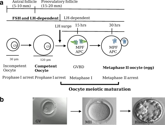
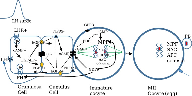
The ovarian follicle luteinizing hormone (LH) signaling molecules that regulate oocyte meiotic maturation have recently been identified. The LH signal reduces preovulatory follicle cyclic nucleotide levels which releases oocytes from the first meiotic arrest. In the ovarian follicle, the LH signal reduces cyclic nucleotide levels via the CNP/NPR2 system, the EGF/EGF receptor network, and follicle/oocyte gap junctions. In the oocyte, reduced cyclic nucleotide levels activate the maturation promoting factor (MPF). The activated MPF induces chromosome segregation and completion of the first and second meiotic divisions. The purpose of this paper is to present an overview of the current understanding of human LH signaling regulation of oocyte meiotic maturation by identifying and integrating the human studies on this topic. We found 89 human studies in the literature that identified 24 LH follicle/oocyte signaling proteins. These studies show that human oocyte meiotic maturation is regulated by the same proteins that regulate animal oocyte meiotic maturation. We also found that these LH signaling pathway molecules regulate human oocyte quality and subsequent embryo quality. Remarkably, in vitro maturation (IVM) prematuration culture (PMC) protocols that manipulate the LH signaling pathway improve human oocyte quality of cultured human oocytes. This knowledge has improved clinical human IVM efficiency which may become a routine alternative ART for some infertile patients.
Keywords: LH follicle signaling; Oocyte meiotic maturation; Oocyte quality.
Conflict of interest statement
The authors declare that they have no conflict of interest.
Figures
Similar articles
-
Multiple pathways mediate luteinizing hormone regulation of cGMP signaling in the mouse ovarian follicle.Biol Reprod. 2014 Jul;91(1):9. doi: 10.1095/biolreprod.113.116814. Epub 2014 Apr 16. Biol Reprod. 2014. PMID: 24740605 Free PMC article.
-
An improved IVM method for cumulus-oocyte complexes from small follicles in polycystic ovary syndrome patients enhances oocyte competence and embryo yield.Hum Reprod. 2017 Oct 1;32(10):2056-2068. doi: 10.1093/humrep/dex262. Hum Reprod. 2017. PMID: 28938744
-
The epidermal growth factor network: role in oocyte growth, maturation and developmental competence.Hum Reprod Update. 2018 Jan 1;24(1):1-14. doi: 10.1093/humupd/dmx029. Hum Reprod Update. 2018. PMID: 29029246 Review.
-
A pre-in vitro maturation medium containing cumulus oocyte complex ligand-receptor signaling molecules maintains meiotic arrest, supports the cumulus oocyte complex and improves oocyte developmental competence.Mol Hum Reprod. 2017 Sep 1;23(9):594-606. doi: 10.1093/molehr/gax032. Mol Hum Reprod. 2017. PMID: 28586460
-
Stops and starts in mammalian oocytes: recent advances in understanding the regulation of meiotic arrest and oocyte maturation.Reproduction. 2005 Dec;130(6):791-9. doi: 10.1530/rep.1.00793. Reproduction. 2005. PMID: 16322539 Review.
Cited by
-
Melatonin Alleviates the Suppressive Effect of Hypoxanthine on Oocyte Nuclear Maturation and Restores Meiosis via the Melatonin Receptor 1 (MT1)-Mediated Pathway.Front Cell Dev Biol. 2021 Apr 15;9:648148. doi: 10.3389/fcell.2021.648148. eCollection 2021. Front Cell Dev Biol. 2021. PMID: 33937242 Free PMC article.
-
The role of amphiregulin in ovarian function and disease.Cell Mol Life Sci. 2023 Feb 7;80(3):60. doi: 10.1007/s00018-023-04709-8. Cell Mol Life Sci. 2023. PMID: 36749397 Free PMC article. Review.
-
Molecular determinants of the meiotic arrests in mammalian oocytes at different stages of maturation.Cell Cycle. 2022 Mar-Mar;21(6):547-571. doi: 10.1080/15384101.2022.2026704. Epub 2022 Jan 24. Cell Cycle. 2022. PMID: 35072590 Free PMC article. Review.
-
Cellular Mechanisms and Regulation of Quiescence.Dev Cell. 2020 Nov 9;55(3):259-271. doi: 10.1016/j.devcel.2020.09.029. Dev Cell. 2020. PMID: 33171109 Free PMC article. Review.
-
HDAC1 in the Ovarian Granulosa Cells of Tan Sheep Improves Cumulus Cell Expansion and Oocyte Maturation Independently of the EGF-like Growth Factors.Biology (Basel). 2022 Oct 6;11(10):1464. doi: 10.3390/biology11101464. Biology (Basel). 2022. PMID: 36290368 Free PMC article.
References
-
- Greenwald GaR SK. Follicular development and its control. In: Knobil E, editor. The physiology of reproduction. 2. New York: Raven Press; 1994. pp. 629–724.
-
- Pincus G. The eggs of mammals. Experimental Biology Monographs. New York, NY; The Macmillian Company; 1936.
-
- Sanchez F, Smitz J. Molecular control of oogenesis. Biochim Biophys Acta. 1822;2012:1896–1912. - PubMed
-
- Coticchio G, Dal Canto M, Mignini Renzini M, Guglielmo MC, Brambillasca F, Turchi D, Novara PV, Fadini R. Oocyte maturation: gamete-somatic cells interactions, meiotic resumption, cytoskeletal dynamics and cytoplasmic reorganization. Hum Reprod Update. 2015;21:427–454. doi: 10.1093/humupd/dmv011. - DOI - PubMed
Publication types
MeSH terms
Substances
LinkOut - more resources
Full Text Sources
Other Literature Sources
Miscellaneous
