CT Imaging Features of 2019 Novel Coronavirus (2019-nCoV)
- PMID: 32017661
- PMCID: PMC7194022
- DOI: 10.1148/radiol.2020200230
CT Imaging Features of 2019 Novel Coronavirus (2019-nCoV)
Abstract
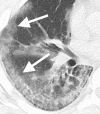
In this retrospective case series, chest CT scans of 21 symptomatic patients from China infected with the 2019 novel coronavirus (2019-nCoV) were reviewed, with emphasis on identifying and characterizing the most common findings. Typical CT findings included bilateral pulmonary parenchymal ground-glass and consolidative pulmonary opacities, sometimes with a rounded morphology and a peripheral lung distribution. Notably, lung cavitation, discrete pulmonary nodules, pleural effusions, and lymphadenopathy were absent. Follow-up imaging in a subset of patients during the study time window often demonstrated mild or moderate progression of disease, as manifested by increasing extent and density of lung opacities.
© RSNA, 2020.
Figures


Similar articles
-
Chest CT Findings in 2019 Novel Coronavirus (2019-nCoV) Infections from Wuhan, China: Key Points for the Radiologist.Radiology. 2020 Apr;295(1):16-17. doi: 10.1148/radiol.2020200241. Epub 2020 Feb 4. Radiology. 2020. PMID: 32017662 Free PMC article. No abstract available.
-
Imaging and clinical features of patients with 2019 novel coronavirus SARS-CoV-2.Eur J Nucl Med Mol Imaging. 2020 May;47(5):1275-1280. doi: 10.1007/s00259-020-04735-9. Epub 2020 Feb 28. Eur J Nucl Med Mol Imaging. 2020. PMID: 32107577 Free PMC article.
-
CT Manifestations of Two Cases of 2019 Novel Coronavirus (2019-nCoV) Pneumonia.Radiology. 2020 Apr;295(1):208-209. doi: 10.1148/radiol.2020200280. Epub 2020 Feb 7. Radiology. 2020. PMID: 32031481 Free PMC article. No abstract available.
-
[Diagnostic imaging findings in COVID-19].Ugeskr Laeger. 2020 Apr 6;182(15):V03200191. Ugeskr Laeger. 2020. PMID: 32286216 Review. Danish.
-
Diagnostic value and key features of computed tomography in Coronavirus Disease 2019.Emerg Microbes Infect. 2020 Dec;9(1):787-793. doi: 10.1080/22221751.2020.1750307. Emerg Microbes Infect. 2020. PMID: 32241244 Free PMC article. Review.
Cited by
-
Advanced federated ensemble internet of learning approach for cloud based medical healthcare monitoring system.Sci Rep. 2024 Oct 30;14(1):26068. doi: 10.1038/s41598-024-77196-x. Sci Rep. 2024. PMID: 39478132
-
Lung field-based severity score (LFSS): a feasible tool to identify COVID-19 patients at high risk of progressing to critical disease.J Thorac Dis. 2024 Sep 30;16(9):5591-5603. doi: 10.21037/jtd-24-544. Epub 2024 Sep 6. J Thorac Dis. 2024. PMID: 39444869 Free PMC article.
-
Role of interleukin 6 polymorphism and inflammatory markers in outcome of pediatric Covid- 19 patients.BMC Pediatr. 2024 Oct 1;24(1):625. doi: 10.1186/s12887-024-05071-9. BMC Pediatr. 2024. PMID: 39354444 Free PMC article.
-
Development and validation of a deep learning-based framework for automated lung CT segmentation and acute respiratory distress syndrome prediction: a multicenter cohort study.EClinicalMedicine. 2024 Jul 26;75:102772. doi: 10.1016/j.eclinm.2024.102772. eCollection 2024 Sep. EClinicalMedicine. 2024. PMID: 39170939 Free PMC article.
-
The top 50 most-cited articles about COVID-19 and the complications of COVID-19: A bibliometric analysis.F1000Res. 2024 May 3;13:105. doi: 10.12688/f1000research.145713.3. eCollection 2024. F1000Res. 2024. PMID: 39149509 Free PMC article.
References
-
- Novel Coronavirus – China. World Health Organization. https://www.who.int/csr/don/12-january-2020-novel-coronavirus-china/en/. Published January 12, 2020.
-
- Novel Coronavirus (2019-nCoV). World Health Organization. https://www.who.int/emergencies/diseases/novel-Coronavirus-2019. Published January 7, 2020.
-
- Summary of probable SARS cases with onset of illness from 1 November 2002 to 31 July 2003. World Health Organization. https://www.who.int/csr/sars/country/table2004_04_21/en/. Published April 21, 2004.
-
- Middle East respiratory syndrome Coronavirus (MERS-CoV). World Health Organization. https://www.who.int/emergencies/mers-cov/en/.
-
- Situation report - 8. World Health Organization. https://www.who.int/docs/default-source/Coronaviruse/situation-reports/2.... Published January 28, 2020.
MeSH terms
LinkOut - more resources
Full Text Sources
Other Literature Sources
Medical

