. 2020 Jan 24;14(1):e0008017.
doi: 10.1371/journal.pntd.0008017. eCollection 2020 Jan.

Burkholderia pseudomallei invades the olfactory nerve and bulb after epithelial injury in mice and causes the formation of multinucleated giant glial cells in vitro

Affiliations

Burkholderia pseudomallei invades the olfactory nerve and bulb after epithelial injury in mice and causes the formation of multinucleated giant glial cells in vitro

Heidi Walkden et al. PLoS Negl Trop Dis. .

Abstract

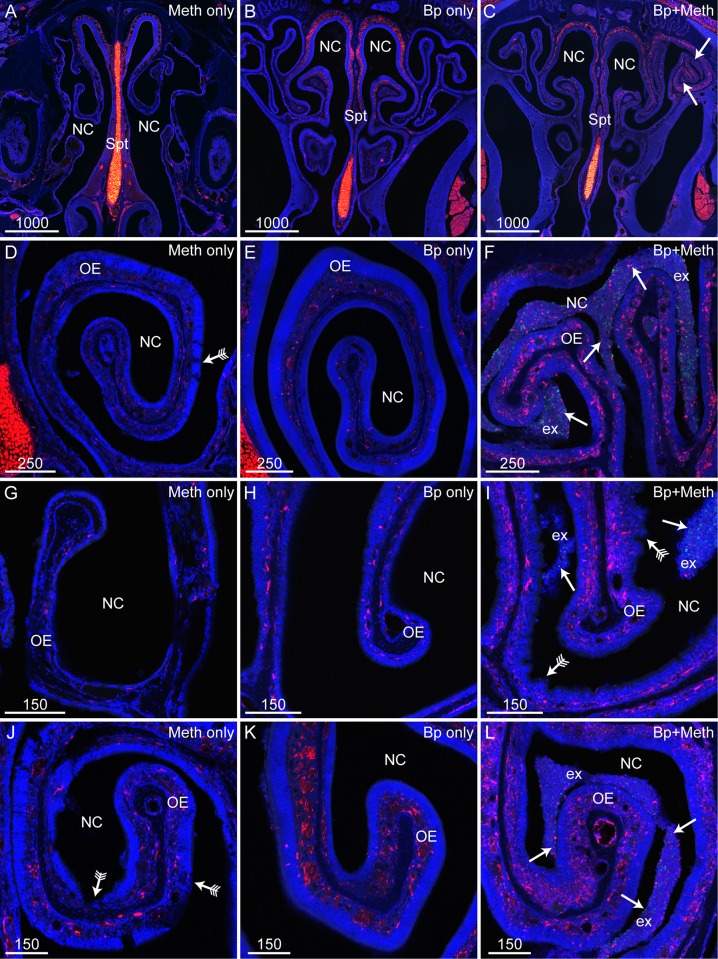
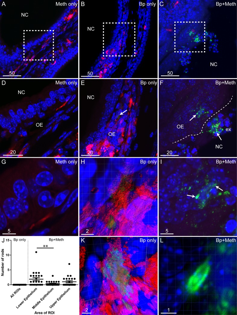
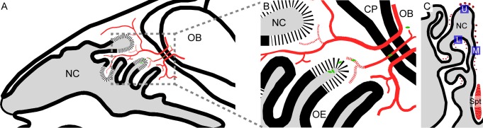
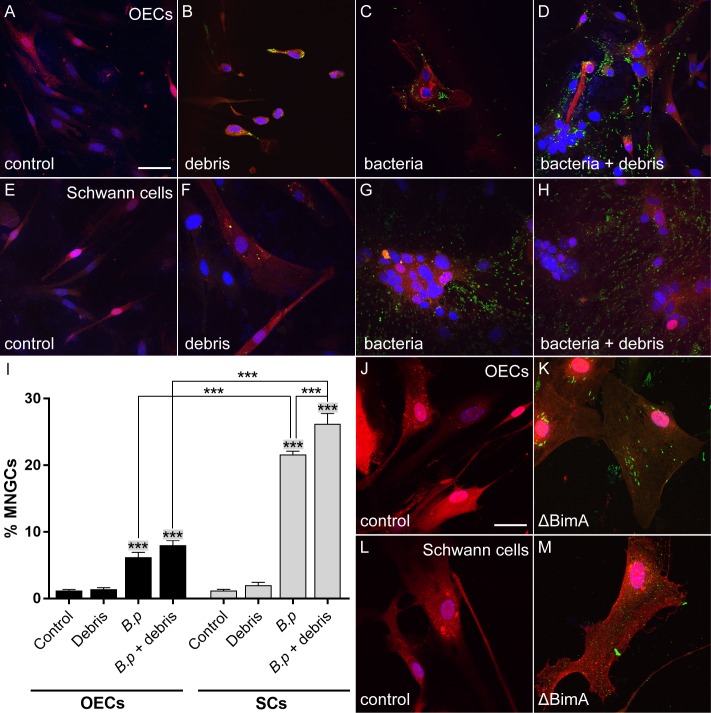
The infectious disease melioidosis is caused by the bacterium Burkholderia pseudomallei. Melioidosis is characterised by high mortality and morbidity and can involve the central nervous system (CNS). We have previously discovered that B. pseudomallei can infect the CNS via the olfactory and trigeminal nerves in mice. We have shown that the nerve path is dependent on mouse strain, with outbred mice showing resistance to olfactory nerve infection. Damage to the nasal epithelium by environmental factors is common, and we hypothesised that injury to the olfactory epithelium may increase the vulnerability of the olfactory nerve to microbial insult. We therefore investigated this, using outbred mice that were intranasally inoculated with B. pseudomallei, with or without methimazole-induced injury to the olfactory neuroepithelium. Methimazole-mediated injury resulted in increased B. pseudomallei invasion of the olfactory epithelium, and only in pre-injured animals were bacteria found in the olfactory nerve and bulb. In vitro assays demonstrated that B. pseudomallei readily infected glial cells isolated from the olfactory and trigeminal nerves (olfactory ensheathing cells and trigeminal Schwann cells, respectively). Bacteria were degraded by some cells but persisted in other cells, which led to the formation of multinucleated giant cells (MNGCs), with olfactory ensheathing cells less likely to form MNGCs than Schwann cells. Double Cap mutant bacteria, lacking the protein BimA, did not form MNGCs. These data suggest that injuries to the olfactory epithelium expose the primary olfactory nervous system to bacterial invasion, which can then result in CNS infection with potential pathogenic consequences for the glial cells.


Conflict of interest statement

The authors have declared that no competing interests exist.

Figures

Fig 1
Fig 1. Effect on olfactory epithelium of methimazole pre-treatment and B. pseudomallei intranasal inoculation.
(A-C) Low power images of the nasal cavity (NC) and olfactory epithelium (OE) from mice treated with either methimazole only (Meth only), B. pseudomallei (Bp only) or methimazole followed by B. pseudomallei (Bp+Meth). Images show both bright-field (grey) and DAPI (blue, nuclear stain) channels. (A) Arrow points to an area of clustered exudate close to the OE. (B) Arrows are pointing to a large collection of stringy exudate within the NC between areas of OE. (C) Arrow points to an area of clumped exudate close to the OE. (D-E) Panels show sections from S100β-DsRed mice in which OECs express DsRed (red), and immunolabelled for beta-tubulin III (white) with nuclei stained with DAPI (blue). Low power images showing a coronal view of the nasal cavity (NC) in mice treated either with B. pseudomallei only (D) or methimazole followed by B. pseudomallei (E). (D) Mice inoculated with B. pseudomallei only showed very little degradation of the OE with negligible degradation to olfactory nerve fascicles (arrows). (E) Mice pre-treated with methimazole then inoculated with B. pseudomallei showed OE degradation, exudate (ex) and damaged peripheral nerve fascicles (arrow with tails). There were also areas of degraded OE where no peripheral nerve fascicles were visible (*). OE degradation, while extensive, was not uniform with some peripheral nerve fascicles remaining intact (arrow without tails). (F) Pre-treatment with methimazole (Meth only and Bp+Meth) causes degradation of the olfactory epithelium. Mice treated with vehicle only (PBS) or B. pseudomallei only did not show extensive olfactory epithelium degradation. 27 data points from three ROIs were measured per mouse (n = 3/group). Graph shows each measured point as a dot with error bar showing the mean plus the standard error of the mean. *** = p <0.001. Scale bars in μm.
Fig 2
Fig 2. Methimazole pre-treatment causes olfactory epithelium degradation and increased exudate production in mice inoculated with B. pseudomallei.
Panels show sections from S100β-DsRed mice; (OECs and chrondrocytes are red), immunolabelled for B. pseudomallei (green) with nuclei stained with DAPI (blue). (A-C) Low power images showing a coronal view of the nasal cavity (NC) and septum (Spt) in mice treated either with methimazole only (A), B. pseudomallei only (B), or both methimazole and B. pseudomallei (C). (C) Large patches of exudate were present within the nasal cavity (arrows) of mice treated with methimazole prior to intranasal inoculation with B. pseudomallei. (D-L) Higher magnification showing coronal views of the NC and OE. (D, G, J) Mice treated with methimazole (Meth only) showed patches of OE degradation (arrows with tails). (E, H, K) Mice intranasally inoculated with B. pseudomallei (Bp only) had intact OE with negligible degradation and exudate present. (F, I, L) Mice first treated with methimazole then intranasally inoculated with B. pseudomallei (Bp+Meth) showed regions of OE crenellation (arrows with tails) and exudate (ex) containing B. pseudomallei (green, arrows). Scale bars in μm.
Fig 3
Fig 3. Methimazole treatment exacerbates B. pseudomallei infection of the olfactory epithelium.
Panels show images of coronal sections of the olfactory mucosa. (A-C) Low power images of the nasal cavity (NC) from S100β-DsRed mice, showing OECs (red) and immunolabellingfor B. pseudomallei (green) with nuclei stained with DAPI (blue). (D) A higher magnification of square in panel A showing the NC and olfactory epithelium (OE) with no immunolabelling seen for B. pseudomallei. (E) Magnified area of square in panel B. B. pseudomallei immunolabelling (green) is seen within the OE (arrow), shown at higher magnification below. (F) A higher magnification of boxed region in panel C. Extensive immunoreactivity for B. pseudomallei (green) is seen in the NC within exudate (ex) and the OE. Arrows show B. pseudomallei rods within areas of associated particles immunoreactive for anti-B. pseudomallei antibodies. (G) A very high magnification of the OE showing no immunoreactivity for B. pseudomallei in mice treated with methimazole alone. (H) A very high magnification and three-dimensional (3D) reconstruction of the B. pseudomallei immunoreactivity seen in panel E (arrow). (I) A very high magnification of B. pseudomallei rods (arrows) within the olfactory epithelium (OE) seen in panel F. Associated particles immunoreactive for anti-B. pseudomallei antibodies can also be seen within the OE. (J) Graph showing the numbers of B. pseudomallei (Bp) rods within the lower, middle and upper olfactory epithelium of mice pre-treated with methimazole prior to B. pseudomallei infection (n = 4) with error bars showing the mean plus the standard error of the mean. Within sections of the olfactory epithelium, six ROIs (440 μm by 440 μm in size with 50 μm depth) were defined (two ROIs each for the lower, middle and upper epithelium). For each mouse, three sections containing these ROIs were analysed. There were significantly more B. pseudomallei rods within the lower epithelium than in the middle epithelium (** = p ≤ 0.01). (K) A rotation of the 3D reconstruction seen in panel H. B. pseudomallei reactivity (green) appears to be localised within an S100β-DsRed positive cell. (L) A very high magnification and 3D reconstruction of the B. pseudomallei rod (green) shown in panel I. Scale bars in μm.
Fig 4
Fig 4. Methimazole pre-treatment causes B. pseudomallei infection of the olfactory bulb via the olfactory nerve.
(A-C) Schematic drawings of a coronally sectioned mouse head showing the nasal cavity (NC), nasal septum (Spt) and turbinates (Tb). Red dots represent peripheral nerve fascicles (olfactory and trigeminal nerves fascicles); the olfactory bulbs are also shown in red (OB). (D-L) All panels show coronal sections from S100β-DsRed mice; D-F were treated with methimazole followed by B. pseudomallei inoculation (Bp+Meth), while G-I were inoculated B. pseudomallei only (no methimazole). Sections show OECs (red), immunolabelling for B. pseudomallei (green), with nuclei stained with DAPI (blue). (D) Location of panel D is shown by the white box in panel A. This is a low power view of the NC showing the olfactory epithelium (OE), exudate (ex), lamina propria (LP) and olfactory nerve (within white box in panel D). (E) Location of panel E is shown by the white box in panel B. Dorsal region of the NC showing the olfactory nerve (ON) passing through the cribriform plate (CP) connecting the OE and the olfactory bulb (OB). (F) Location of panel F is shown by the white box in panel C. This is a low power coronal view of the OB. (G) Location of panel G represented by the white box in panel A. Zoomed image of the ON from a mouse inoculated with B. pseudomallei only. No B. pseudomallei was detected within the ON. (H) Location of panel H represented by the white box in panel B. Magnified view of the ON and CP from a mouse inoculated with B. pseudomallei only. No B. pseudomallei was detected within the ON. (I) Location of panel I represented by the white box in panel C. Magnified view of the OB from a mouse inoculated with B. pseudomallei only. No B. pseudomallei was found within the OB. (J) A zoomed image of the ON shown within the white box in panel D. Arrows point to B. pseudomallei bacteria (green) present within the ON. (K) A zoomed image of the ON shown within the white box of panel E with an arrow pointing to a B. pseudomallei (green) rod. (L) A zoomed image of the white box in panel F showing the outer layer of the OB. The arrow indicates B. pseudomallei (green) present within the OB. (J-L) Smaller images within each panel show a very high magnification of B. pseudomallei with the scale bar representing 2.5 μm. Scale bars in μm.
Fig 5
Fig 5. Schematic drawings summarising B. pseudomallei invasion of the olfactory bulb in mice pre-treated with methimazole.
(A-B) A schematic drawing of a sagittal mouse head section showing the nasal cavity (NC), olfactory epithelium (OE), cribriform plate (CP), olfactory bulb (OB) and the olfactory nerve (red). (A) Low power view showing the NC and location of the olfactory nerve (red). The olfactory nerve projects from the OB into the OE. (B) A magnified view of the boxed region in panel A. B. pseudomallei rods (green) are shown within the nasal cavity (NC) close to degraded olfactory epithelium (OE; degradation depicted as segmented lines). The olfactory nerve (red) projects from the olfactory bulb (OB) into the olfactory epithelium (OE) via the cribriform plate (CP). B. pseudomallei (green) is shown to invade the degraded olfactory nerve (red; degradation depicted as segmented lines) and penetrate the olfactory bulb (OB). (C) A schematic drawing of a coronal mouse head showing the nasal cavity (NC) and nasal septum (Spt). Red dots represent peripheral nerve fascicles (olfactory and trigeminal nerves fascicles). Blue squares indicate representative anatomical locations for the regions of interest (ROIs) used for B. pseudomallei rod quantification; lower epithelium (L), middle epithelium (M) and upper epithelium (U).
Fig 6
Fig 6. B. pseudomallei can infect OECs and TgSCs, causing the formation of multinucleated cells.
Panels A-B show OECs (red) isolated from olfactory nerve fascicles within the lamina propria (LP), panels C-F show OECs (red) isolated from the olfactory bulb (OB) and panels G-L show TgSCs (red) infected by B. pseudomallei (MOI 75:1). Cells were infected for 24 h. Nuclei are stained with DAPI (blue) and B. pseudomallei immunolabelling is shown in green. (A) LP-OECs (red) infected by B. pseudomallei (green). Whole B. pseudomallei rods (green; arrow) and degraded bacteria (green; arrow with tails) can be seen. (B) The same image as shown in panel A without the red fluorescence. (C) Multinucleation of OB-OECs (red) after infection with B. pseudomallei (green). (D) OB-OECs (red) infected by B. pseudomallei (green); bacteria can also be seen attached to filopodia (zoomed images shown in panels E-F). (E-F) Magnified images of panel D showing OB-OECs (red) with filpodia (double-headed arrows) attached to B. pseudomallei bacteria (green). (G) Multinucleation of TgSCs (red) after infection with B. pseudomallei (green). (H) Magnified view of the TgSC shown in panel G infected with B. pseudomallei (green). In this image, only blue (DAPI; cell nuclei) and green (B. pseudomallei) fluorescence is shown. The cell has three nuclei. (I) The same image as in panel H, here showing staining of nuclei only (DAPI; grey; arrows). In addition to staining the nuclei of TgSCs, DAPI labels DNA within B. pseudomallei (arrows). (J) The TgSCs shown (red) appears to have degraded some of the B. pseudomallei bacteria (green). Whole B. pseudomallei rods (green; arrow) and degraded bacteria (green; arrow with tails) can be seen within cells. (K) Another example image showing multinucleation of TgSCs (red) after B. pseudomallei (green) infection. The cell has three nuclei. (L) Magnified image of panel K showing a membrane protrusion (double-headed arrow) of the TgSC (red) attached to B. pseudomallei (green). Scale bars in μm. Shown are representative images from two biological and three technical repeats.
Fig 7
Fig 7. B. pseudomallei-induced multinucleation of OECs and TgSCs in the absence and presence of axon-derived debris, and example images of glia cultured with B. pseudomallei lacking BimA.
Cells were cultured in the absence/ presence of axonal debris/B. pseudomallei for 24 h. Panels A-D show lamina propria-derived OECs (red), panels E-H show TgSCs (SCs; red); nuclei are stained with DAPI (blue). (A) OECs (red) without debris/bacteria (control). (B) OECs cultured with debris derived from ZsGreen-expressing axons (green); the debris was phagocytosed by the cells. (C) OECs cultured with B. pseudomallei (MOI 75:1) (green). (D) OECs cultured with a combination of B. pseudomallei and cell debris (both bacteria and debris are green). (E) TgSCs in the absence of debris/bacteria (control). (F) TgSCs with axonal debris. (G) TgSCs with B. pseudomallei. (H) TgSCs with a combination of B. pseudomallei and cell debris. Scale bar in A is 15 μm for A-H. (I) Bar graphs show the percentages of multinucleated giant cells in the different conditions (control, debris, B. pseudomallei (Bp) and Bp + debris) for OECs (black bars) and TgSCs (grey bars). ***significantly different from the control group and from the debris group, p ≤ 0.001. ***significantly different from each other, p ≤ 0.001. N = five fields of view each comprising 50–70 cells (derived from three S100β-DsRed mice); p values are adjusted p values from one-way ANOVA with Tukey’s multiple comparison post-hoc test. (J) OECs (red) cultured without debris/bacteria (control). (K) OECs (red) cultured with B. pseudomallei ΔBimA (green) for 48 h. (L) TgSCs (red) in the absence of bacteria/debris. (M) TgSCs (red) cultured with B. pseudomallei ΔBimA (green). Scale bar in J is 15 μm for J-M.

Similar articles

Cited by

References

    1. Parameswaran U, Baird RW, Ward LM, Currie BJ. Melioidosis at Royal Darwin Hospital in the big 2009–2010 wet season: comparison with the preceding 20 years. The Medical journal of Australia. 2012;196(5):345–8. 10.5694/mja11.11170 . - DOI - PubMed
    1. Cheng AC, Currie BJ. Melioidosis: epidemiology, pathophysiology, and management. Clinical microbiology reviews. 2005;18(2):383–416. 10.1128/CMR.18.2.383-416.2005 - DOI - PMC - PubMed
    1. Limmathurotsakul D, Golding N, Dance DA, Messina JP, Pigott DM, Moyes CL, et al. Predicted global distribution of Burkholderia pseudomallei and burden of melioidosis. Nat Microbiol. 2016;1(1):15008 Epub 2016/02/16. 10.1038/nmicrobiol.2015.8 - DOI - PubMed
    1. Chaowagul W, White NJ, Dance DA, Wattanagoon Y, Naigowit P, Davis TM, et al. Melioidosis: a major cause of community-acquired septicemia in northeastern Thailand. The Journal of infectious diseases. 1989;159(5):890–9. 10.1093/infdis/159.5.890 . - DOI - PubMed
    1. Inglis TJ, Sousa AQ. The public health implications of melioidosis. Braz J Infect Dis. 2009;13(1):59–66. Epub 2009/07/07. 10.1590/s1413-86702009000100013 . - DOI - PubMed

Publication types

MeSH terms

Grants and funding

This study was supported by an Australian Research Council Discovery grant (DP150104495) to JE, KB and JSJ (https://www.arc.gov.au/grants/discovery-program/discovery-projects), a Clem Jones Foundation grant to JE and JS (https://experts.griffith.edu.au/project/ndf0b8caf8de786e2416acec7fc2c92b7), a Menzies Health Institute Queensland Capacity Grant to JE, AC and KB (https://www.griffith.edu.au/menzies-health-institute-queensland), a Goda Foundation grant to JE and JSJ, an Australian Government Research Training Program Scholarship to HW (https://www.education.gov.au/research-training-program), and a Griffith University International Postgraduate Research Scholarship to AD (https://www.griffith.edu.au/research-study/scholarships/guiprs). The funders had no role in study design, data collection and interpretation, or the decision to submit the work for publication.