. 2020 Jan 21;13(615):eaay7315.
doi: 10.1126/scisignal.aay7315.

Spatial and temporal alterations in protein structure by EGF regulate cryptic cysteine oxidation

Affiliations

Spatial and temporal alterations in protein structure by EGF regulate cryptic cysteine oxidation

Jessica B Behring et al. Sci Signal. .

Abstract

Stimulation of plasma membrane receptor tyrosine kinases (RTKs), such as the epidermal growth factor receptor (EGFR), locally increases the abundance of reactive oxygen species (ROS). These ROS then oxidize cysteine residues in proteins to potentiate downstream signaling. Spatial confinement of ROS is an important regulatory mechanism of redox signaling that enables the stimulation of different RTKs to oxidize distinct sets of downstream proteins. To uncover additional mechanisms that specify cysteines that are redox regulated by EGF stimulation, we performed time-resolved quantification of the EGF-dependent oxidation of 4200 cysteine sites in A431 cells. Fifty-one percent of cysteines were statistically significantly oxidized by EGF stimulation. Furthermore, EGF induced three distinct spatiotemporal patterns of cysteine oxidation in functionally organized protein networks, consistent with the spatial confinement model. Unexpectedly, protein crystal structure analysis and molecular dynamics simulations indicated widespread redox regulation of cryptic cysteine residues that are solvent exposed only upon changes in protein conformation. Phosphorylation and increased flux of nucleotide substrates served as two distinct modes by which EGF specified the cryptic cysteine residues that became solvent exposed and redox regulated. Because proteins that are structurally regulated by different RTKs or cellular perturbations are largely unique, these findings suggest that solvent exposure and redox regulation of cryptic cysteine residues contextually delineate redox signaling networks.


Conflict of interest statement

Competing Interests: The authors declare that they have no competing interests.

Figures

Figure 1.
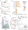
Figure 1.. The OxRAC workflow to globally profile cysteine oxidation and overview of results.
A) Serum-starved A431 cells were left untreated (0 min) or stimulated with 100ng/mL EGF for the times indicated prior to lysis. B) OxRAC workflow schematic in which free cysteine residues are trapped with NEM and oxidized thiols are enriched by thiopropyl sepharose resin and trypsin digested on-resin. The oxidized cysteine residues remain bound during washing, then are eluted by reduction and labeled with iodoacetamide (IAC) to differentiate oxidized (IAC-labeled) from non-oxidized (NEM-labeled) cysteine residues. Peptides are analyzed by data-dependent acquisition (DDA) to identify peptides and data-independent acquisition (DIA) mass spectrometry for quantification purposes based on high resolution MS2 scans. C) DIA-MS2 scans of the pyruvate kinase (PKM) peptide CCSGAIIVLTK. The site defining y10 fragment ion (red line) between the two labeled cysteine residues confirms peak identity. (D and E) Time-dependent changes in the relative oxidation of PKM Cys423 and Cys424 (D), and of cysteine residues in procollagen-lysine 2-oxoglutarate 5-dioxygenase 1 (PLOD1), catenin delta-1 (CTNND1), and dihydrolipoyl dehydrogenase (DLD) in response to EGF stimulation. *P < 0.05, **P <0.01, based on one-way ANOVA with Dunnett’s post-hoc test. Error bars are SEM for N=3 independent biological replicates. Fold change is log2 transformed. F) Enumeration of the functional annotation, disulfide bond types, and number of cysteine residue per peptide in the dataset.
Figure 2.
Figure 2.. EGF-dependent regulation of cysteine redox networks cluster into three distinct temporal profiles associated with unique subcellular locations and biological processes.
A) Average log2 fold change of all peptides compared to baseline (n=3 independent biological replicates). Lines indicate normal distributions and the red line indicates the fold change used for normalization. B) Heatmap of all significantly oxidized cysteine-containing peptides (Q < 0.05, ANOVA corrected by Benjamini-Hochberg) clustered by K-means (1 minus Pearson correlation, K = 3) of relative oxidation levels. C) Fuzzy c-means clustering of significantly oxidized peptides and selected Gene Ontology (GO) and Reactome annotations. P-values are from Panther. Fold change is log2 transformed.
Figure 3.
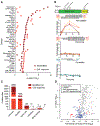
Figure 3.. Cysteine residues in all major organelles are oxidized by EGF but location influences the temporal dynamics.
A) Membrane orientation of 182 modified sites on either the cytoplasmic, extracellular, or lumenal side presented as percentage significant over time in response to EGF. Significance per time point is based on Q < 0.05, ANOVA corrected by Benjamini-Hochberg. N = 3 independent biological replicates. B) Differential response over time of the extracellular and cytoplasmic side of the transmembrane protein Plexin-2B. C) The percentage of cysteine residues in 942 sentinel proteins detected by OxRAC and annotated to a single cellular compartment that have at least one cysteine residue significantly oxidized by EGF (ANOVA corrected by the Benjamini-Hochberg method). D) Estimated percent oxidation of all cysteine residues in sentinel proteins. E) Selected examples of differentially oxidized peptides containing 2 cysteine residuess. A disulfide bond in EGFR between Cys248 and Cys251 becomes reduced upon endocytosis in response to EGF. Examples of two previously unknown functional cysteine residues in TXNDC5 which are potentially disulfide linked. 1Ox and 2Ox indicate singly or doubly oxidized forms of the peptide, respectively. F) Linear regression (r, Pearson) of the fold-change over time of the singly oxidized (1Ox) forms compared to the variation (standard deviation, SD) between the two singly oxidized sites. Sites annotated as disulfide linked, active, or metal binding are indicated. Includes differentially oxidized sites identified in peptides spanning two cysteine residues.
Figure 4.
Figure 4.. Synchronized redox regulation of cysteine residues throughout canonical EGF signaling pathways at 15 and 30 minutes.
Select enriched canonical pathways from IPA downstream of EGFR are pictured. All genes with a significantly regulated cysteine residue are colored green. P < 0.05 based one-way ANOVA with Dunnett’s post-hoc test, N=3 independent biological replicates. Proteins detected but not significantly oxidized by EGF are filled in grey and those undetected, but important for continuity of a pathway, were left unfilled. EGF redox dynamics over 60 minutes for significantly changing peptides in each pathway are represented in heatmaps.
Figure 5.
Figure 5.. Protein domains redox regulated by EGF stimulation.
A) Enrichment of protein domains detected in the entire dataset ‘all identified’ or significantly oxidized by EGF, ‘EGF response’, as compared to all domains in the human proteome. P-values determined by two-tailed Fisher’s exact test. B) Domain organization of PRDX1 and locations and function of its cysteine residues. The relative oxidation of the resolving, peroxidatic and non-catalytic cysteine residues in PRDXs over time in response to EGF stimulation. *P < 0.05, **P <0.01, based one-way ANOVA with Dunnett’s post-hoc test. Error bars are SEM for N=3 independent biological replicates. C) Functional annotation of all cysteine residues detected compared to those significantly regulated in response to EGF. D) The average ratio of all peptides assigned to the most enriched domains compared to their overall significance (q), overlaid with functional annotation when available. Closed circles: enriched sites in response to EGF, open circles: not enriched in response to EGF. Adjusted p-values (q) were calculated from ANOVA results by applying the Benjamini-Hochberg method to correct for multiple comparisons.
Figure 6.
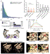
Figure 6.. EGF-dependent redox regulation of buried cysteine residues in 14-3-3, small GTPase proteins, and ERK2.
A) Density plot of relative solvent accessibility (RSA) that compares cysteine residues significantly and not regulated by EGF. B) RSA for all cysteine residues in proteins identified compared to all cysteine residues specifically quantified and those significantly regulated. Sites >0.25 are considered solvent accessible. * p< 0.05, *** p <0.001 by one-way ANOVA. For the box-whisker plot: center line is median, limits are upper and lower quartiles, and whiskers are 5 and 95 percentiles. C) RSA prediction for sulfenated cysteine residues in (24). D) Amino acid sequence conservation of the cysteine residues identified in 14-3-3 proteins. Structure of dimerized 14-3-3 sigma (PDB: 4DAU) with helix 4 shown in purple and locations of the remaining oxidized cysteine residues shown in black. Purple arrows point to the conserved cysteine residue in Helix 4, represented as a sphere. Relative surface accessibility (RSA) is indicated for each cysteine site or the average across 14-3-3 family members. E) Time-dependent changes in the oxidation of 14-3-3 cysteine residue. *P < 0.05, **P <0.01, based one-way ANOVA with Dunnett’s post-hoc test. Error bars are SEM for N=3 independent biological replicates. F) Unmodified and phosphorylated (Thr183 and Tyr185) ERK2 crystal structures (PDB: 1ERK and 2ERK, respectively) with Cys65 highlighted in red and solvent as a cyan sphere. G) Crystal structures of the GTP binding pocket in CDC42 (PDBID: 5CJP) and Rac1 (PDBID: 5O33 GTP analogue). Cys157 is highlighted in red. Yellow structure is the bound GTP nucleotide analogue. Cyan spheres indicate solvent molecules. H) Representative crystal structure of KRAS (left, PDB: 5VQ2) in which Cys80 (red) is solvent inaccessible as well as a representative structure from MD simulations of KRAS (right) in which Cys80 is solvent accessible. Amino acid side chains within 7 angstroms of Cys80 are shown as spheres, and Cys80 is shown in red as a ball-and-stick.

Similar articles

Cited by

References

    1. Bae YS, Kang SW, Seo MS, Baines IC, Tekle E, Chock PB, Rhee SG, Epidermal Growth Factor (EGF)-induced Generation of Hydrogen Peroxide, J. Biol. Chem 272, 217–221 (1997). - PubMed
    1. Kwon J, Lee S-R, Yang K-S, Ahn Y, Kim YJ, Stadtman ER, Rhee SG, Reversible oxidation and inactivation of the tumor suppressor PTEN in cells stimulated with peptide growth factors, Proc. Natl. Acad. Sci 101, 16419–16424 (2004). - PMC - PubMed
    1. Paulsen CE, Truong TH, Garcia FJ, Homann A, Gupta V, Leonard SE, Carroll KS, Peroxide-dependent sulfenylation of the EGFR catalytic site enhances kinase activity, Nat. Chem. Biol 8, 57–64 (2012). - PMC - PubMed
    1. Winterbourn CC, Reconciling the chemistry and biology of reactive oxygen species, Nat. Chem. Biol 4, 278–286 (2008). - PubMed
    1. Mahadev K, Zilbering A, Zhu L, Goldstein BJ, Insulin-stimulated Hydrogen Peroxide Reversibly Inhibits Protein-tyrosine Phosphatase 1B in Vivo and Enhances the Early Insulin Action Cascade, J. Biol. Chem 276, 21938–21942 (2001). - PubMed

Publication types

MeSH terms