. 2020 Jan 17;11(1):365.
doi: 10.1038/s41467-019-14245-4.

Bacterial Hsp70 resolves misfolded states and accelerates productive folding of a multi-domain protein

Affiliations

Bacterial Hsp70 resolves misfolded states and accelerates productive folding of a multi-domain protein

Rahmi Imamoglu et al. Nat Commun. .

Abstract

The ATP-dependent Hsp70 chaperones (DnaK in E. coli) mediate protein folding in cooperation with J proteins and nucleotide exchange factors (E. coli DnaJ and GrpE, respectively). The Hsp70 system prevents protein aggregation and increases folding yields. Whether it also enhances the rate of folding remains unclear. Here we show that DnaK/DnaJ/GrpE accelerate the folding of the multi-domain protein firefly luciferase (FLuc) ~20-fold over the rate of spontaneous folding measured in the absence of aggregation. Analysis by single-pair FRET and hydrogen/deuterium exchange identified inter-domain misfolding as the cause of slow folding. DnaK binding expands the misfolded region and thereby resolves the kinetically-trapped intermediates, with folding occurring upon GrpE-mediated release. In each round of release DnaK commits a fraction of FLuc to fast folding, circumventing misfolding. We suggest that by resolving misfolding and accelerating productive folding, the bacterial Hsp70 system can maintain proteins in their native states under otherwise denaturing stress conditions.


Conflict of interest statement

The authors declare no competing interests.

Figures

Fig. 1
Fig. 1. Dnak/DnaJ/GrpE reaction cycle.
DnaJ captures the substrate protein and transfers it to DnaK in the ATP-bound state. DnaJ and substrate synergistically trigger ATP hydrolysis by DnaK, thereby generating a stable complex between the substrate and DnaK in the ADP-bound state. Catalysis of ADP-ATP exchange by GrpE stimulates client release and regenerates DnaK-ATP for another round of client engagement. Figure modified from ref. .
Fig. 2
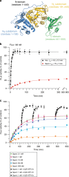
Fig. 2. The KJE chaperone system accelerates FLuc folding.
a Structure of FLuc (PDB ID: 1LCI) with domains indicated. b Spontaneous and chaperone-assisted folding of 100 nM FLuc was assayed upon dilution from 5 M GuHCl into buffer without or with KJE-ATP, respectively, by monitoring luminescence activity. KJE-mediated folding was performed with 3 µM DnaK, 1 µM DnaJ, 1.5 µM GrpE and 5 mM ATP. Error bars represent s.d. (n = 10). c Concentration dependence of FLuc refolding. FLuc was diluted from denaturant to different final concentrations (0.1–200 nM) and refolding assayed as in (b). Rescue of spontaneous folding reactions was assayed by adding KJE-ATP (0.3 µM DnaK, 0.1 µM DnaJ, 0.5 µM GrpE and 5 mM ATP) after 2, 4 or 8 h. Error bars represent s.d. (n = 3). Ass., KJE-ATP-assisted; Spont., spontaneous folding. Source data are provided as a Source Data file.
Fig. 3
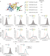
Fig. 3. Slow spontaneous folding of FLuc is caused by inter-domain misfolding.
a Structure of FLuc (PDB ID: 1LCI) showing fluorophore labelling positions. Mutations introduced to allow site-specific cysteine labelling are indicated, and estimated distances between dye pairs are given. FLuc variants were labelled with Atto532 (donor) and Atto647N (acceptor). be spFRET efficiency (fE) histograms of the native proteins: FLucNC (b), FLucN (c), FLucNS (d) and FLucC (e). fi, fE histograms measured during spontaneous refolding of 50 pM denatured FLuc. spFRET was recorded for 1 h, either immediately (0 h) or at the indicated folding time points. j, k, spFRET efficiency (fE) histogram of the NS-subdomain (FLuc190) (j) and spontaneous folding of 50 pM denatured FLuc190 (k). Data were recorded for 30 min. Reactions b–k contained 5 mM ATP and 50 µM phenobenzothiazine (PBT). The fE corresponding to the native state for each construct is indicated by a dashed vertical red line. Representative measurements of three independent repeats are shown. Spont., spontaneous folding. Source data are provided as a Source Data file.
Fig. 4
Fig. 4. KJE reverses misfolding and commits bound protein to fast folding.
ad spFRET efficiency histograms measured during KJE-ATP assisted folding of FLucNC (a), FLucN (b), FLucNS (c) or FLucC (d). GuHCl-denatured FLuc was diluted to 50 pM in buffer containing 0.3 µM DnaK, 0.1 µM DnaJ, 5 mM ATP and 50 µM PBT, and the fE distribution of DnaK-bound FLuc was recorded, followed by initiation of folding with 0.5 µM GrpE. spFRET was recorded for 15 min either immediately (0 min) or after 60 min of folding. Representative measurements of three independent repeats are shown. e KJ-ATP converts compact, misfolded (MF) intermediates of FLuc to the DnaK-bound, expanded state. GuHCl-denatured FLucNC was diluted to 50 pM in buffer containing ATP and PBT, and the distance between N- and C-domains probed by spFRET as in (a). KJ-ATP were added after 1 h to generate DnaK-bound FLuc, followed by GrpE to initiate folding. Representative measurements of three independent repeats are shown. Spont., spontaneous folding. Source data are provided as a Source Data file.
Fig. 5
Fig. 5. Conformational dynamics of native and DnaK-bound FLuc.
a Scheme of equilibrium H/DX-MS experiment. bc, Peptide-level deuterium exchange of native FLuc (N-FLuc) (b) or DnaK-bound FLuc (K-bound) (c) after exposure to deuterium for 10-300 s. Relative fractional deuterium exchange for each peptide is mapped onto the structure of FLuc (PDB ID: 1LCI) as a gradient from blue (0%) to red (75%). d Difference in deuterium exchange between DnaK-bound and N-FLuc at peptide level. Deuteration differences are scaled from blue (−50%) to red (+50%). Regions coloured red are deprotected when bound to DnaK. H/DX data are the average of three independent repeats.
Fig. 6
Fig. 6. KJE-assisted folding of FLuc at peptide resolution.
a Scheme of H/DX-MS experiment. DnaK is shown schematically in grey and DnaJ in orange. b Fraction of native deuterium exchange reached by FLuc peptides after 10 s folding. D uptake at 10 s was normalized to the difference in D uptake between K-bound and N-FLuc for each peptide. Only peptides exhibiting a >1 Da difference between the DnaK-bound and native states are shown. Peptides are characterized as fast (upper third, blue), medium (middle third, gold) or slow (lower third, red) folders, based on their relative protection compared to N-FLuc. Error bars represent s.d. (n = 3). Source data are provided as a Source Data file. c Location of the peptides colour-coded as in (b), indicated on the structure of FLuc. Dotted line indicates the NS–NL domain interface.
Fig. 7
Fig. 7. DnaK commits a fraction of bound FLuc to fast folding.
Folding of FLuc in a single-round of chaperone action. Rebinding of FLuc to DnaK was inhibited using excess NR peptide (125 µM). For single round folding, GuHCl-denatured FLuc was diluted to 100 pM in buffer containing KJ-ATP, followed by addition of NR and initiation of folding with GrpE (red triangles). As controls, NR was either omitted (black circles) or added to KJ-ATP before FLuc (green squares), or after 5 min of chaperone-assisted folding (white circles). Error bars represent s.d. (n = 3). Ass., KJE-ATP-assisted. Source data are provided as a Source Data file.
Fig. 8
Fig. 8. KJE-accelerated folding maintains the native state of FLuc.
a Luminescence activity of 10 pM FLuc was monitored at 37 oC, in the presence or absence of KJE-ATP. Time-dependent loss of FLuc activity was monitored by shifting N-FLuc from 25 oC to 37 oC (blue circles). 1 µM DnaK, 0.33 µM DnaJ and 1.5 µM GrpE were included during temperature upshift of N-FLuc (orange circles), or added after 1 h incubation of N-FLuc at 37 oC (magenta diamonds), or after 1 h of spontaneous refolding from denaturant at 37oC (green triangles). Assisted (Ass., black squares) and spontaneous (Spont., red squares) refolding are also shown. Error bars represent s.d. (n = 5). Ass., KJE-ATP-assisted; Spont., spontaneous folding. b N-FLucNC (10 pM) in folding buffer with PBT and ATP was shifted from 25 oC to 37 oC in the presence or absence of KJ. spFRET was recorded for 30 min after 2 h without chaperones. Upon heating in the presence of KJ-ATP, spFRET was recorded either immediately (0 min) or after 90 min. GrpE was added after 2 h to initiate refolding of FLuc from the DnaK-bound state. Representative measurements of three independent repeats are shown. Source data are provided as a Source Data file.
Fig. 9
Fig. 9. Model of spontaneous and KJE-ATP assisted folding of FLuc.
a MF, misfolded compact intermediate; Agg., aggregated protein; US, unfolded state in the absence of chaperones; IS, folding intermediate of spontaneous folding; UC, unfolded state in complex with DnaK; IC, intermediate committed to fast folding; N, native state. Lengths of arrows reflects approximate relative rates. b Simplified energy diagrams to illustrate the effect of KJE-ATP in stabilizing the native state of FLuc at elevated temperature by accelerating refolding of MF.

Similar articles

Cited by

References

    1. Balchin D, Hayer-Hartl M, Hartl FU. In vivo aspects of protein folding and quality control. Science. 2016;353:aac4354. doi: 10.1126/science.aac4354. - DOI - PubMed
    1. Mayer MP, Gierasch LM. Recent advances in the structural and mechanistic aspects of Hsp70 molecular chaperones. J. Biol. Chem. 2019;294:2085–2097. doi: 10.1074/jbc.REV118.002810. - DOI - PMC - PubMed
    1. Rosenzweig R, Nillegoda NB, Mayer MP, Bukau B. The Hsp70 chaperone network. Nat. Rev. Mol. Cell Biol. 2019;20:665–680. doi: 10.1038/s41580-019-0133-3. - DOI - PubMed
    1. Zhu X, et al. Structural analysis of substrate binding by the molecular chaperone DnaK. Science. 1996;272:1606–1614. doi: 10.1126/science.272.5268.1606. - DOI - PMC - PubMed
    1. Szabo A, et al. The ATP hydrolysis-dependent reaction cycle of the Escherichia coli Hsp70 system DnaK, DnaJ, and GrpE. Proc. Natl Acad. Sci. USA. 1994;91:10345–10349. doi: 10.1073/pnas.91.22.10345. - DOI - PMC - PubMed

Publication types

MeSH terms