. 2020 Feb;5(2):304-313.
doi: 10.1038/s41564-019-0640-1. Epub 2020 Jan 6.

Commensal Bacteroidetes protect against Klebsiella pneumoniae colonization and transmission through IL-36 signalling

Affiliations

Commensal Bacteroidetes protect against Klebsiella pneumoniae colonization and transmission through IL-36 signalling

Richard P Sequeira et al. Nat Microbiol. 2020 Feb.

Abstract

The microbiota primes immune defences but the identity of specific commensal microorganisms that protect against infection is unclear. Conversely, how pathogens compete with the microbiota to establish their host niche is also poorly understood. In the present study, we investigate the antagonism between the microbiota and Klebsiella pneumoniae during colonization and transmission. We discover that maturation of the microbiota drives the development of distinct immune defence programmes in the upper airways and intestine to limit K. pneumoniae colonization within these niches. Immune protection in the intestine depends on the development of Bacteroidetes, interleukin (IL)-36 signalling and macrophages. This effect of Bacteroidetes requires the polysaccharide utilization locus of their conserved commensal colonization factor. Conversely, in the upper airways, Proteobacteria prime immunity through IL-17A, but K. pneumoniae overcomes these defences through encapsulation to effectively colonize this site. Ultimately, we find that host-to-host spread of K. pneumoniae occurs principally from its intestinal reservoir, and that commensal-colonization-factor-producing Bacteroidetes are sufficient to prevent transmission between hosts through IL-36. Thus, our study provides mechanistic insight into when, where and how commensal Bacteroidetes protect against K. pneumoniae colonization and contagion, providing insight into how these protective microorganisms could be harnessed to confer population-level protection against K. pneumoniae infection.


Conflict of interest statement

Competing Interests

The authors declare no competing interests.

Figures

Extended Data Figure 6
Extended Data Figure 7
Figure 1
Figure 1. The adult microbiota protects against colonization by antibiotic-resistant Klebsiella pneumoniae in the intestine but not upper airway.
(a) Experimental scheme for (b-j): “ABX” = metronidazole, neomycin, vancomycin and ampicillin (MNVA) in drinking water; “Kp” = K. pneumoniae oral or intranasal inoculation; and “Sample” = upper airway or fecal sampling day. Days relative to K. pneumoniae inoculation. (b,c) K. pneumoniae (OXA-48) burden in the (b) upper airway (n=8 animals) or (c) colon of neonatal mice (n=8 animals). (d-f) K. pneumoniae (OXA-48) (n=8 animals) (d), K. pneumoniae (B5055) (n=8,9 animals) (e), and K. pneumoniae (ST258) (n=7 animals) (f) burden in upper airways of adult mice. (g-j) K. pneumoniae (OXA-48) (n=6 animals) (g), K. pneumoniae (B5055) (n=8 animals) (h), K. pneumoniae (ST258) (n=7 animals) (i) and K. quasipneumoniae subsp. similipneumoniae (n=5 animals) (j) fecal burden in adult mice. (k) Experimental scheme for (l,m): “ABX” = MNVA in drinking water; “MT” = microbiota transfer by oral and intranasal inoculation; “Kp” = K. pneumoniae oral inoculation; and “Sample” = fecal sampling day. K. pneumoniae (OXA-48) (n=5 animals) (l) and K. pneumoniae (ST258) (n=6 animals) (m) fecal burden in adult mice. Upper airway colonization data displayed in red and intestinal colonization data in blue. All statistical comparisons were made by Mann-Whitney test (two-tailed), horizontal lines indicate median values, ND is none detected (limit of detection for K. pneumoniae in feces = 103 CFU/g).
Figure 2
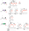
Figure 2. Intestinal Bacteroidetes protect against Klebsiella pneumoniae colonization.
(a,b) Relative abundance of bacterial phyla (a) and orders (b) in (adult feces n=5 animals) and large intestine of neonatal mice (n=6 animals). Each bar represents a single mouse. (c) Experimental scheme for (d-g): “ABX” = MNVA in drinking water; “GF” = germ-free mice; “CC” = commensal consortia inoculation by oral gavage (see Materials and Methods); “Kp” = K. pneumoniae oral inoculation; and “Sample” = fecal sampling day. Days relative to K. pneumoniae inoculation. (d-f) K. pneumoniae (OXA-48) (n=7,8 animals) (d), K. pneumoniae (B5055) (n=8 animals) (e), and K. pneumoniae (ST258) (n=7,8 animals) (f) fecal burden in adult antibiotic treated mice. (g) K. pneumoniae (ST258) fecal burden in germ-free mice (n=5 animals). (h) Experimental scheme for (i-k): acronyms as in (c). K. pneumoniae (OXA-48) (n=5 animals) (i) and K. pneumoniae (ST258) (n=7 animals) (j) fecal burden in adult mice. K. pneumoniae (ST258) (k) fecal burden in neonatal mice (n=6,7 animals). All statistical comparisons were made by Kruskal-Wallis test with Dunn’s correction for multiple comparisons. Horizontal lines indicate median values.
Figure 3
Figure 3. Bacteroidetes protect against Klebsiella pneumoniae colonization in the intestine through IL-36 signalling and macrophages.
(a) Experimental scheme for (b-e): “ABX” = MNVA in drinking water; “CC” = commensal consortia oral inoculation; “Kp” = K. pneumoniae oral inoculation; and “Sample” = fecal sampling day; “Antibody treatment” = antibody, or isotype control, treatment. Days relative to K. pneumoniae inoculation. (b-e) K. pneumoniae (OXA-48) burden in the feces of adult mice (n=5,7,8 animals). (e) K. pneumoniae (ST258) burden in the feces of adult mice (n=6 animals). (f) Experimental scheme for (g-i): acronyms as in (a), days on which recombinant IL-36γ was administered by intraperitoneal injection are indicated. (g,h) K. pneumoniae (OXA-48) burden (n=6,7 animals) (g), and K. pneumoniae (ST258) burden (n=6 animals) (h) in the feces of adult mice. (i) Intestinal cytokine levels 3 days after oral inoculation with indicated commensal consortia (n=5 animals). (j) Experimental plan for (k-n), acronyms in experimental plan as in (a), “Clodronate” = day of clodronate liposome, or empty liposome treatment. (k,l) K. pneumoniae (OXA-48) burden (n=7,8 animals) (k), and K. pneumoniae (ST258) burden (n=5,6 animals) (l) in the feces of adult mice. (m) Intestinal cytokine levels 3 days after oral inoculation with indicated commensal consortia (n=5). (n) K. pneumoniae (OXA-48) fecal burden in adult mice (n=6,7 animals). Statistical comparisons were made by Kruskal–Wallis test with Dunn’s correction for multiple comparisons (b-e, i-l and n), and by Mann-Whitney test (two-tailed) (g,h,m). Horizontal lines indicate median values, error bars are standard deviation.
Figure 4
Figure 4. Bacteroidetes require their commensal colonization factors to protect against Klebsiella pneumoniae colonization in the intestine.
(a) Experimental scheme: “ABX” = MNVA in drinking water; “CC” = commensal consortia oral inoculation; “Kp” = K. pneumoniae oral inoculation; and “Sample” = fecal sampling day. Days relative to K. pneumoniae inoculation. (b) K. pneumoniae (OXA-48) fecal burden in adult mice (n=5 animals). Mice were orally inoculated with either a consortium of wild-type or isogenic ΔCCF Bacteroidetes (B. fragilis and B. vulgatus). (d) Bacteroidetes levels in feces of mice from (b). (d) Intestinal cytokine levels 3 days after oral inoculation of Bacteroidetes consortia (n=5 animals). (e,f) K. pneumoniae (OXA-48) (n=6 animals) (e) and K. pneumoniae (ST258) (n=6 animals) (f) fecal burden in adult mice. Indicated mice were orally inoculated with either WT B. fragilis, ΔCCF B. fragilis or ΔCCF::CCF B. fragilis. (g) Intestinal cytokine levels 3 days after oral inoculation with either WT B. fragilis, ΔCCF B. fragilis or ΔCCF::CCF B. fragilis (n=5). (h) Mucosal association of WT B. fragilis, ΔCCF B. fragilis and ΔCCF::CCF B. fragilis (n=5,6 animals). (i) B. fragilis levels in feces (n=5,6 animals). Statistical comparisons were made by Kruskal–Wallis test with Dunn’s correction for multiple comparisons (b-c,e-i), and by Student’s t-test (two-tailed) (d). Horizontal lines indicate median values, error bars are standard deviation.
Figure 5
Figure 5. Proteobacteria primes upper airway defences through IL-17A but encapsulation allows Klebsiella pneumoniae to overcome these defences.
(a) Experimental scheme for (b): “ABX” = MNVA in drinking water; “MT” = microbiota transfer; “Kp” = K. pneumoniae intranasal inoculation; and “Sample” = upper airway lavage sampling; “Dexa” = dexamethasone treatment. Days relative to K. pneumoniae inoculation. (b) Upper airway K. pneumoniae burden (WT B5055), unencapsulated K. pneumoniae (ΔCPS B5055), and complemented K. pneumoniae (ΔCPS::CPS B5055) in adult mice (n=7,9 animals). (c) Experimental plan for (d-f), acronyms as in (a), “Antibody treatment” = antibody, or isotype control, treatment. Days relative to K. pneumoniae inoculation. (d-f) K. pneumoniae (WT and ΔCPS B5055) upper airway burden in adult mice (n=5,7,8,9 animals). (g) Experimental scheme for (h): acronyms as in (a), days of rIL-17A, or vehicle control, administration are indicated. (h) Upper airway burden of unencapsulated K. pneumoniae (ΔCPS B5055), and complemented K. pneumoniae (ΔCPS::CPS B5055) in adult mice (n=6 animals). (i) Experimental plan for (j-m), acronyms in experimental plan as in (a,c), “CC” = indicated commensal consortia inoculation by intranasal inoculation (see Materials and Methods). (j-m) Upper airway K. pneumoniae burden (WT B5055), unencapsulated K. pneumoniae (ΔCPS B5055), and complemented K. pneumoniae (ΔCPS::CPS B5055) in adult mice (n=5,7,8 animals). Mice were antibiotic treated in (j) and (l) and germ-free in (k). Statistical comparisons were made by Kruskal–Wallis test with Dunn’s correction for multiple comparisons (b,f,h,j-m) and by Mann-Whitney test (two-tailed) (d,e). Horizontal lines are median values

Similar articles

Cited by

References

    1. Baumler AJ, Sperandio V. Interactions between the microbiota and pathogenic bacteria in the gut. Nature. 2016;535:85–93. doi: 10.1038/nature18849. - DOI - PMC - PubMed
    1. Brown RL, Clarke TB. The regulation of host defences to infection by the microbiota. Immunology. 2017;150:1–6. doi: 10.1111/imm.12634. - DOI - PMC - PubMed
    1. Honda K, Littman DR. The microbiome in infectious disease and inflammation. Annu Rev Immunol. 2012;30:759–795. doi: 10.1146/annurev-immunol-020711-074937. - DOI - PMC - PubMed
    1. Clarke TB. Microbial programming of systemic innate immunity and resistance to infection. PLoS Pathog. 2014;10:e1004506. doi: 10.1371/journal.ppat.1004506. - DOI - PMC - PubMed
    1. Keith JW, Pamer EG. Enlisting commensal microbes to resist antibiotic-resistant pathogens. J Exp Med. 2019;216:10–19. doi: 10.1084/jem.20180399. - DOI - PMC - PubMed

Publication types

MeSH terms