

Tumor microenvironment differences between primary tumor and brain metastases
- PMID: 31900168
- PMCID: PMC6941297
- DOI: 10.1186/s12967-019-02189-8
Tumor microenvironment differences between primary tumor and brain metastases
Abstract
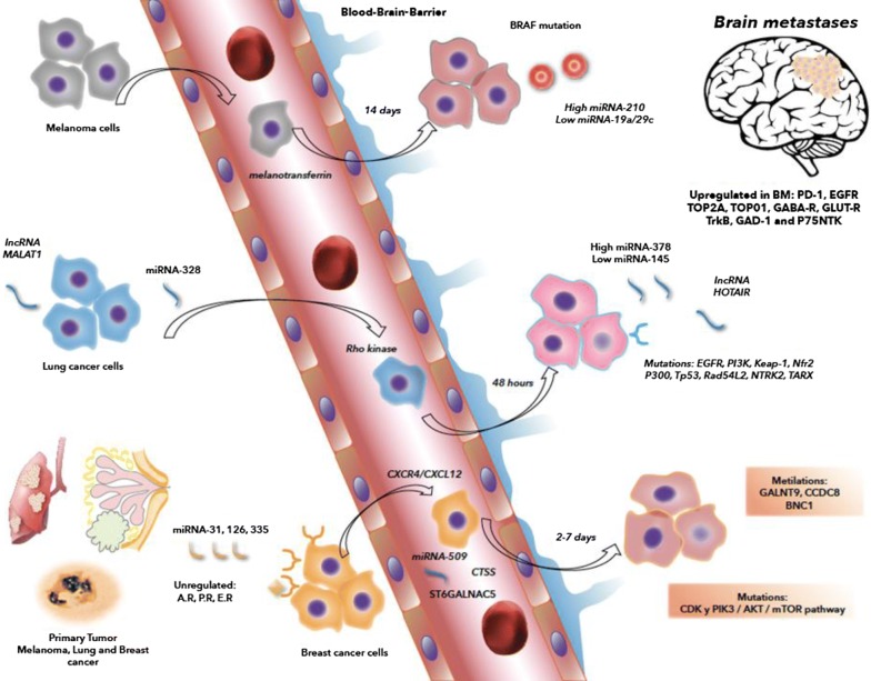
The present review aimed to discuss contemporary scientific literature involving differences between the tumor microenvironment (TME) in melanoma, lung cancer, and breast cancer in their primary site and TME in brain metastases (BM). TME plays a fundamental role in the behavior of cancer. In the process of carcinogenesis, cells such as fibroblasts, macrophages, endothelial cells, natural killer cells, and other cells can perpetuate and progress carcinogenesis via the secretion of molecules. Oxygen concentration, growth factors, and receptors in TME initiate angiogenesis and are examples of the importance of microenvironmental conditions in the performance of neoplastic cells. The most frequent malignant brain tumors are metastatic in origin and primarily originate from lung cancer, breast cancer, and melanoma. Metastatic cancer cells have to adhere to and penetrate the blood-brain barrier (BBB). After traversing BBB, these cells have to survive by producing various cytokines, chemokines, and mediators to modify their new TME. The microenvironment of these metastases is currently being studied owing to the discovery of new therapeutic targets. In these three types of tumors, treatment is more effective in the primary tumor than in BM due to several factors, including BBB. Understanding the differences in the characteristics of the microenvironment surrounding the primary tumor and their respective metastasis might help improve strategies to comprehend cancer.
Keywords: Brain metastases; Cancer; Tumor microenvironment.
Conflict of interest statement
The authors declare that they have no competing interests.
Figures
Similar articles
-
The Microenvironment of Lung Cancer and Therapeutic Implications.Adv Exp Med Biol. 2016;890:75-110. doi: 10.1007/978-3-319-24932-2_5. Adv Exp Med Biol. 2016. PMID: 26703800 Review.
-
Matrix Metalloproteinases' Role in Tumor Microenvironment.Adv Exp Med Biol. 2020;1245:97-131. doi: 10.1007/978-3-030-40146-7_5. Adv Exp Med Biol. 2020. PMID: 32266655 Review.
-
Angiogenesis and the tumor microenvironment: vascular endothelial growth factor and beyond.Semin Oncol. 2014 Apr;41(2):235-51. doi: 10.1053/j.seminoncol.2014.02.007. Epub 2014 Feb 28. Semin Oncol. 2014. PMID: 24787295 Review.
-
Microenvironmental Heterogeneity in Brain Malignancies.Front Immunol. 2019 Oct 1;10:2294. doi: 10.3389/fimmu.2019.02294. eCollection 2019. Front Immunol. 2019. PMID: 31632393 Free PMC article. Review.
-
Single-cell sequencing reveals the landscape of the human brain metastatic microenvironment.Commun Biol. 2023 Jul 21;6(1):760. doi: 10.1038/s42003-023-05124-2. Commun Biol. 2023. PMID: 37479733 Free PMC article.
Cited by
-
The Role of Tumor Microenvironment in Cancer Metastasis: Molecular Mechanisms and Therapeutic Opportunities.Cancers (Basel). 2021 Apr 23;13(9):2053. doi: 10.3390/cancers13092053. Cancers (Basel). 2021. PMID: 33922795 Free PMC article. Review.
-
Therapeutic Opportunities for Biomarkers in Metastatic Spine Tumors.Cancers (Basel). 2024 Sep 14;16(18):3152. doi: 10.3390/cancers16183152. Cancers (Basel). 2024. PMID: 39335124 Free PMC article. Review.
-
Ultrasound-mediated disruption of the blood tumor barrier for improved therapeutic delivery.Neoplasia. 2021 Jul;23(7):676-691. doi: 10.1016/j.neo.2021.04.005. Epub 2021 Jun 14. Neoplasia. 2021. PMID: 34139452 Free PMC article. Review.
-
Exosome proteomes reveal glycolysis-related enzyme enrichment in primary canine mammary gland tumor compared to metastases.Proteome Sci. 2024 Feb 28;22(1):4. doi: 10.1186/s12953-023-00226-5. Proteome Sci. 2024. PMID: 38419074 Free PMC article.
-
Neoadjuvant immune checkpoint inhibition in the management of glioblastoma: Exploring a new frontier.Front Immunol. 2023 Feb 17;14:1057567. doi: 10.3389/fimmu.2023.1057567. eCollection 2023. Front Immunol. 2023. PMID: 36875096 Free PMC article. Review.
References
-
- Wei SC, Duffy CR, Allison JP. Fundamental mechanisms of immune checkpoint blockade therapy. Cancer Discov. 2018;8(9):1069–1086. doi: 10.1158/2159-8290.CD-18-0367. - DOI - PubMed
Publication types
MeSH terms
LinkOut - more resources
Full Text Sources
Medical
