

Apoptosis and autophagy in polycystic kidney disease (PKD)
- PMID: 31881325
- PMCID: PMC7127985
- DOI: 10.1016/j.cellsig.2019.109518
Apoptosis and autophagy in polycystic kidney disease (PKD)
Abstract
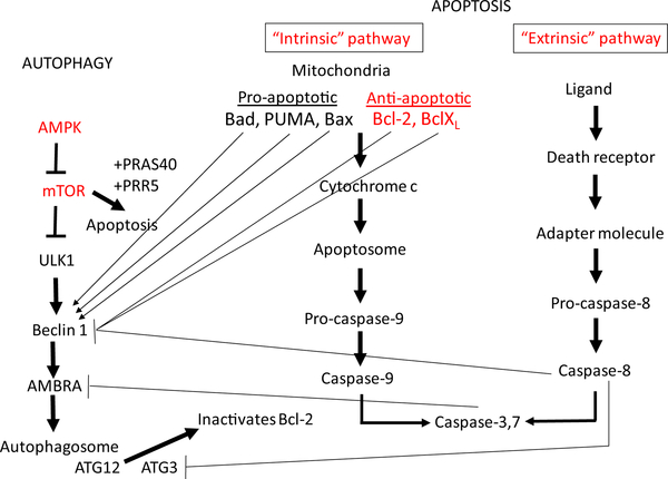
Apoptosis in the cystic epithelium is observed in most rodent models of polycystic kidney disease (PKD) and in human autosomal dominant PKD (ADPKD). Apoptosis inhibition decreases cyst growth, whereas induction of apoptosis in the kidney of Bcl-2 deficient mice increases proliferation of the tubular epithelium and subsequent cyst formation. However, alternative evidence indicates that both induction of apoptosis as well as increased overall rates of apoptosis are associated with decreased cyst growth. Autophagic flux is suppressed in cell, zebra fish and mouse models of PKD and suppressed autophagy is known to be associated with increased apoptosis. There may be a link between apoptosis and autophagy in PKD. The mammalian target of rapamycin (mTOR), B-cell lymphoma 2 (Bcl-2) and caspase pathways that are known to be dysregulated in PKD, are also known to regulate both autophagy and apoptosis. Induction of autophagy in cell and zebrafish models of PKD results in suppression of apoptosis and reduced cyst growth supporting the hypothesis autophagy induction may have a therapeutic role in decreasing cyst growth, perhaps by decreasing apoptosis and proliferation in PKD. Future research is needed to evaluate the effects of direct autophagy inducers on apoptosis in rodent PKD models, as well as the cause and effect relationship between autophagy, apoptosis and cyst growth in PKD.
Keywords: Apoptosis; Autophagy; Polycystic kidney disease.
Copyright © 2020 Elsevier Inc. All rights reserved.
Figures
Similar articles
-
What is the role of tubular epithelial cell apoptosis in polycystic kidney disease (PKD)?Cell Cycle. 2005 Nov;4(11):1550-4. doi: 10.4161/cc.4.11.2185. Epub 2005 Nov 17. Cell Cycle. 2005. PMID: 16258272 Review.
-
Autophagy induction promotes renal cyst growth in polycystic kidney disease.EBioMedicine. 2020 Oct;60:102986. doi: 10.1016/j.ebiom.2020.102986. Epub 2020 Sep 16. EBioMedicine. 2020. PMID: 32949996 Free PMC article.
-
Polycystic kidney disease: a case of suppressed autophagy?Semin Nephrol. 2014 Jan;34(1):27-33. doi: 10.1016/j.semnephrol.2013.11.005. Epub 2013 Nov 22. Semin Nephrol. 2014. PMID: 24485027 Review.
-
Apoptosis in polycystic kidney disease: involvement of caspases.Am J Physiol Regul Integr Comp Physiol. 2000 Mar;278(3):R763-9. doi: 10.1152/ajpregu.2000.278.3.R763. Am J Physiol Regul Integr Comp Physiol. 2000. PMID: 10712299
-
Caspase inhibition reduces tubular apoptosis and proliferation and slows disease progression in polycystic kidney disease.Proc Natl Acad Sci U S A. 2005 May 10;102(19):6954-9. doi: 10.1073/pnas.0408518102. Epub 2005 Apr 29. Proc Natl Acad Sci U S A. 2005. PMID: 15863619 Free PMC article.
Cited by
-
Identification and Validation of the miR/RAS/RUNX2 Autophagy Regulatory Network in AngII-Induced Hypertensive Nephropathy in MPC5 Cells Treated with Hydrogen Sulfide Donors.Antioxidants (Basel). 2024 Aug 7;13(8):958. doi: 10.3390/antiox13080958. Antioxidants (Basel). 2024. PMID: 39199205 Free PMC article.
-
Defects of renal tubular homeostasis and cystogenesis in the Pkhd1 knockout.iScience. 2024 Mar 11;27(4):109487. doi: 10.1016/j.isci.2024.109487. eCollection 2024 Apr 19. iScience. 2024. PMID: 38550996 Free PMC article.
-
Non-Coding RNAs in Hereditary Kidney Disorders.Int J Mol Sci. 2021 Mar 16;22(6):3014. doi: 10.3390/ijms22063014. Int J Mol Sci. 2021. PMID: 33809516 Free PMC article. Review.
-
Long-term Cu exposure alters CYP450s activity and induces jejunum injury and apoptosis in broilers.Biometals. 2024 Apr;37(2):421-432. doi: 10.1007/s10534-023-00559-w. Epub 2023 Nov 22. Biometals. 2024. PMID: 37991682
-
Protective impact of Spirulina platensis against γ-irradiation and thioacetamide-induced nephrotoxicity in rats mediated by regulation of micro-RNA 1 and micro-RNA 146a.Toxicol Res (Camb). 2021 Apr 28;10(3):453-466. doi: 10.1093/toxres/tfab037. eCollection 2021 May. Toxicol Res (Camb). 2021. PMID: 34141159 Free PMC article.
References
Publication types
MeSH terms
Substances
Grants and funding
LinkOut - more resources
Full Text Sources
Miscellaneous
